


Abstract
Background: The fact that lateral malleolar fracture is accompanied by posterior malleolar fracture may adversely affect syndesmosis malreduction rates. We aimed to compare syndesmosis malreduction rates determined on postoperative radiographs between isolated lateral malleolar fractures and lateral malleolar fractures accompanied by posterior malleolar fractures. Methods: We retrospectively examined 128 operative patients: 73 with isolated lateral malleolar fractures (group L) and 55 with lateral + posterior malleolar fractures (group LP). In group LP, no patients received posterior fragment fixation. In both groups, indirect syndesmosis fixation was performed with a single screw after open reduction and internal fixation of the lateral malleolus. Patient age, sex, fracture side, fracture type (Lauge-Hansen and Danis-Weber classifications), Kellgren-Lawrence osteoarthritis classification, syndesmotic incongruency on postoperative radiographs, syndesmotic malreduction of postoperative fibula fracture, fracture union time, complication rates, accompanying injuries, and preoperative and postoperative radiographic syndesmotic measurements (tibiofibular overlap, tibiofibular clear space, medial clear space) were recorded, and the groups were compared. Results: Mean ± SD age was 44.32 ± 15.66 years in group L and 48.93 ± 14.03 years in group LP (P = .087). There were no significant differences in preoperative and postoperative tibiofibular distance, tibiofibular overlap, and medial clear space values between groups (P > .05). The prevalence of grade 2 fractures according to the Kellgren-Lawrence classification was significantly higher in group LP (P = .047). Postoperative syndesmosis malreduction was detected in 12 patients in group L and in nine in group LP (P = .991). Conclusions: In lateral malleolar fractures accompanied by small-fragment posterolateral or avulsion-type posterior malleolar fractures, closed syndesmotic screw fixation does not cause syndesmosis malreduction.
Posterior malleolar fractures accompany nearly 46% of Danis-Weber type B and C lateral malleolar fractures.[1] The effect of posterior malleolar fragment fixation on syndesmosis stability and reduction of the distal fibula to the fibular notch has already been shown.[2,3,4] It is thought that the posterior inferior tibiofibular ligament (PITFL) contributes highly to the stability of the ankle syndesmosis.[2] Clanton et al,[5] in a biomechanical analysis of individual syndesmotic ligaments, showed that the PITFL is an important structure to control internal rotation. Because the PITFL is attached to the primary fracture fragment, when the posterior fragment is fixed by reduction, a proportion of these fractures may require no further syndesmotic stabilization.[6] Furthermore, some studies suggest that the fixation of the posterior malleolar fragment provides greater stability than a syndesmotic screw.[2,4]
In the literature, syndesmotic incongruences determined on postoperative imaging are a problem that may be seen after lateral malleolar fracture surgery.[7,8,9] In most patients with postoperative syndesmosis malreduction, the fibula is in internal rotation into the incisura.[10]
The indications for open reduction and internal fixation of posterior malleolar fractures are controversial.[1] Recent studies have emphasized the importance of fixation of posterior fracture fragments by considering the size of the posterior fragment as well as whether the fracture extends to the fibula incisura, whether it involves the medial malleolus, and syndesmotic stability.[1,2,3,4,6,9] However, these studies are biomechanical studies or studies where the posterior fragment has been fixed in all cases.[2,4] There is not a sufficient number of clinical studies to demonstrate the efficacy of posterior malleolar fracture fixation on syndesmosis repair.
In light of this information, it may be concluded that in the presence of posterior malleolar fracture accompanying lateral malleolar fracture, if the syndesmotic screw is applied without fixing the posterior malleolus, the PITFL will have no effect on posterior syndesmotic stability during syndesmotic reduction with a malleolar clamp and, thus, the fibula may have a higher tendency to be in internal rotation into the incisura. Therefore, this study aimed to compare the syndesmosis malreduction rates determined on postoperative radiographs between isolated lateral malleolar fractures and lateral malleolar fractures accompanied by posterior malleolar fractures. The results of this study suggest that postoperative syndesmotic incongruences may be higher in cases in which posterior malleolar fracture cannot be determined.
Patients and Methods
The files of 269 patients who were operated on owing to ankle fracture between January 1, 2011, and December 31, 2020, were examined retrospectively. In our clinic, posterior fragment fixation is applied to cases whose posterior fracture fragment size is greater than 25%, those with fracture displacement greater than 2 mm, those with a displaced fracture involving the fibular incisura, and those with a posterior malleolar fracture involving the medial malleolus. In total, 128 adult patients were included in the study. Pediatric patients, patients who underwent a two-stage surgical procedure, those with injuries accompanied by a medial malleolar fracture, those in whom fixation has been applied to the posterior malleolus, and those whose preoperative computed tomographic (CT) images could not be obtained were not included in the study. Sixty-two patients who were operated on owing to bimalleolar fracture (medial + lateral malleolar), 37 patients who were operated on owing to trimalleolar fracture, 30 patients with lateral + posterior malleolar fracture with the use of a plate for fixing the posterior malleolus, and 12 patients with nonremoved syndesmotic screws were excluded. Consequently, 73 patients with isolated lateral malleolar fractures (group L) and 55 patients with lateral + posterior malleolar fractures (group LP) were examined.
Based on the preoperative CT images, the cases were categorized by two surgeons (A.Y. and M.Y.) according to the Lauge-Hansen classification. In the case of disagreement between the two surgeons, a decision was made after examination of the images by two additional observers (A.M. and T.O.B.).[11] Cases for which the surgeons could not make a joint decision were not included in the study.
All of the patients were operated on by a lateral approach in the supine position. In all of the patients, an anatomical lateral distal fibula plate manufactured by Acumed LLC (Hillsboro, Oregon) was used. The syndesmosis reduction was performed with a malleolar clamp with the ankle in 10° of dorsiflexion in patients with incongruences in the external rotation test under fluoroscopy after fracture fixation. The fibula and the tibia were drilled using a 3-mm drill leaving a 30° angle from posterior to anterior in parallel with the joint. Syndesmosis fixation was performed by inserting a 3.5-mm partially threaded syndesmotic screw through the appropriate unlocked screw hole 2 to 5 cm proximal to the joint level over the plate. In all of the patients, closed syndesmosis reduction was performed under fluoroscopy. Then, the operation was ended by checking the stability of the syndesmosis with external rotation testing. In all of the cases, the syndesmotic screw engaged three cortices, and a single syndesmotic screw was used. The patients were allowed nonweightbearing walking for up to 6 weeks after the operation; then they were allowed weightbearing as tolerated. Full weightbearing was allowed by gradually increasing the weight. In patients who completed 12 weeks of follow-up after the operation, the syndesmotic screw was removed. The patients were operated on by four different surgeons (A.Ç.T., S.S.D., Y.İ. and H.G.). The surgeons had more than 5 years of experience in trauma surgery. The number of ankle fracture operations performed was more than 20 per year.
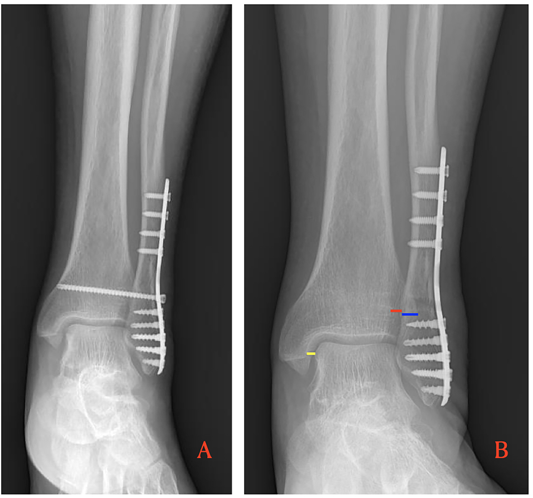
The preoperative fluoroscopic images and postoperative radiographs were evaluated based on the criteria of tibiofibular clear space (TCS) (reference range, <6 mm), anteroposterior tibiofibular overlap (TFO) (reference range, >6 mm), and medial clear space (MCS) (reference range, <5 mm) (Figure 1).[7]
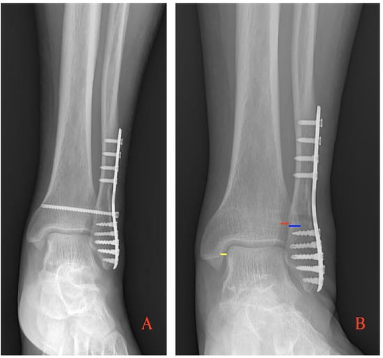
Figure 1.
A, Ankle anteroposterior radiograph of a 34-year-old man with lateral malleolus fracture. B, Measurements on an ankle anteroposterior radiograph of a 34-year-old man who was operated on for lateral malleolus fracture. Medial clear space is shown with a yellow line, tibiofibular overlap with a blue line, and tibiofibular clear space with a red line.
The monitoring software INFINITT PACS (Picture Archiving and Communication Systems) Version 3.0.11.4(BN13) (INFINITT North America Inc, Phillipsburg, New Jersey), which was available in our hospital, was used for the postoperative radiographic measurements. The measurements were performed by two experienced orthopedic surgeons (A.Y. and M.Y.) who did not perform the operations, and then they were averaged. The numerical data obtained based on these measurements were used as data in statistical calculations. The radiographic measurements were performed based on the radiographs taken during the polyclinic follow-up in postoperative month 6. The Kellgren-Lawrence osteoarthritis classification was made based on the radiographs in postoperative year 1.[12] All of the radiographs were taken after the syndesmotic screw was removed.
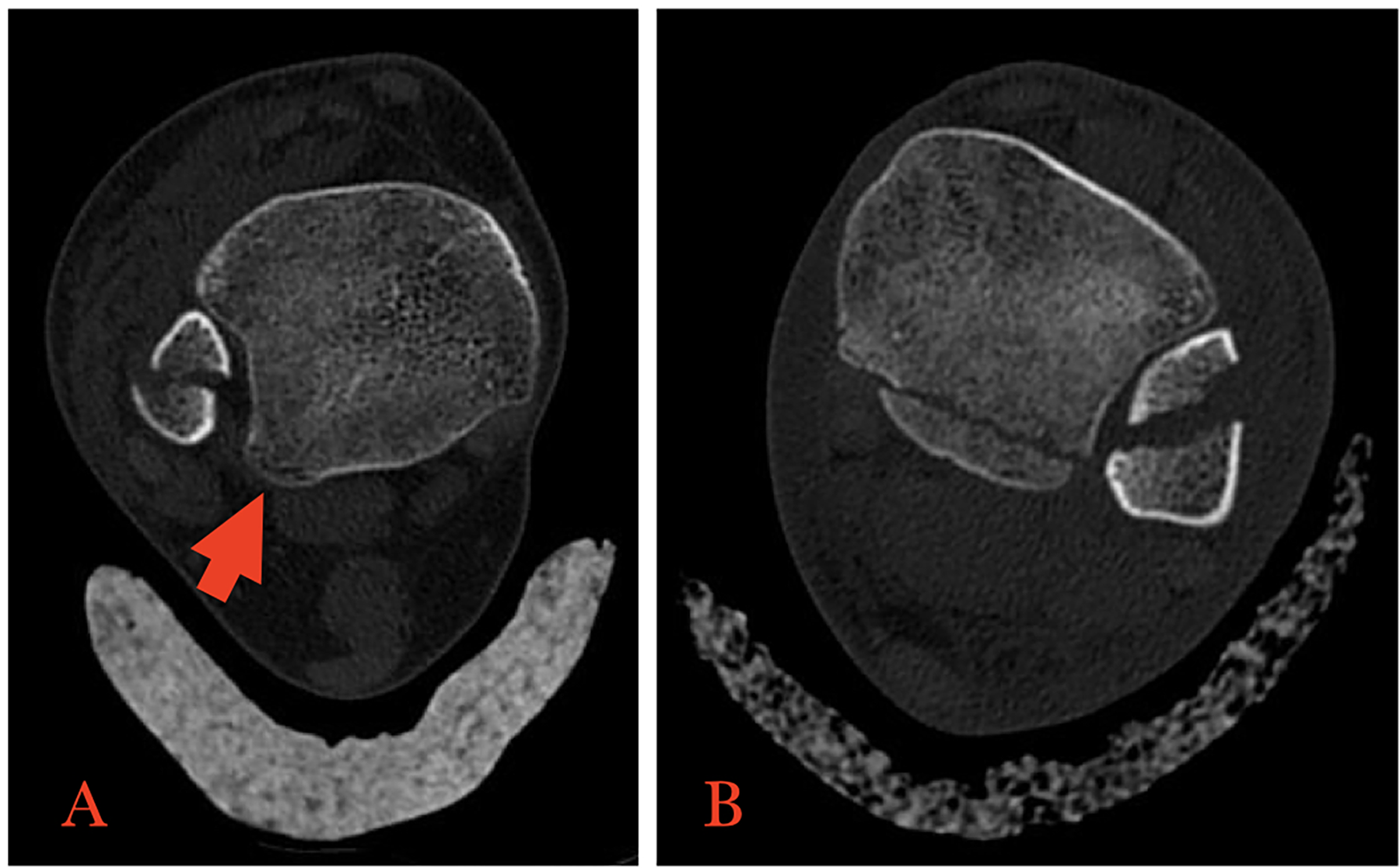
When classifying the posterior malleolar fractures according to the CT images, the Mason and Molloy classification was used.[6] According to the Mason and Molloy classification, 50 patients had type 1 (posterior avulsion) and five had type 2A (posterolateral) fractures (Figure 2). In all of the patients, the posterior fracture fragment involved less than 25% of the distal tibia. There was no displacement greater than 2 mm and no large posterolateral fracture fragment involving more than one-third of the incisura fibula.
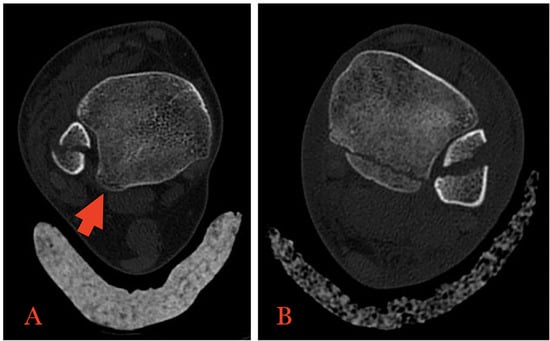
Figure 2.
Axial computed tomographic images of a 29-year-old woman with a type 1 posterior malleolar fracture (arrow) (A) and a 20-year-old man with a type 2A posterior malleolus fracture (B).
Patient age, sex, fracture side, fracture type according to the Lauge-Hansen and Danis-Weber classifications, Kellgren-Lawrence osteoarthritis classification, syndesmotic incongruency in postoperative radiographs, syndesmotic malreduction of postoperative fibula fracture, fracture union time, complication rates, accompanying injuries, and preoperative and postoperative radiographic syndesmotic measurements (TFO, TCS, MCS) were recorded, and comparisons between groups were made.
As a descriptive statistic, mean ± SD values are given for the numeric variables and number and percentage values are given for the categorical variables. The conformity of the data with normal distribution was tested with the Shapiro-Wilk test; the Student t test was used to compare the parameters with normal distribution between groups L and LP, and the Mann-Whitney U test was used for the parameters without normal distribution between groups L and LP. The relationships of categorical variables were analyzed by the Pearson and exact χ2 tests. The statistical analyses were performed using IBM SPSS Statistics for Windows, Version 23.0 (IBM Corp, Armonk, New York), and a result with P < .05 was considered statistically significant.
Results
The mean ± SD age of patients in group L was 44.32 ± 15.66 years and of those in group LP was 48.93 ± 14.03 years (P = .087). In group L there were 36 men (49.3%) and 37 women (50.7%); in group LP there were 22 men (40%) and 33 women (60%) (P = .295). The distribution of cases by the Lauge-Hansen, Danis-Weber, and Kellgren-Lawrence classifications and the distribution of fracture sides by group are summarized in Table 1. It was seen that the prevalence of grade 2 fractures according to the Kellgren-Lawrence classification was significantly higher in group LP (P = .047).

Table 1.
Distribution of Fracture Side, Sex, and Radiologic Classifications by Group
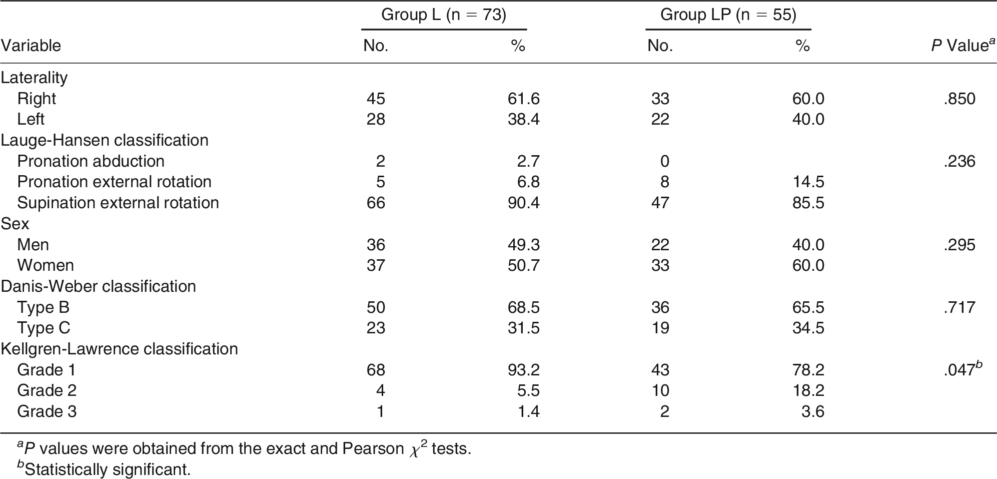
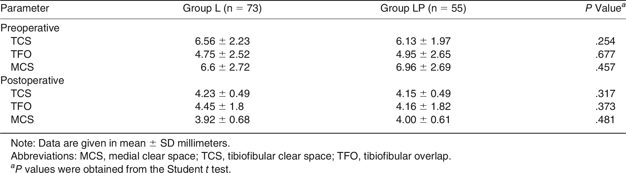
There were no statistically significant differences in preoperative tibiofibular distance, TFO, and MCS and postoperative tibiofibular distance, TFO, and MCS values between the two groups (Table 2).
Table 2.
Preoperative and Postoperative Radiologic Measurement Parameters
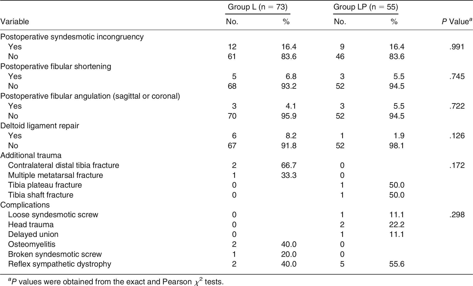
Rates of postoperative fibular shortening, postoperative fibular angulation, deltoid ligament repair, postoperative syndesmotic incongruence, postoperative MCS impairment, additional trauma, and postoperative complications were similar between the two groups (Table 3). The mean ± SD fracture union time was 7.33 ± 1.97 weeks in group L and 7.62 ± 1.47 weeks in group LP (P = .362).
Table 3.
Comparison of Qualitative Variables Between Groups
There was no significant difference in syndesmotic screw removal time between group L (13 weeks [range, 12–20 weeks]) and group LP (13 weeks [range, 12–19 weeks]) (P = .872).
Discussion
The effect of posterior malleolar fracture fixation on repair of the fibular incisura and syndesmotic stability has been emphasized in recent studies.[1,2,3,4,6] There are few studies on postoperative syndesmotic malreduction or stability of the posterior malleolar fracture. In these studies, the positive effects of the fixation of posterior malleolar fracture on syndesmosis were discussed, and fixation was recommended. However, these suggestions were based only on the increased rates of syndesmotic malreduction accompanying posterior malleolar fracture on postoperative CT, studies where the posterior malleolar fracture was determined in all of the cases, or biomechanical studies.[2,4,7,10] Also, the size and characteristics of the posterior malleolar fracture were not considered in these studies.[4,10] There is no comparative study showing that failure to fix the posterior malleolar fracture resulted in increased rates of syndesmotic malreduction. The present study is different in this respect because the presence of nonfixed posterior malleolar fracture caused no increase in postoperative syndesmotic malreduction rates. This was different from the findings of a few studies in the literature. On the other hand, the present study included avulsion fracture of the posterior malleolus or posterolateral fractures involving less than one-third of the fibular incisura. Making a fixation decision according to the size and characteristics of the posterior malleolus fragment may be a correct method in terms of its effect on the reduction of the postoperative syndesmosis.
Up to 15% of postoperative syndesmosis malreductions can be detected by direct radiography.[10] Syndesmosis malreduction may occur depending on the position of the malleolar clamp or overcompression during the closed reduction of syndesmosis.[13,14,15] Also, in most postoperative syndesmotic incongruences, the fibula is in internal rotation.[4,7,10] So, the presence of posterior malleolar fracture means that the posterior syndesmotic stability has been impaired. Thus, when performing syndesmosis reduction with a malleolar clamp, the fibula may move into internal rotation. Moreover, overcompression with a clamp may displace the posterior malleolar fracture, causing an increase in postoperative syndesmotic malreductions. However, the present study results suggest that there was no increase in syndesmotic malreduction rates according to the postoperative radiographs after closed reduction and screw fixation of syndesmosis in patients with a posterior malleolar fracture.
Open reduction and internal fixation of the displaced posterior fragment in ankle fractures restores the joint surface of the distal tibia, the stability of the tibiotalar joint, and the integrity of the fibular notch. It also facilitates reduction of the distal fibula in the fibular notch and restores the stability of tibiofibular syndesmosis.[1] The posterior tubercle of the distal tibia also functions as the strong superior, obliquely oriented fibers of the posterior tibiofibular ligament in the shape of a trapezoid. The inferiorly and more horizontally oriented fibers of the PITFL originate from the edge of the joint surface of the distal tibia, and medial to the posterior tubercle. The upper and lower fibers of the ligament merge at distal toward the posterior fibula, and these fibers attach to the malleolar fossa.[16] Gardner et al[2] suggested that the PIFTL was intact after the posterior malleolar fracture in the magnetic resonance images, and the syndesmosis stability was higher after posterior fixation compared with indirect reduction using a syndesmotic screw. Another study by the same authors reported that 44% of poorly reduced syndesmosis was associated with a nonfixed posterior malleolar fracture.[7] A cadaver study by Jayatilaka et al[17] reported that the PITFL attached to significantly greater and wider areas compared with the posterior malleolar fracture fragments of average size. Therefore, they suggested that a posterior malleolar fracture can cause posterior syndesmotic instability only in the presence of a ligamentous injury.[17] The reason why syndesmotic malreduction rates were not higher when the posterior malleolar fracture was not fixed in the present study may be the fact that the PITFL injury was independent from the posterior fracture fragment, as suggested by Jayatilaka et al.[17]
Miller et al[4] did not find a statistically significant difference in postoperative radiographic measurements between the group that received posterior malleolar fixation and the group that received screw fixation for syndesmosis injury with the posterior malleolus intact. Only in the latter group, the postoperative TCS had a higher average. They suggested that posterior malleolar fixation causes syndesmosis stability as much as syndesmotic screw fixation, and similar functional and radiologic results. However, in this study, the size and characteristics of the fixed posterior fragment were not considered. Also, the comparison groups were too small. In the present study, there was no statistically significant difference in postoperative TCS measurement results between the cases where the posterior malleolus was intact and those where the posterior malleolus was fractured. Even the group with the posterior malleolus intact had a lower average value. However, we think that this difference of less than 1 mm is a minimal difference that might even result from measurement errors.
Miller et al[10] found no statistically significant difference in syndesmotic malreduction rates between the group where the posterior malleolar fracture was fixed (regardless of its size) and the group where the syndesmosis was repaired using screws by directly seeing that the posterior malleolus was intact, based on the postoperative CT images. They suggested that the fibula may rotate from articulation to posterior without any fixing support at the posterior incisura, and plain radiography may fail to determine this. In the example picture that they added for this suggestion in their study, there was an intramedullary tibial nail. The syndesmotic screw was further posterior of the tibia as it should be due to the nail and passed from the fracture line at posterior. Also, they did not consider the size of the posterior malleolar fracture.[10] It may be difficult to detect syndesmotic malreductions in external rotation by direct radiography.[18] However, most syndesmotic malreductions are seen in internal rotation. In cases in which the posterior fragment has not been displaced, if the fracture line corresponds to the insertion zone of the syndesmotic screw, the fibula may be fixed in external rotation, displacing the posterior fragment while applying a drill or screw. This may not be detected on plain radiographs. In large posterior fragments, the fracture line may extend to the middle one-third region of the incisura (the region through which the syndesmotic screw should pass). This may mean that the syndesmotic screw passes through the fracture line, displacing the posterior fragment, and the incisura structure is more likely to be impaired (the fibula is more likely to be in external rotation). On the other hand, in small-fragment fractures, where the posterior malleolar fracture does not include the incisura or includes only a small portion of the incisura, external rotation may not cause an increase in syndesmotic malreductions.
There is evidence that the presence of a posterior malleolar fragment has an adverse effect on postoperative outcomes.[19] In the present study, although there was no difference in syndesmotic malreductions, stage 2 arthrosis findings were significantly higher than in the group without posterior malleolar fracture in the first year according to the Kellgren-Lawrence classification. This may be interpreted as meaning that the presence of posterior malleolar fracture, regardless of syndesmosis malreduction, may be a precursor of increased ankle arthrosis and poor functional outcomes in the long term.
Tricortical screws are less tight in the tibia than quadricortical screws. Therefore, it is thought that they slowly loosen as the weight is increased. This theoretically leads to less interference and less screw breaking during micromotions that normally occur in weightbearing syndesmosis.[20,21] Less rigid syndesmotic fixation can activate micromotions that could theoretically lead to a shorter healing time and reduced stiffness.[20] Syndesmotic screws may restrict the normal movement between the tibia and the fibula. Removal, breakage, or loosening of the screw serves to restore motion; however, if it occurs before the ligamentous healing, it may result in the loss of syndesmotic reduction.[22] The presence of a syndesmotic screw may cause an increase in postoperative syndesmotic incongruences. Removal of the screw may contribute to a decrease in syndesmotic malreduction rates.[23] In the present study, a tricortical screw was used in all of the cases, and, thus, radiographic measurements were made after the syndesmotic screw was removed. The fact that the measurement results were similar between the two groups may indicate that sufficient time has been given for healing of the posterior malleolar fracture as well as syndesmotic healing until the syndesmotic screw was removed, or that an accompanying posterior malleolar fracture had no adverse effect on syndesmosis healing time.
Postoperative CT is more valuable to show postoperative syndesmotic incongruences.[7] This may mean that the information on postoperative minimal syndesmotic incongruences in the present study is insufficient. However, the effect of these postoperative minimal incongruences on functional results as determined in the studies on CT is unclear.[7,8,24,25] Plain radiography has some advantages, such as being easily accessible, fast, and cheap.[26] The shortcomings of the present study include the possibility of failure to detect syndesmotic malreductions in external rotation by direct radiography[18] and the fact that these syndesmotic malreductions could not be shown with CT. However, the present study was retrospective. Also, postoperative CT could not be performed routinely for every patient for financial reasons and concerns about radiation exposure. In most patients with a postoperative CT image, CT was performed because of a clinical complaint or symptom. Because the evaluation of only postoperative CT images will cause a bias, we evaluated only the postoperative roentgenograms in the present study. Other shortcomings of this study may include its retrospective nature; the lack of a third group to which posterior fixation was applied (because in all of the cases in which the posterior malleolar fracture was fixed the posterior fragment was >25%, involved more than one-third of the incisura, and included the medial malleolus or multipart fractures, this group was not comparable with the other two groups); the inclusion of only radiographic parameters; and not providing information about functional results.
Conclusions
In lateral malleolar fractures accompanied by small-fragment posterolateral or avulsion-type posterior malleolar fractures, the closed syndesmosis fixation with screws alone is not associated with an increased syndesmosis malreduction rate compared with isolated lateral malleolar fixation.
Financial Disclosure
None reported.
Conflicts of Interest
None reported.
References
- Bartoníček J, Rammelt S, Kostlivý K, et al: Anatomy and classification of the posterior tibial fragment in ankle fractures. Arch Orthop Trauma Surg 135: 505, 2015.
- Gardner MJ, Brodsky A, Briggs SM, et al: Fixation of posterior malleolar fractures provides greater syndesmotic stability. Clin Orthop Relat Res 447: 165, 2006.
- Bartoníček J, Rammelt S, Tuček M, et al: Posterior malleolar fractures of the ankle. Eur J Trauma Emerg Surg 41: 587, 2015.
- Miller AN, Carroll EA, Parker RJ, et al: Posterior malleolar stabilization of syndesmotic injuries is equivalent to screw fixation. Clin Orthop Relat Res 468: 1129, 2010.
- Clanton TO, Williams BT, Backus JD, et al: Biomechanical analysis of the ındividual ligament contributions to syndesmotic stability. Foot Ankle Int 38: 66, 2017.
- Mason LW, Marlow WJ, Widnall J, et al: Pathoanatomy and associated ınjuries of posterior malleolus fracture of the ankle. Foot Ankle Int 38: 1229, 2017.
- Gardner MJ, Demetrakopoulos D, Briggs SM, et al: Malreduction of the tibiofibular syndesmosis in ankle fractures. Foot Ankle Int 27: 788, 2006.
- Hovis WD, Kaiser BW, Watson JT, et al: Treatment of syndesmotic disruptions of the ankle with bioabsorbable screw fixation. J Bone Joint Surg Am 84: 26, 2002.
- Weening B, Bhandari M: Predictors of functional outcome following transsyndesmotic screw fixation of ankle fractures. J Orthop Trauma 19: 102, 2005.
- Miller AN, Carroll EA, Parker RJ, et al: Direct visualization for syndesmotic stabilization of ankle fractures. Foot Ankle Int 30: 419, 2009.
- Warner SJ, Garner MR, Hinds RM, et al: Correlation between the Lauge-Hansen classification and ligament ınjuries in ankle fractures. J OrthopTrauma 29: 574, 2015.
- Holzer N, Salvo D, Marijnissen ACA, et al: Radiographic evaluation of posttraumatic osteoarthritis of the ankle: the Kellgren-Lawrence scale is reliable and correlates with clinical symptoms. Osteoarthritis Cartilage 23: 363, 2015.
- Miller AN, Barei DP, Iaquinto JM, et al: Iatrogenic syndesmosis malreduction via clamp and screw placement. J Orthop Trauma 27: 100, 2013.
- Kennedy MT, Carmody O, Leong S, et al: A computed tomography evaluation of two hundred normal ankles, to ascertain what anatomical landmarks to use when compressing or placing an ankle syndesmosis screw. Foot (Edinb) 24: 157, 2014.
- Tornetta P III, Spoo JE, Reynolds FA, et al: Overtightening of the ankle syndesmosis: is it really possible? J Bone Joint Surg Am 83: 489, 2001.
- Bartoníček J, Rammelt S, Tuček M: Posterior malleolar fractures: changing concepts and recent developments. Foot Ankle Clin 22: 125, 2017.
- Jayatilaka MLT, Philpott MDG, Fisher A, et al: Anatomy of the insertion of the posterior inferior tibiofibular ligament and the posterior malleolar fracture. Foot Ankle Int 40: 1319, 2019.
- Marmor M, Hansen E, Han HK, et al: Limitations of standard fluoroscopy in detecting rotational malreduction of the syndesmosis in an ankle fracture model. Foot Ankle Int 32: 616, 20111.
- Bali N, Aktselis I, Ramasamy A, et al: An evolution in the management of fractures of the ankle: safety and efficacy of posteromedial approach for Haraguchi type 2 posterior malleolar fractures. Bone Joint J 99-B: 1496, 2017.
- Bauer AS, Bluman EM, Wilson MG, et al: Injuries of the distal lower extremity syndesmosis. Curr Orthop Pract 20: 111, 2009.
- Moore JA Jr, Shank JR, Morgan SJ, et al: Syndesmosis fixation: a comparison of three and four cortices of screw fixation without hardware removal. Foot Ankle Int 27: 567, 2006.
- Manjoo A, Sanders DW, Tieszer C, et al: Functional and radiographic results of patients with syndesmotic screw fixation: implications for screw removal. J Orthop Trauma 24: 2, 2010.
- Song DJ, Lanzi JT, Groth AT, et al: The effect of syndesmosis screw removal on the reduction of the distal tibiofibular joint: a prospective radiographic study. Foot Ankle Int 35: 543, 2014.
- Weening B, Bhandari M: Predictors of functional outcome following transsyndesmotic screw fixation of ankle fractures. J Orthop Trauma 19: 102, 2005.
- Warner SJ, Fabricant PD, Garner MR, et al: The measurement and clinical importance of syndesmotic reduction after operative fixation of rotational ankle fractures. J Bone Joint Surg Am 97: 1935, 2015.
- Rasi AM, Kazemian G, Omidian MM, et al: Syndesmotic malreduction after ankle ORIF; is radiography sufficient? Arch Bone Joint Surg 1: 98, 2013.
© 2023 American Podiatric Medical Association