


Abstract
Background: Diabetes-related peripheral neuropathy (DPN) and limited joint mobility of the foot and ankle are implicated in the development of increased plantar pressures and diabetes-related foot ulcers. The extent of this relationship has not been conclusively established. We aimed to determine the relationship between ankle joint and first metatarsophalangeal joint dorsiflexion range of motion and DPN using a cross-sectional observational study design. Methods: Primary outcomes were DPN status, ankle joint range of motion (extended and flexed knee lunge tests), and nonweightbearing first metatarsophalangeal joint range of motion. Correlations were performed using Pearson r, and hierarchical regression analyses were undertaken to determine the independent contribution of DPN to the variance in dorsiflexion range of motion of ankle and first metatarsophalangeal joints using standardized β regression coefficients, controlling for age, sex, body mass index, diabetes duration, and hemoglobin A1c level. Results: One hundred one community-dwelling participants (mean ± SD age, 65.0 ± 11.2 years; 55 men; 97% type 2 diabetes; mean ± SD diabetes duration, 8.7 ± 7.8 years; 23% with DPN) were recruited. Diabetes-related peripheral neuropathy demonstrated significant correlations with reduced range of motion at the ankle joint (knee extended: r = –0.53; P < .001 and knee flexed: r = –0.50; P < .001) and the first metatarsophalangeal joint (r = –0.37; P < .001). Also, DPN made significant, unique contributions to the regression models for range of motion at the ankle joint (knee extended: r2 change = 0.121; β = –0.48; P < .001 and knee flexed: r2 change = 0.109; β = –0.45; P < .001) and first metatarsophalangeal joint (r2 change = 0.037; β = –0.26; P = .048). Conclusions: These findings suggest that DPN contributes to reduced ankle and first metatarsophalangeal joint range of motion. Due to the established link between reduced ankle and first metatarsophalangeal joint range of motion and risk of diabetes-related foot ulcer, we recommend that clinicians assess dorsiflexion range of motion at these joints as part of routine foot assessment in people with diabetes, especially those with DPN.
Globally, approximately 436 million adults aged 20 to 79 years are living with diabetes.[1] Diabetes is the leading cause of lower-limb amputation and is associated with a lifetime incidence of diabetes-related foot ulcer (DFU) of up to 34%.[2] Diabetes-related peripheral neuropathy (DPN) affects approximately 30% to 50% of people with diabetes[3] and is one of the most significant risk factors for the development of DFU and amputation.[4] Diabetes-related peripheral neuropathy occurs as a result of neural ischemia and perineural edema causing neural demyelination, affecting nerve conductivity.[5] In the presence of DPN, intrinsic foot muscle wasting can lead to the development of foot deformities such as digital clawing, which, when coupled with structural and functional changes to the skin, make it less resistant to shear forces and further increase plantar pressure and risk of DFU.[6,7].
Limited joint mobility of the foot often coexists with DPN.[8] Nonenzymatic glycosylation of collagen in tendons and ligaments reduces joint range of motion, further contributing to the development of foot deformities, elevated plantar pressure, and risk of DFU.[9,10,11,12,13,14] Although limited joint mobility and DPN have independently demonstrated strong relationships with increased plantar pressures and DFU risk,[15,16,17,18] as well as with long-standing diabetes and hyperglycemia,[19,20] the link between DPN and limited joint mobility has not yet been conclusively established.
Studies investigating the relationship between DPN and ankle joint dorsiflexion range of motion are limited by small sample sizes,[8] the use of unreliable measurement techniques,[21] or nonweightbearing measurements that may not reflect dynamic function.[22] Furthermore, the limited research investigating the relationship between DPN and first metatarsophalangeal joint range of motion has not reported the individual contribution of DPN to joint range of motion.[23] There is, therefore, scope to investigate the relationship between DPN and simple, chairside range-of-motion tests, such as a weightbearing lunge test and a nonweightbearing first metatarsophalangeal joint measurement. The aim of this study was to investigate the relationship between DPN (present or absent) and dorsiflexion range of motion at the ankle and first metatarsophalangeal joints in adults with diabetes. The secondary aim was to investigate the relationship between demographic and medical history variables (age, sex, body mass index [BMI], diabetes duration, and hemoglobin A1c [HbA1c] level) and dorsiflexion range of motion at the ankle and first metatarsophalangeal joints.
Methods
Study Design
A cross-sectional observational study of clinical assessment of DPN and limited joint mobility of the ankle and first metatarsophalangeal joint in people with diabetes was undertaken between December 1, 2014, and May 30, 2017. All of the measures were undertaken at The University of Newcastle podiatry clinics at Wyong or Newcastle, Australia. All of the measures were performed by a podiatric physician with more than 10 years of clinical experience. Ethics approval was granted by The University of Newcastle Human Research Ethics Committee before data collection. Written informed consent was obtained from participants before their inclusion.
Participant Selection
People 18 years and older with diabetes attending The University of Newcastle podiatric medical services were recruited on the same day as receiving regular foot care. Volunteers were excluded if they had any of the following criteria bilaterally: existing DFU, wound or infection in the lower leg, any surgery to the foot or lower limb involving fixation of a joint, any recent injury to the foot or ankle that may be exacerbated or result in pain due to movement of the ankle or first metatarsophalangeal joint, or foot amputation preventing range-of-motion measurements.
Primary Outcome Measures
Neurologic Assessment.
A 10-g 5.07 Semmes-Weinstein monofilament (North Coast Medical, Morgan Hill, California) and a neurothesiometer (Wilford Industrial, Nottingham, England) were used to determine loss of protective sensation and vibration perception threshold (VPT), respectively. The monofilament was applied to ten sites on the foot in the method described by Zhang et al.[24] The VPT was determined by placing the stylus of the neurothesiometer on the plantar surface of the hallux, and minimum perceived voltage was recorded. For DPN to be present, participants had to score less than 7 of 10 for the monofilament test[25] and greater than 25 V for the neurothesiometer.[6]
Ankle Joint and First Metatarsophalangeal Joint Dorsiflexion Range of Motion.
Weightbearing ankle joint dorsiflexion range of motion was measured with the knee of the testing limb in a flexed (lunge)[26] and extended (modified lunge) position.[27] For both tests, participants were required to place their hands on the wall, lunge forward on the nontesting limb, and keep the heel of the testing limb on the ground and aligned with the sagittal plane. An inclinometer (Digital Protractor 82201B-00, Guilin GemRed Sensor Technology Co, Ltd, Guangxi, China) was then placed on the anterior border of the tibia, approximately 15 cm distal to the tibial tuberosity to measure the angle relative to vertical. Both measurements have been shown to have adequate reliability.[27,28]
First metatarsophalangeal joint range of dorsiflexion was measured nonweightbearing with a tractograph (Briggate Medical Co, Melbourne, Australia) in a method that has previously demonstrated high reliability.[29,30] Specifically, the measurement was performed with the participant in a supine position and the foot placed in subtalar joint neutral, with a dorsiflexion force applied by the podiatric physician to the fourth and fifth metatarsals to stabilize the midtarsal joints. The hallux was then maximally dorsiflexed, and the angle between the bisection of the medial aspect of the first metatarsal shaft and the medial aspect of the proximal phalanx of the hallux was measured. All of the joint range-of-motion measurements were taken three times, and an average of the three measurements was used for the analysis.
Secondary Outcome Measures
Demographic characteristics, anthropometric measures, and medical history were included as secondary outcome measures. Age, sex, and DFU history were self-reported by the participants. Height and weight were measured with a stadiometer (Tanita HR-200 wall-mounted height rod; Tanita Corp, Arlington Heights, Illinois) and a digital platform scale (Tanita BC-418 body composition analyzer; Tanita Corp, Tokyo, Japan), respectively, and BMI was calculated as weight in kilograms divided by height in meters squared. Medical records were retrieved for confirmation of diagnosis and diabetes duration, smoking status, chronic disease history (including cardiovascular disease), microvascular disease (nephropathy or retinopathy), and glycemic control via the most recent blood glucose levels (fasting plasma glucose and HbA1c levels).
Data Analysis
It was determined a priori that age, sex, BMI, diabetes duration, HbA1c level, and DPN would be the independent variables, with ankle and first metatarsophalangeal joint range of motion the dependant variables. Therefore, based on a sample size calculation of 50 + (8 × the 6 independent variables), a sample size of 98 participants was required.[31] Data from one limb per person were used in the analysis to satisfy the assumption of independence of data.[32] The most abnormal neurologic findings of the left and right limbs were reported for each participant and were correlated with range-of-motion measurements in that foot.
Statistical analyses were conducted with IBM SPSS Statistics for Windows, Version 25.0 (IBM Corp, Armonk, New York). Data were assessed for normality using histograms, box plots, and the Shapiro-Wilk test. Mean ± SD and frequency (percentage), where appropriate, are reported for participant demographic characteristics and medical conditions, as well as for foot joint range of motion and presence or absence of DPN. For analysis, DPN was dichotomized as present or absent, and ankle and first metatarsophalangeal joint ranges of motion were treated as continuous variables. Independent-samples t tests were performed to investigate the differences in participant characteristics between those with and without DPN, along with Levene tests for the equality of variances.
For the primary outcomes, the Pearson correlation coefficient was used to identify associations between DPN status and ankle joint (knee extended and flexed) and first metatarsophalangeal joint dorsiflexion range of motion. To investigate this relationship further, separate hierarchical regression analyses were performed to determine the independent contribution of DPN to ankle and first metatarsophalangeal joint range of motion using standardized β regression coefficients. Age, sex, BMI, diabetes duration, and HbA1c level were included in the first step to account for potential confounding effects. An independent-samples t test was performed to compare ankle and first metatarsophalangeal joint dorsiflexion range-of-motion values between those with and without DPN. Effect sizes were calculated as Cohen d and interpreted as 0.2 = small effect, 0.5 = medium effect, and 0.8 = large effect.[33] In addition, participants were grouped into those with and those without a weightbearing ankle equinus, with equinus defined as weightbearing ankle dorsiflexion less than 30° for the extended knee lunge test.[34] In the absence of normative values for a flexed knee lunge test, we used the extended knee lunge test only. First metatarsophalangeal joint range of dorsiflexion was similarly classified as restricted if there was less than 65° of dorsiflexion.[35] The likelihood of a history of DFU occurring in participants with equinus compared with those without was determined using the χ2 analysis to calculate the unadjusted odds ratio (95% confidence intervals) between weightbearing equinus and nonequinus and DFU history or no DFU history. The odds ratios for first metatarsophalangeal joint dorsiflexion restriction and history of DFU could not be calculated because the data violated the minimum expected cell frequency, with all of the participants with a history of DFU having restricted first metatarsophalangeal joint dorsiflexion.
For the secondary outcomes, the Pearson correlation coefficient was performed to explore the associations among age, sex, BMI, diabetes duration, HbA1c level, and ankle (knee flexed and extended) and first metatarsophalangeal joint range of motion. The strength of the correlations was interpreted in accordance with Cohen,[33] where 0.10 to 0.29 indicates poor/weak strength, 0.30 to 0.49 indicates moderate strength, and 0.50 to 1.0 indicates strong strength. Assumptions for all of the analyses were met.
Results
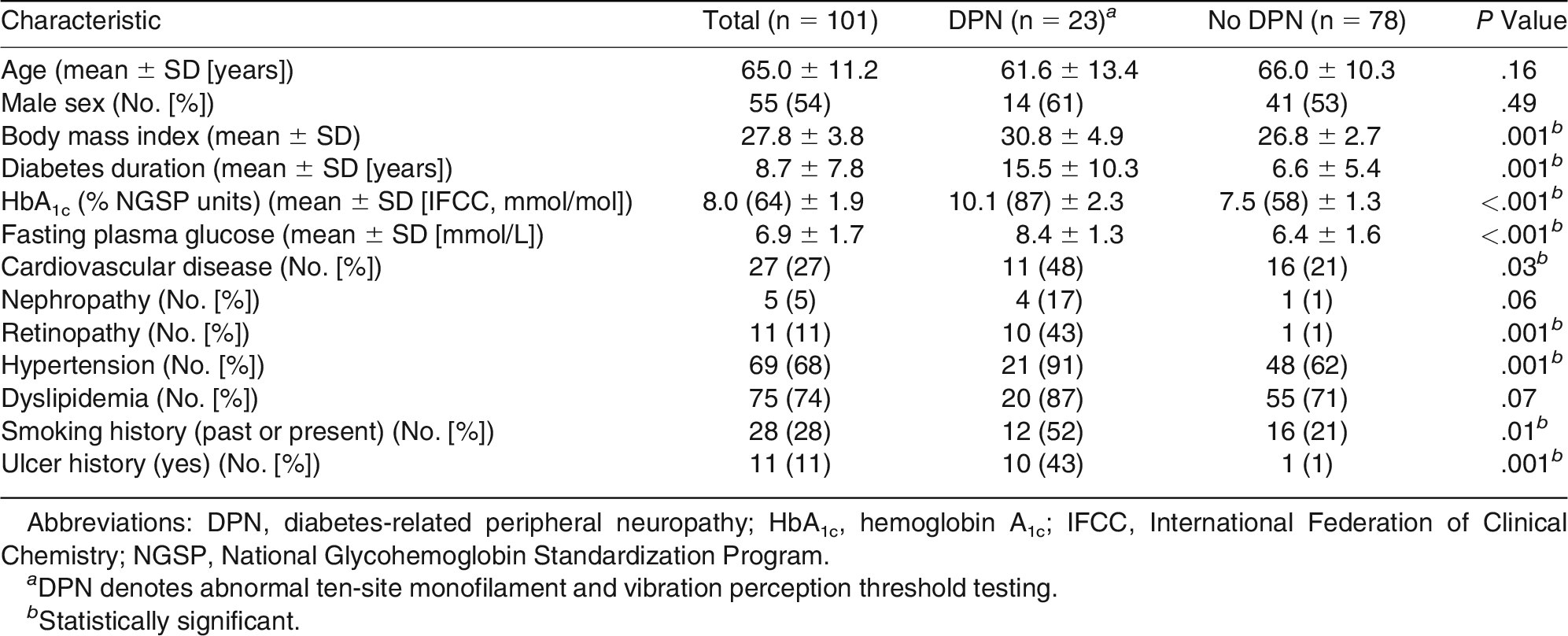
A convenience sample of 101 participants (mean ± SD age, 65.0 ± 11.2 years; 54% male; 97% type 2 diabetes; mean ± SD diabetes duration, 8.7 ± 7.8 years) were recruited (Table 1). The DPN group demonstrated statistically significantly higher BMI, HbA1c level, and incidence of cardiovascular disease, hypertension, retinopathy, smoking, and DFU, as well as longer diabetes duration.

Table 1.
Participant Characteristics
Primary Outcomes
Hierarchical regression analyses are presented in Table 2. Diabetes-related peripheral neuropathy had a significant, large correlation with reduced knee extended (r = –0.53, n = 101; P < .001) and knee flexed (r = –0.50, n = 101; P < .001) ankle joint range of motion and a significant, moderate correlation with reduced first metatarsophalangeal joint range of motion (r = –0.37; n = 101; P < .001).
Table 2.
Regression Analyses for Ankle Joint Dorsiflexion Range of Motion (Extended and Flexed Knee) and First Metatarsophalangeal Joint Dorsiflexion Range of Motion
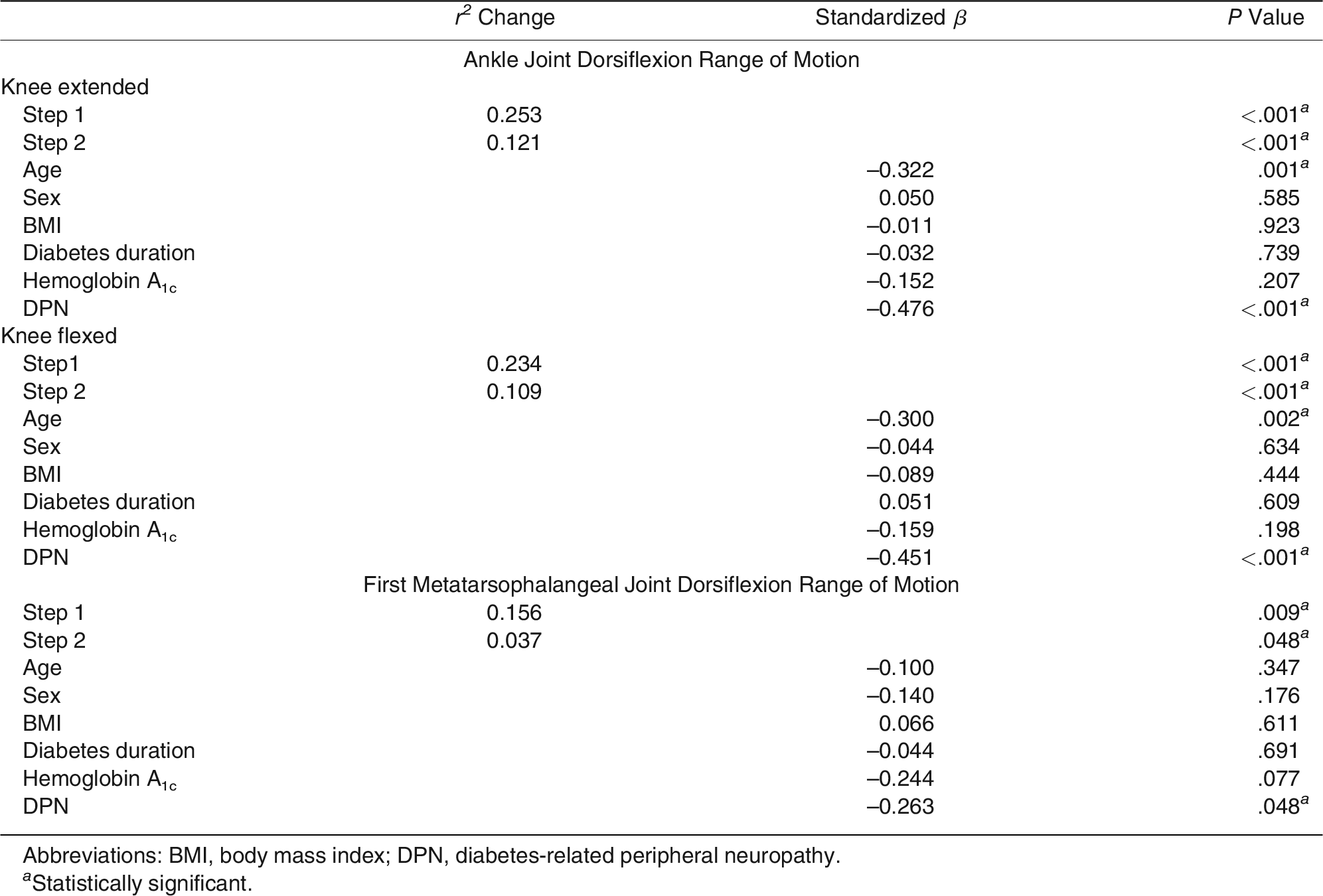
A hierarchical regression analysis was used to investigate the amount of variance in ankle joint range of motion (knee extended) explained by DPN after controlling for age, sex, BMI, diabetes duration, and HbA1c level. The first step explained 25.3% of the variance in ankle joint range of motion. Diabetes-related peripheral neuropathy was entered at the second step, and the model as a whole explained 37.4% of the variance (r2 change = 0.12; F6,88 = 8.76; P < .001); DPN made a significant, unique contribution to the model (β = –0.48; P < .001).
A hierarchical regression analysis was used to investigate the amount of variance in ankle joint range of motion (knee flexed) explained by DPN after controlling for age, sex, BMI, diabetes duration, and HbA1c level. The first step explained 23.4% of the variance in ankle joint range of motion. Diabetes-related peripheral neuropathy was entered at the second step, and the model as a whole explained 34.2% of the variance (r2 change = 0.11; F6,88 = 7.63; P < .001); DPN made a significant, unique contribution to the model (β = –0.45; P < .001).
A hierarchical regression analysis was used to investigate the amount of variance in first metatarsophalangeal joint range of motion explained by DPN after controlling for age, sex, BMI, diabetes duration, and HbA1c level. The first step explained 15.6% of the variance in ankle joint range of motion. Diabetes-related peripheral neuropathy was entered at the second step, and the model as a whole explained 19.3% of the variance (r2 change = 0.04; F6,88 = 3.50; P = .004); DPN made a significant, unique contribution to the model (β = –0.26; P = .048).
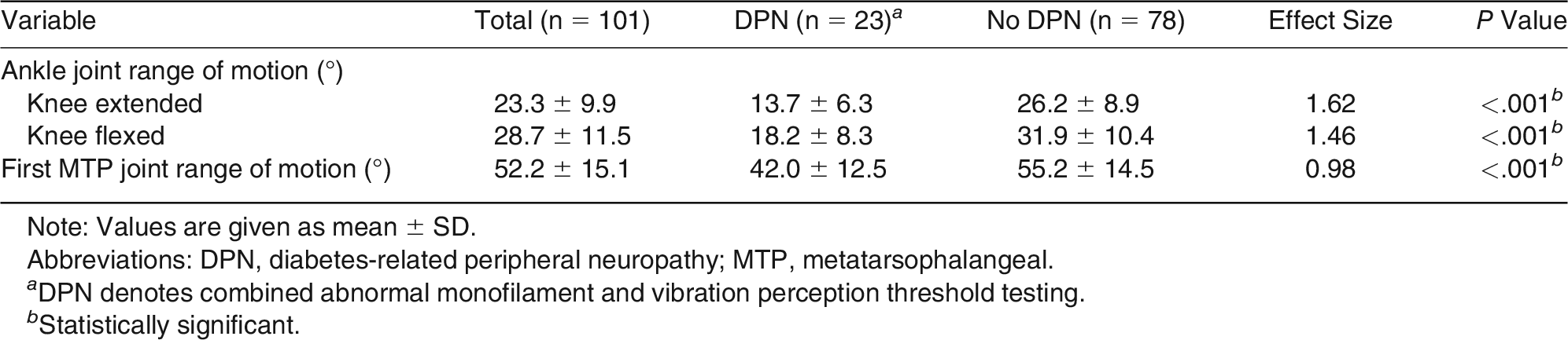
Participants with DPN were found to have significantly reduced ankle and first metatarsophalangeal joint dorsiflexion range of motion compared with those without DPN (Table 3). The odds ratio for history of DFU and weightbearing ankle equinus with the modified lunge test (odds ratio = 3.75; 95% CI, 0.4–30.88; P = .18) was not statistically significant.
Table 3.
Ankle and First MTP Joint Range of Motion Between Groups
Secondary Outcomes
Table 4.
Correlations with Ankle and First MTP Joint Range of Motion Using Pearson r
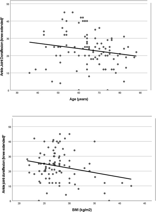
Figure 1.
Scatterplots demonstrating correlations with ankle joint dorsiflexion range of motion (knee extended) and the continuous variables (age [A], body mass index [BMI] [B], diabetes duration [C], and hemoglobin A1c [HbA1c] level [D]). NGSP, National Glycohemoglobin Standardization Program.
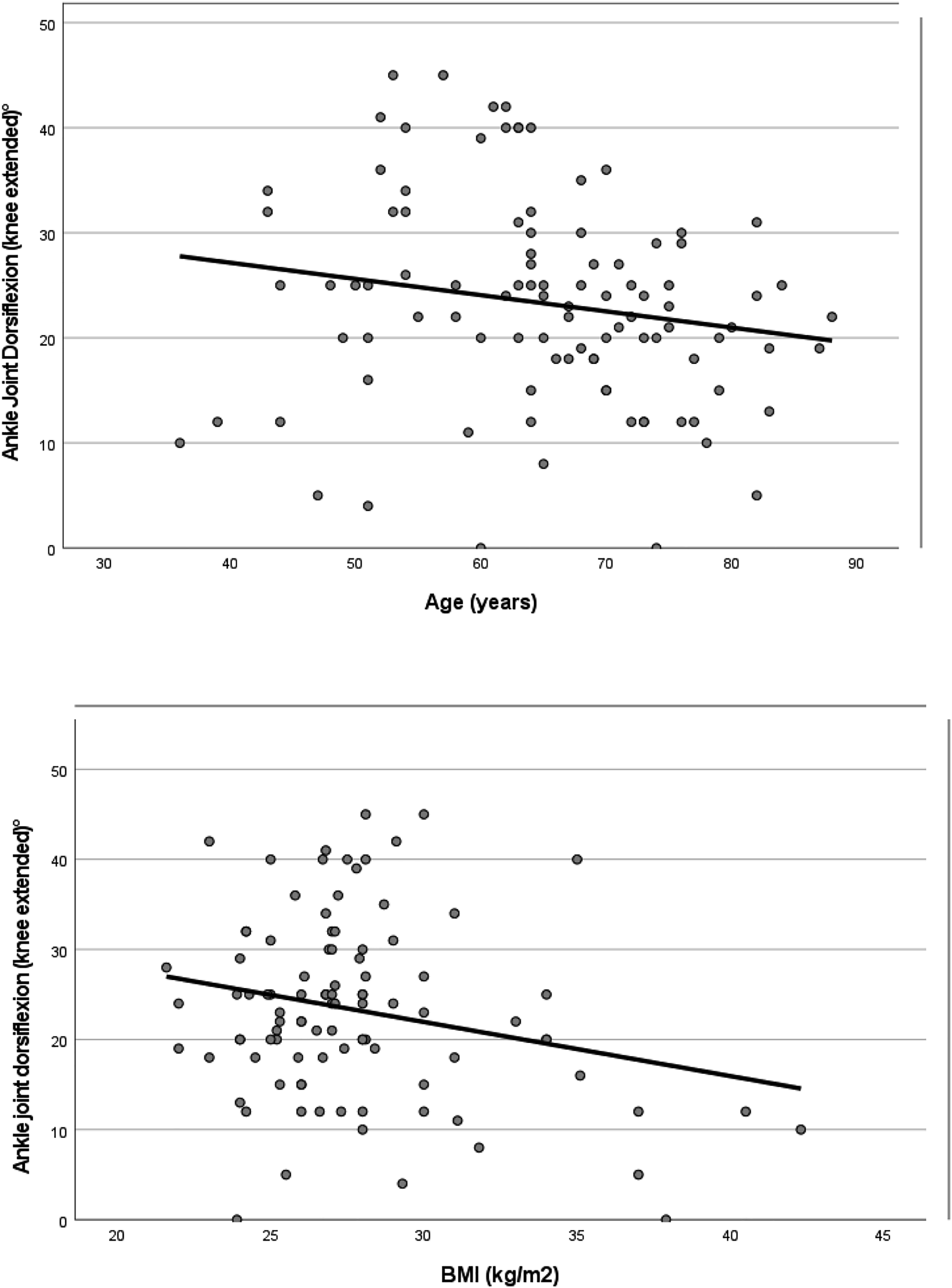
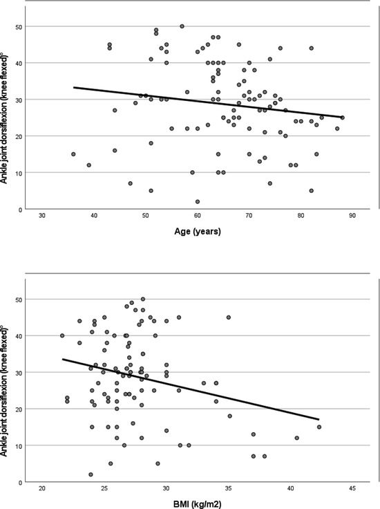
Figure 2.
Scatterplots demonstrating correlations with ankle joint dorsiflexion range of motion (knee flexed) and the continuous variables (age [A], body mass index [BMI] [B], diabetes duration [C], and hemoglobin A1c level [HbA1c] [D]). NGSP, National Glycohemoglobin Standardization Program.
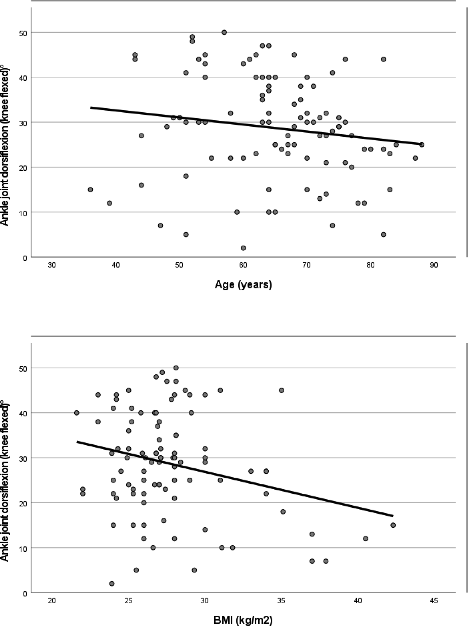
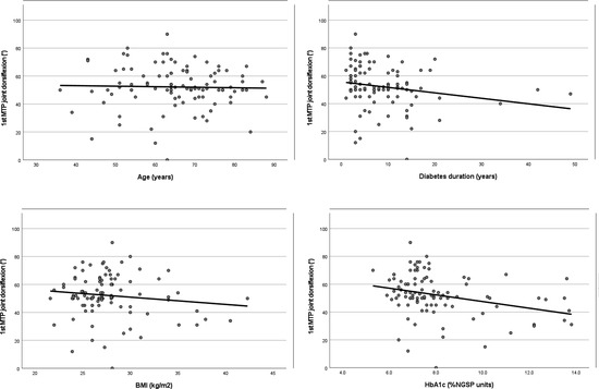
Figure 3.
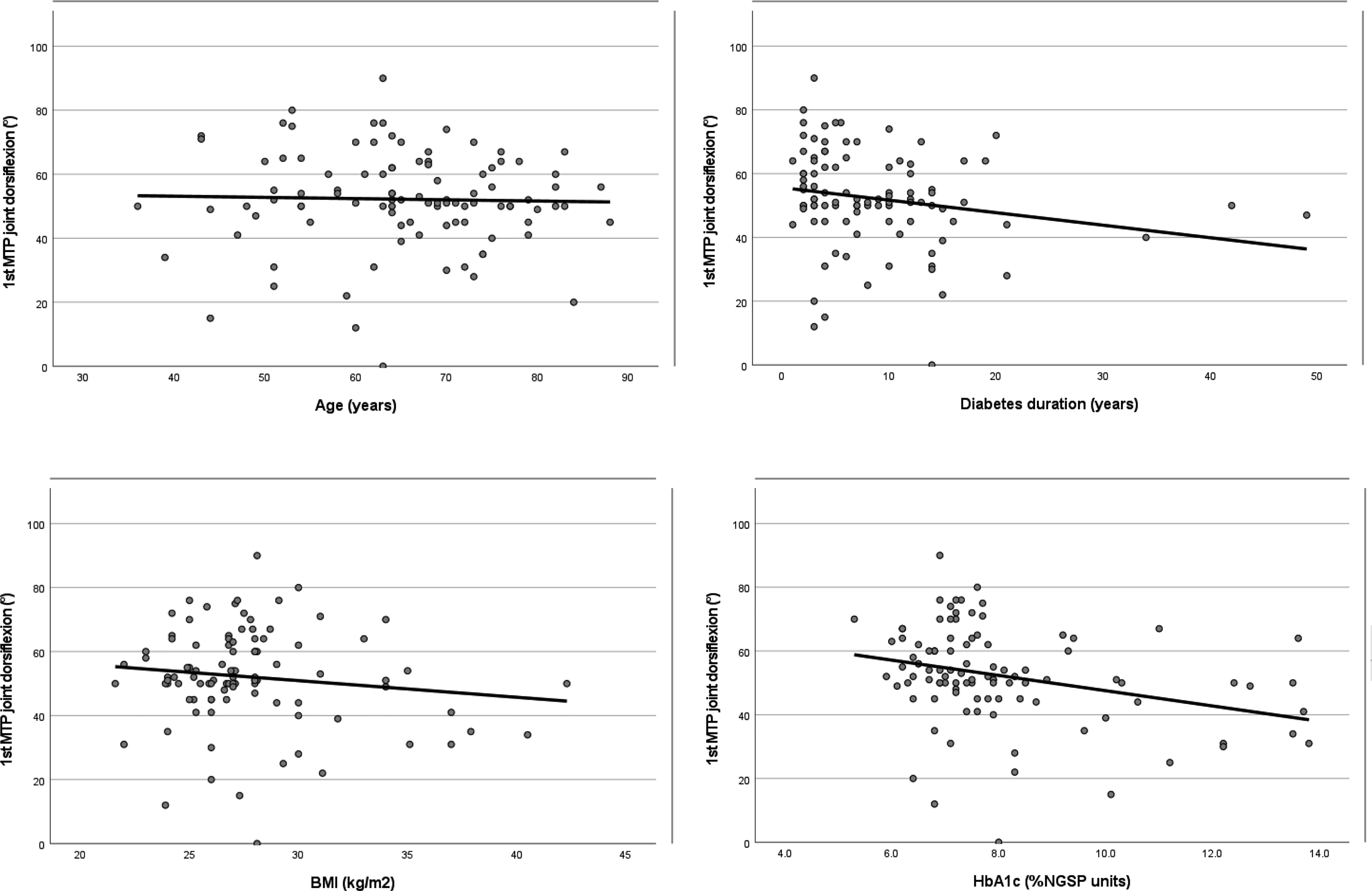
Scatterplots demonstrating correlations with first metatarsophalangeal (MTP) joint dorsiflexion range of motion and the continuous variables (age [A], body mass index [BMI] [B], diabetes duration [C], and hemoglobin A1c [HbA1c] level [D]). NGSP, National Glycohemoglobin Standardization Program.
Discussion
The aim of this study was to investigate the relationship between clinically diagnosed DPN and ankle and first metatarsophalangeal joint dorsiflexion range of motion in adults with diabetes. We found that DPN significantly predicted the amount of variance in ankle (knee extended and flexed) and first metatarsophalangeal joint range of motion after controlling for age, sex, BMI, diabetes duration, and HbA1c level. We also found that participants with DPN had significantly reduced ankle (knee extended and flexed) and first metatarsophalangeal joint range of motion compared with those without DPN. There was no evidence of increased likelihood of those with a weightbearing ankle equinus (knee extended) having a history of DFU. This is likely due to the high proportion of participants with an equinus (n = 74) and the low proportion with a history of DFU (n = 11). In addition, in people with diabetes, we found that higher BMI and HbA1c levels and longer diabetes duration were significantly associated with reduced ankle (knee flexed and extended) joint range of motion. Similarly, longer diabetes duration and higher HbA1c level were significantly associated with reduced first metatarsophalangeal joint range of motion.
The present findings suggest that DPN makes an independent contribution to reduced ankle joint dorsiflexion range of motion in particular but also to reduced first metatarsophalangeal joint dorsiflexion range of motion. Limited joint mobility in the foot and ankle has been linked to presence of chronic hyperglycemia[19] and subsequent changes in function leading to increased plantar pressure and associated complications such as ulceration.[17] However, the findings of the present study support additional joint stiffness developing in the presence of DPN. The mean ± SD ankle joint dorsiflexion range of motion (weightbearing lunge, extended knee) in this study (23.3° ± 9.9°; n = 101) is lower than that previously reported in similar populations with diabetes by Tang et al[36] (27° ± 7°; n = 74) and Searle et al[27] (34.2° ± 6.5° to 35.1° ± 6.8°, n = 30), which suggests that this may be more reflective of the general population with diabetes. Limited joint mobility seen in the present cohort with DPN might be attributable to nonenzymatic glycosylation as this has previously been implicated in the development of DPN[37]; however, it could also be independent of this process. These findings are consistent with previous research demonstrating increased ankle stiffness during the late stance phase of gait and prolonged forefoot loading in people with DPN compared with those with diabetes alone.[38] Although we did not investigate the effect of limited joint range of motion on plantar pressure, previous research suggests that in combination, DPN and limited joint mobility are more likely to result in DFU via elevated plantar pressure.[8,21] Based on the present findings, clinicians should assess foot joint range of motion and neurologic function in people with diabetes and consider prophylactic management to reduce plantar pressure and risk of DFU. Recent evidence suggests that stretching exercises may have limited capacity to effectively reduce plantar pressures in people with diabetes.[39] Therefore, further consideration of the safety and efficacy of other therapies, such as foot orthoses and footwear modifications, to reduce the elevated plantar pressure associated with limited ankle and first metatarsophalangeal joint range of motion in people with diabetes and DPN is warranted.
We also demonstrated that longer diabetes duration and higher HbA1c level were significantly correlated with decreased ankle joint range of motion, performed with the knee extended and flexed, and with reduced nonweightbearing first metatarsophalangeal joint range of motion. In a study by Searle et al[22] including 136 people with diabetes, significantly higher HbA1c level but not longer diabetes duration was found in individuals with a nonweightbearing ankle equinus (<5° dorsiflexion) compared with those without. The mixed findings in relation to diabetes duration are likely to be related to variability in time to diagnosis. Up to 35% of people with newly diagnosed type 2 diabetes have been found to have complications associated with chronic hyperglycemia and long-standing disease.[40] In addition, it is estimated in the United States that 20% to 25% of adults with diabetes are undiagnosed,[41] and in Australia three in ten adults have undiagnosed disease.[42] Nevertheless, these findings suggest that reduced ankle joint range of motion is likely to occur with higher HbA1c level and presence of DPN. We also found that increasing BMI correlated with reduced ankle joint range of motion performed with the knee extended (P < .05) and flexed (P < .01). Higher BMI in the presence of reduced ankle joint range of motion could serve to exacerbate elevations in plantar pressures independently associated with BMI[43] and ankle joint restriction.[17] This relationship would amplify the need to address weight loss as part of a treatment plan aimed at reducing the risk of DFU.
The present study, with a relatively large sample size, included a range of potential confounding medical and demographic variables and clinical measures. However, there are several limitations that need to be considered when interpreting the findings. Although the study population could be considered reflective of community-dwelling people with diabetes typically presenting for foot care (>65 years, overweight), the study is generalizable to people with type 2 diabetes only. In addition, we used chairside testing (VPT and ten-site monofilament) to diagnose DPN. Although these methods have previously demonstrated predictive capacity for determining DFU risk,[25,44,,46] the protocol for these measures varies across international position statements.[47,,49] It is possible that we underestimated the number of people with DPN in the present study given that the prevalence of 23% is lower than the reported range in people with diabetes (30%–50%).[3] This may be attributable to the conservative results of a ten-site monofilament test compared with a four-site test, which requires only one absent site for diagnosis.[6] Furthermore, given that these forms of testing focus on large-fiber DPN and require the disease to cause sensory loss for it to be detected, it is possible that the participants in the present study were likely to have had more advanced disease. It is, therefore, likely that this testing technique did not identify participants with earlier-stage DPN, which may have affected the strength of the relationship we found between DPN and limited joint mobility.
Although the present findings show that DPN is associated with significantly reduced ankle and first metatarsophalangeal joint range of motion, the effect of these changes on peak pressure and pressure-time integral were not investigated in this study. In addition, we did not perform a weightbearing first metatarsophalangeal joint test because it has not been shown to be related to dynamic gait.[50] Similarly, we chose not to investigate nonweightbearing ankle joint range of motion due to concerns surrounding its reliability and ability to reflect dynamic function[51,52] and hence cannot comment on these commonly used clinical tests regarding their relationship to DPN. It is unclear whether the weightbearing lunge test is truly indicative of ankle joint dynamic function in gait because subtalar or other tarsal joint motion may be encompassed by this measure. Regardless, this measure was used in the present study due to its independence from clinician-applied torque and being more likely to reflect physiologic torque of gait.[53] Furthermore, the present study focused on primary sagittal plane joints largely implicated in propulsive mechanics; however, there is a need to consider how DPN may relate to motion at other foot joints, such as the subtalar or midtarsal joints.
The present findings suggest that adults with type 2 diabetes and DPN are likely to have significantly reduced ankle and first metatarsophalangeal joint range of motion compared with those without DPN, independent of age, sex, BMI, diabetes duration, or HbA1c level. Clinical assessment of foot and ankle joint range of motion should, therefore, be undertaken in patients with DPN because limited joint mobility may be more severe and contribute to increased risk of diabetes-related foot disease, including DFU. Early identification of limited joint mobility in the foot will enable timely intervention with treatment modalities aimed at reducing DFU risk by alleviating plantar pressures, such as with education, foot orthoses, and modifications to footwear. Further research with more precise neurologic testing (eg, nerve conduction) and more extensive joint range-of-motion assessment is needed to conclusively establish this relationship. In addition, the most effective management strategies for reducing the effects of limited joint mobility in people with DPN requires further investigation.
Acknowledgments
We thank the participants for their involvement in this research.
Financial Disclosure
None reported.
Conflicts of Interest
None reported.
References
- Saeedi P, Salpea P, Karuranga S, et al: Mortality attributable to diabetes in 20–79 years old adults, 2019 estimates: results from the International Diabetes Federation Diabetes Atlas, 9th edition. Diabetes Res Clin Pract 162: 108086, 2020.
- Armstrong DG, Boulton AJ, Bus SA: Diabetic foot ulcers and their recurrence. N Engl J Med 376: 2367, 2017.
- Tesfaye S, Vileikyte L, Rayman G, et al: Painful diabetic peripheral neuropathy: consensus recommendations on diagnosis, assessment and management. Diabetes Metab Res 27: 629, 2011.
- Lazzarini PA, Pacella RE, Armstrong DG, et al: Diabetes-related lower-extremity complications are a leading cause of the global burden of disability. Diabet Med 35: 1297, 2018.
- Hampton K, Alani S, Wilson J, et al: Resistance to ischaemic conduction failure in chronic hypoxaemia and diabetes. J Neurol Neurosurg Psychiatry 52: 1303, 1989.
- Boulton A, Armstrong D, Albert S, et al: Comprehensive foot examination and risk assessment. Endocr Pract 14: 576, 2008.
- Lazzarini PA, Crews RT, van Netten JJ, et al: Measuring plantar tissue stress in people with diabetic peripheral neuropathy: a critical concept in diabetic foot management. J Diabetes Sci Technol 13: 869, 2019.
- Sacco I, Hamamoto A, Gomes A, et al: Role of ankle mobility in foot rollover during gait in individuals with diabetic neuropathy. Clin Biomech 24: 687, 2009.
- Armstrong DG, Stacpoole-Shea S, Nguyen H, et al: Lengthening of the Achilles tendon in diabetic patients who are at high risk for ulceration of the foot. J Bone Joint Surg Am 81: 535, 1999.
- Barry D, Sabacinski K, Habershaw G, et al: Tendo Achillis procedures for chronic ulcerations in diabetic patients with transmetatarsal amputations. JAPMA 83: 96, 1993.
- Hastings MK, Mueller MJ, Sinacore DR, et al: Effects of a tendo-Achilles lengthening procedure on muscle function and gait characteristics in a patient with diabetes mellitus. J Orthop Sport Phys 30: 85, 2000.
- Lin SS, Lee TH, Wapner KL: Plantar forefoot ulceration with equinus deformity of the ankle in diabetic patients: the effect of tendo-Achilles lengthening and total contact casting. Orthopedics 19: 465, 1996.
- Mueller MJ, Diamond JE, Delitto A, et al: Insensitivity, limited joint mobility, and plantar ulcers in patients with diabetes mellitus. Phys Ther 69: 453, 1989.
- Wyatt LH, Ferrance RJ: The musculoskeletal effects of diabetes mellitus. J Can Chiropr Assoc 50: 43, 2006.
- Boulton AJ, Hardisty CA, Betts RP, et al: Dynamic foot pressure and other studies as diagnostic and management aids in diabetic neuropathy. Diabetes Care 6: 26, 1983.
- Caselli A, Pham H, Giurini JM, et al: The forefoot-to-rearfoot plantar pressure ratio is increased in severe diabetic neuropathy and can predict foot ulceration. Diabetes Care 25: 1066, 2002.
- Fernando DJ, Masson EA, Veves A, et al: Relationship of limited joint mobility to abnormal foot pressures and diabetic foot ulceration. Diabetes Care 14: 8, 1991.
- Veves A, Murray H, Young M, et al: The risk of foot ulceration in diabetic patients with high foot pressure: a prospective study. Diabetologia 35: 660, 1992.
- Abate M, Schiavone C, Salini V, et al: Management of limited joint mobility in diabetic patients. Diabetes Metab Syndr Obes 6: 197, 2013.
- Gerrits EG, Landman GW, Nijenhuis-Rosien L, et al: Limited joint mobility syndrome in diabetes mellitus: a minireview. World J Diabetes 6: 1108, 2015.
- Amemiya A, Noguchi H, Oe M, et al: Elevated plantar pressure in diabetic patients and its relationship with their gait features. Gait Posture 40: 408, 2014.
- Searle A, Spink M, Chuter V: Prevalence of ankle equinus and correlation with foot plantar pressures in people with diabetes. Clin Biomech 60: 39, 2018.
- Turner D, Helliwell PS, Burton AK, et al: The relationship between passive range of motion and range of motion during gait and plantar pressure measurements. Diabet Med 24: 1240, 2007.
- Zhang Q, Yi N, Liu S, et al: Easier operation and similar power of 10 g monofilament test for screening diabetic peripheral neuropathy. J Intern Med Res 46: 3278, 2018.
- Armstrong DG, Lavery LA, Vela SA, et al: Choosing a practical screening instrument to identify patients at risk for diabetic foot ulceration. Arch Intern Med 158: 289, 1998.
- Rabin A, Kozol Z: Weightbearing and nonweightbearing ankle dorsiflexion range of motion: are we measuring the same thing? JAPMA 102: 406, 2012.
- Searle A, Spink M, Chuter V: Weight bearing versus non-weight bearing ankle dorsiflexion measurement in people with diabetes: a cross sectional study. BMC Musculoskel Dis 19: 1, 2018.
- Menz HB, Tiedemann A, Kwan MM-S, et al: Reliability of clinical tests of foot and ankle characteristics in older people. JAPMA 93: 380, 2003.
- Hopson MM, McPoil T, Cornwall MW: Motion of the first metatarsophalangeal joint. Reliability and validity of four measurement techniques. JAPMA 85: 198, 1995.
- Sánchez-Gómez R, Becerro-de-Bengoa-Vallejo R, Losa-Iglesias ME, et al: Reliability study of diagnostic tests for functional hallux limitus. Foot Ankle Int 41: 457, 2020.
- Stevens JP: Applied Multivariate Statistics for the Social Sciences, Routledge, Philadelphia, 2012.
- Menz HB: Two feet, or one person? problems associated with statistical analysis of paired data in foot and ankle medicine. Foot 14: 2, 2004.
- Cohen J: Statistical Power Analysis for the Behavioral Sciences, 2nd Ed, p 13, Lawrence Erlbaum Associates Inc, Hillsdale, NJ, 1988.
- Searle A, Spink MJ, Chuter VH: Validation of a weight bearing ankle equinus value in older adults with diabetes. J Foot Ankle Res 11: 1, 2018.
- Joseph J: Range of movement of the great toe in men. J Bone Joint Surg Br 36: 450, 1954.
- Tang UH, Zügner R, Lisovskaja V, et al: Foot deformities, function in the lower extremities, and plantar pressure in patients with diabetes at high risk to develop foot ulcers. Diabet Foot Ankle 6: 27593, 2015.
- Thornalley PJ: Glycation in diabetic neuropathy: characteristics, consequences, causes, and therapeutic options. Int Rev Neurobiol 50: 37, 2002.
- Williams DB, Brunt D, Tanenberg RJ: Diabetic neuropathy is related to joint stiffness during late stance phase. J Appl Biomech 23: 251, 2007.
- Searle A, Spink MJ, Oldmeadow C, et al: Calf muscle stretching is ineffective in increasing ankle range of motion or reducing plantar pressures in people with diabetes and ankle equinus: a randomised controlled trial. Clin Biomech 69: 52, 2019.
- Gedebjerg A, Almdal TP, Berencsi K, et al: Prevalence of micro-and macrovascular diabetes complications at time of type 2 diabetes diagnosis and associated clinical characteristics: a cross-sectional baseline study of 6958 patients in the Danish DD2 cohort. J Diabetes Complications 32: 34, 2018.
- Fang M: Trends in the prevalence of diabetes among US adults: 1999–2016. Am J Prev Med 55: 497, 2018.
- Sainsbury E, Shi Y, Flack J, et al: Burden of diabetes in Australia: its time for more action. Available at: https://www.sydney.edu.au/content/dam/corporate/documents/faculty-of-medicine-and-health/research/centres-institutes-groups/burden-of-diabetes-its-time-for-more-action-report.pdf.
- Butterworth PA, Urquhart DM, Landorf KB, et al: Foot posture, range of motion and plantar pressure characteristics in obese and non-obese individuals. Gait Posture 41: 465, 2015.
- Mayfield JA, Sugarman JR: The use of the Semmes-Weinstein monofilament and other threshold tests for preventing foot ulceration and amputation in persons with diabetes. J Fam Pract 49: S17, 2000.
- Singh N, Armstrong DG, Lipsky BA: Preventing foot ulcers in patients with diabetes. JAPMA 293: 217, 2005.
- Young MJ, Breddy JL, Veves A, et al: The prediction of diabetic neuropathic foot ulceration using vibration perception thresholds: a prospective study. Diabetes Care 17: 557, 1994.
- Ibrahim A: IDF clinical practice recommendation on the diabetic foot: a guide for healthcare professionals. Diabetes Res Clin Pract 127: 285, 2017.
- Pop-Busui R, Boulton AJ, Feldman EL, et al: Diabetic neuropathy: a position statement by the American Diabetes Association. Diabetes Care 40: 136, 2017.
- Schaper NC, van Netten JJ, Apelqvist J, et al: Practical guidelines on the prevention and management of diabetic foot disease (IWGDF 2019 update). Diabetes Metab Res Rev 36: e3266, 2020.
- Halstead J, Redmond AC: Weight-bearing passive dorsiflexion of the hallux in standing is not related to hallux dorsiflexion during walking. J Orthop Sports Phys Ther 36: 550, 2006.
- Gatt A, Chockalingam N: Clinical assessment of ankle joint dorsiflexion: a review of measurement techniques. JAPMA 101: 59, 2011.
- Kim PJ, Peace R, Mieras J, et al: Interrater and intrarater reliability in the measurement of ankle joint dorsiflexion is independent of examiner experience and technique used. JAPMA 101: 407, 2011.
- Baumbach SF, Brumann M, Binder J, et al: The influence of knee position on ankle dorsiflexion: a biometric study. BMC Musculoskelet Disord 15: 246, 2014.
© 2023 American Podiatric Medical Association