


Abstract
A 64-year-old woman presents with wounds to her left ankle. Although her soft-tissue cultures and arterial Doppler and duplex studies were unremarkable, her venous reflux studies showed right and left small saphenous vein insufficiency. After 8 weeks of standard treatment, her wounds to the left ankle did not improve, and she developed a wound to her right anterior leg. Her left ankle wound healed 8 months after initial presentation, and her right leg wound healed in 3 months. Thereafter she underwent an endovenous ablation of her left small saphenous vein, without apparent complications. Two weeks after surgery, she developed an incision site scab that worsened and ulcerated. This case report highlights pathergy from endovenous ablation for lower-extremity venous disease.
In the past decade, less invasive endovenous ablation procedures have gained in popularity for the treatment of lower-extremity varicosities. These minimally invasive techniques have been shown to reduce symptoms, improve quality of life, and reduce rates of reoperation.[1] Previous studies, such as Effect of Surgery and Compression on Healing and Recurrence (ESCHAR) and Early Venous Reflux Ablation (EVRA), have helped guide treatments of superficial venous reflux with venous ulcers.[2,3] The ESCHAR study showed less recurrence of venous leg ulceration in patients treated with ablative superficial venous surgery with compression than in those treated with compression alone (12% versus 28%).[2] The EVRA trial highlighted the importance of promptly conducting endovenous ablation with compression by showing a shorter healing time of venous leg ulcers versus compression alone with delayed intervention (56 days versus 82 days).[3] These recent improvements in clinical practice suggest that endovenous ablation surgery could reduce the recurrence of ulcers and provide earlier healing time for chronic venous disease.
However, literature reviews of endovascular laser treatments have reported surgically induced ecchymosis and pain with or without induration (100%), skin burns (<1%), dysesthesias (0%–22%), superficial thrombophlebitis (0%–25%), deep venous thrombosis (0%–6%), nerve injury (<1%), and hematomas were reported much less.[4] Many publications have focused on superficial thrombophlebitis and deep venous thrombosis after endovenous ablation as primary studies. From a literature search, we found only two case reports of pathergy.
Pathergy is defined as minor trauma–induced skin lesions. Lesions that arise from pathergy tend to be nonspecific papules or pustules that may develop into ulcers.[5] The Koebner phenomenon is the appearance of new skin lesions on unaffected skin after trauma.[5] These lesions are both clinically and histologically identical to the patient’s skin disease. Unlike the Koebner phenomenon, the site of pathergy will not reflect the underlying skin disease of the patient.[5] Currently, it is thought that pathergy is a T-cell–mediated reaction that parallels the delayed hypersensitivity reaction.[6] The pathergy phenomenon aggravates and complicates wound healing by forming satellite lesions at skin manipulation sites.[7] Pathergy is most commonly associated with pyoderma gangrenosum, Sweet syndrome, lichen planus, and Behcet disease.[6]
Although pathergy is well documented as being induced by surgery, there are only a few case reports highlighting pathergy after lower-extremity endovenous ablation. In 2020, Kartal et al[8] reported a case of a patient with no comorbidities in whom a lesion at the site of incision occurred on day 12 after lower-extremity endovenous laser ablation and varicose vein excision. Antibiotic drug therapy and recurrent surgical debridements were started. There were no improvements of the wound sites; in fact, there was rapid deterioration, expansion of the lesion, and ulceration. Skin biopsy results revealed an extensive neutrophilic inflammation and dermal lysis in the upper dermis, and a diagnosis of pyoderma gangrenosum was made.
Other cases have helped add to the understanding of the pathergy phenomenon induced by endovenous laser treatment. In 2004, Sichlau and Ryu[9] reported cutaneous thermal injury after endovenous laser energy ablation that led to a 10 × 13–mm blistered area that later ulcerated at the medial midthigh. Ultrasound findings showed fat surrounding the superficial thrombosed tributaries, and it was concluded that the cutaneous thermal injury came from the heated blood traveling from the great saphenous vein to the superficial tributaries, directly beneath the skin site.[9] Mekako and Chetter[10] described a brown pigmentation that persisted for 6 weeks after endovenous laser therapy. Although the cause of the persistent hyperpigmentation was not known, the article suggested that direct thermal damage, thrombophlebitis, and migration of melanin pigment may have been the cause.[10]
The purpose of this case report is to highlight a case of pathergy after endovascular ablation and to emphasize caution when treating venous disease with lower-extremity endovenous ablation.
Case Report
A 64-year-old woman initially presented to urgent care, where she was prescribed doxycycline and underwent debridement of a wound to her left ankle (Figure 1A). She subsequently developed a second wound adjacent to the first. She then presented to our office 4 weeks after development of the first wound. She attributed the wound to a spider bite but did not observe the actual spider or the bite. She relayed a history of a similar wound 4 years earlier that was resistant to healing but immediately healed after a blood transfusion for an unrelated anemia.
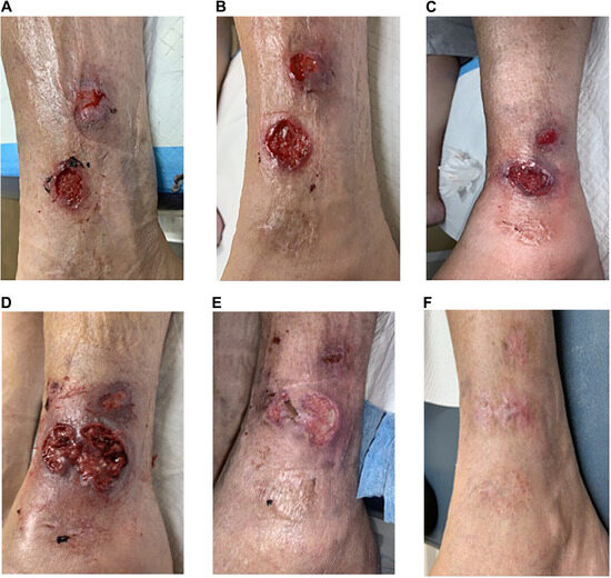
Figure 1.
Lateral wounds of the left ankle, which were the patient’s first wounds. A, Initial presentation (May 18, 2020). B, Scar with hyperpigmentation noted distal to the wounds (June 1, 2020). C, No improvements noted (June 22, 2020). D, Adjacent red patch ulcerated (June 29, 2020). E, Improvement of wounds (July 27, 2020). F, Full healing (November 25, 2020).
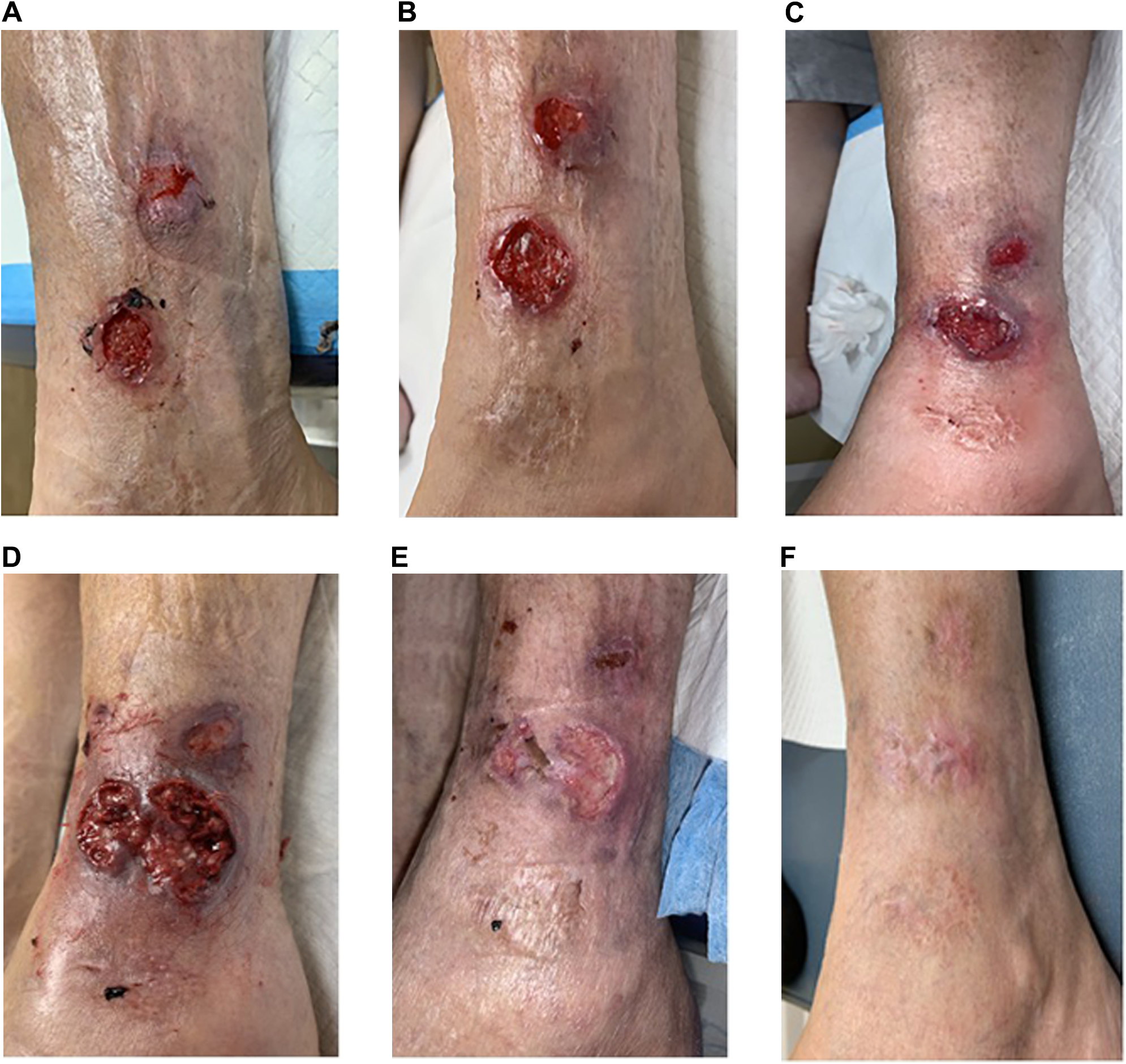
Her medical history was significant for obesity, osteopenia, and anemia. Her surgical history included hysterectomy and gastric bypass. She denied tobacco or alcohol use. On physical examination, her light touch sensation, vibratory sensation, deep tendon reflexes, and protective sensation were all intact. Her dorsalis pedis and posterior tibial pulses were palpable bilaterally. She had localized edema to the bilateral legs. The dermatologic examination showed a 1.0 × 1.0 × 0.4–cm ulceration with a scalloped border at the left lateral ankle 6 cm proximal to the distal tip of the fibula. Distal and anterior to the ulceration was a patch of erythema measuring 3 × 1.5 cm with minimal break in the skin. Just distal to the two lesions was a 3 × 3–cm stellate scar with surrounding hyperpigmentation (Figure 1B).
Treatment included excisional debridement of the ulceration through the subcutaneous tissue, with healthy bleeding. A soft-tissue culture was obtained. Arterial Doppler and duplex ultrasounds were ordered. Venous reflux duplex insufficiency ultrasound was ordered. The wound was dressed with a multilayer compression dressing, and a high-protein diet was recommended.
Based on clinical appearance, the working differential diagnosis included venous wound versus insect bite versus abscess. Her arterial ultrasounds were unremarkable, with triphasic waveforms, an ankle-brachial index of 1.10 bilaterally, and a toe-brachial index of 1.09 on the right and 1.12 on the left. Her venous insufficiency study showed right small saphenous vein incompetence with 1.4 sec of reflux and a diameter of 0.38 cm, and left small saphenous vein incompetence with 1.0 sec of reflux and a diameter of 0.27 cm. She was referred to a vein doctor for evaluation and possible endovenous ablation. The wound culture had no growth; however, she was taking doxycycline at the time the culture was obtained.
Subsequently, the wound did not improve. She was given a course of clindamycin. A second wound culture was obtained, with no growth. We performed an incision and drainage to the wound because the tissue appeared friable and began breaking down, but no purulence was identified. At this point we obtained two soft-tissue biopsy samples from the area, which were sent to a dermatopathology laboratory. These specimens showed fibrosing granulation tissue formation, cutaneous ulceration with surface purulence, and fibrin deposition. We obtained laboratory values, including a complete blood cell count, which was unremarkable; a basic metabolic panel, which showed a glucose level elevated to 144 mg/dL; a hemoglobin A1c level of 7.6%, an erythrocyte sedimentation rate (ESR) of 34 mm/hr; and a C-reactive protein (CRP) level of 9 mg/dL. She was not previously diabetic and was referred to primary care for evaluation of her newly diagnosed diabetes.
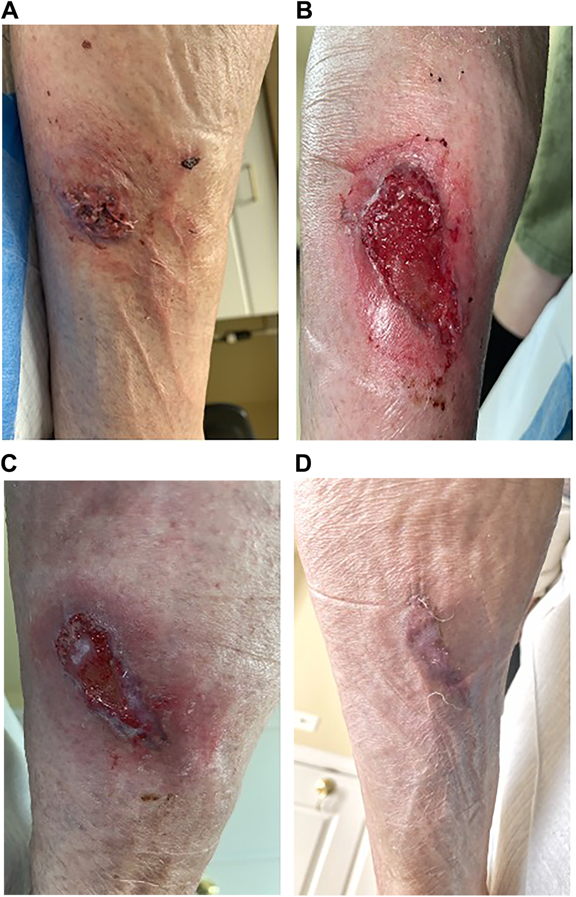
The treatment continued for 8 weeks and there were no improvements for 8 weeks. Figure 1C shows the wound at 5 weeks. The wound continued to worsen, and the adjacent red patch broke down into a full-thickness wound as well (Figure 1D). She developed an additional wound to the right anterior leg that began as a red spot, with the center eventually breaking down and ulcerating (Figure 2). At this point there was concern for pyoderma gangrenosum. Debridements were stopped. She was prescribed methylprednisolone (Medrol Dosepak; Pfizer, New York, New York), and the wound was treated topically with a dressing (Hydrofera Blue; Hydrofera LLC, Manchester, Connecticut) soaked with 1 mL of dexamethasone. She was referred to dermatology, infectious disease, and rheumatology. Whereas the dermatology and infectious disease referrals were futile, rheumatology work showed an ESR of 68 mm/hr, a CRP level of 12, and elevated antineutrophil cytoplasmic antibodies of 1:320 (reference range, <1:20). At this point her wounds were improving, and rheumatology held high-dose corticosteroids (Figure 1E).
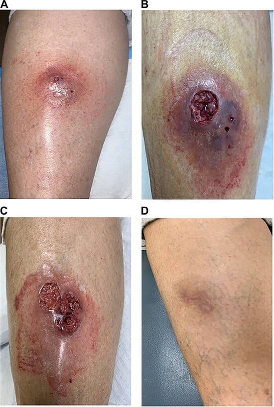
Figure 2.
Anterior wound of the right leg that began as a red spot that ulcerated and eventually healed. A, Initial presentation (June 22, 2020). B, Breakdown and ulceration (June 29, 2020). C, Worsening (July 6, 2020). D, Full healing (November 25, 2020).
Her left ankle wounds healed 8 months after initial presentation (Figure 1F), and the right leg wound overlying the great saphenous vein healed in only 3 months (Figure 2D). Owing to the speed at which the right leg healed as opposed to the left, it was still postulated that the small saphenous vein reflux was contributing to the delayed healing of the left ankle wounds, and the patient was recommended to have endovenous ablation of the left small saphenous vein, which she underwent without complication.
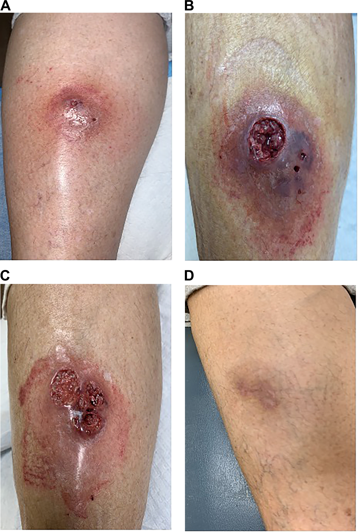
Two weeks after the ablation, she still had a small scab to the entrance site (Figure 3A). At 4 weeks this scab turned into a red patch measuring 1.6 × 1.0 cm. This site continued to worsen, with the largest measurement being 5.0 × 2.5 × 0.3 cm 2 months after the ablation (Figure 3B). Initially thought to be a thermal injury from the ablation, this site continued to turn red, with the center skin breaking down, forming a scalloped violaceous border (Figure 3C). She was referred back to rheumatology. Laboratory tests showed antineutrophil cytoplasmic antibodies now 1:640, an ESR of 21 mm/hr, and a CRP level of 13. She was started on hydroxychloroquine sulfate (Plaquenil; Concordia Pharmaceuticals, Fort Lauderdale, Florida) 200 mg daily. The wound was again treated with no debridement and application of Hydrofera Blue embedded with 1 mL of dexamethasone phosphate with a multilayer compression dressing, changed weekly. She went on to successfully heal the wound after 16 weeks of treatment (Figure 3D).
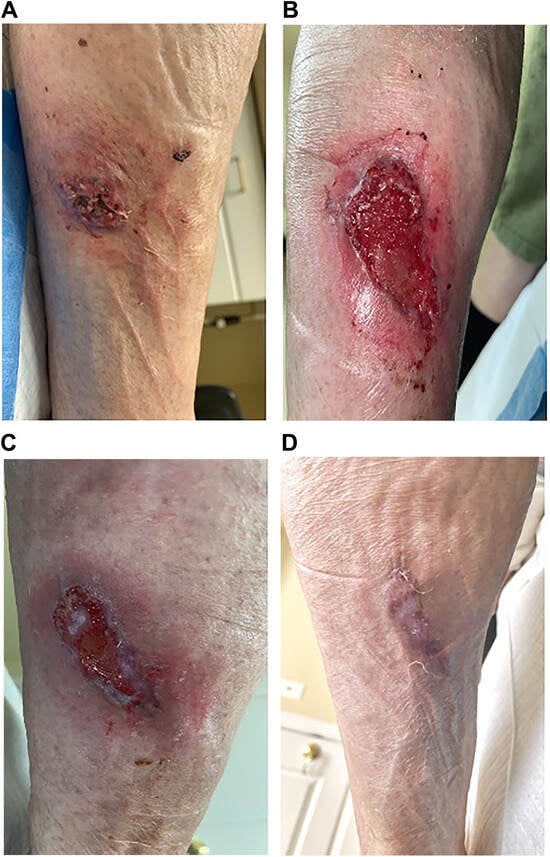
Figure 3.
Lateral wound of the left leg that developed after the left small saphenous vein endovenous ablation surgery. A, Two weeks after ablation showing the first lesion appearance (January 18, 2021). B, Two months after ablation, the sight continued to worsen, with the largest measurements (March 10, 2021). C, Three months after ablation showing further skin breakdown and scalloped violaceous borders (March 24, 2021). D, Full healing (May 5, 2021).
Discussion
This case showed the rare complication of pathergy after endovenous ablation for venous disease. The proposed diagnostic criteria set by Su et al[11] suggest pyoderma gangrenosum. We suspect this pathergy that occurred after the endovenous ablation was due to the patient’s underlying pyoderma gangrenosum.
First, on initial presentation, the patient attributed her initial left ankle wounds to a spider bite, but she did not observe the actual bite. Often patients with the onset of pyoderma gangrenosum complain of being bitten by a spider without noticing the spider actually biting them.[12] Patients with pyoderma gangrenosum are often initially treated for an infection with antibiotic drugs and debridement of the wound.[12] Surgical debridement is contraindicated in patients with pyoderma gangrenosum.[7] In fact, 25% to 50% of patients with pyoderma gangrenosum experience pathergy.[7] In the present patient, the left ankle wound was initially treated with antibiotics and surgical debridement with wound culture, and arterial Doppler ultrasound, arterial duplex ultrasound, venous duplex ultrasound, and a venous insufficiency study were ordered. With no improvement, worsening of the wound, and the development of a right anterior leg wound, pyoderma gangrenosum was of concern, and excisional debridement was stopped. With the right leg healing faster than the left leg and abnormal venous reflux studies, it was thought that the small saphenous vein reflux was a contributing factor to the delayed healing of the left leg wound, and a left small saphenous vein endovenous ablation was recommended. Following the results of the ESCHAR and EVRA trials, an endovenous ablation was performed to reduce recurrence and provide earlier healing time.
We believe that the differential diagnosis of pyoderma gangrenosum should have been recognized and acknowledged before the endovenous ablation procedure. As we currently know, pyoderma gangrenosum is determined usually through a diagnosis of exclusion. In the present case, pathergy was demonstrated through surgical debridement before the endovenous ablation. Another clue for the diagnosis of pyoderma gangrenosum was the ragged edges and violaceous borders present for both wounds before the endovenous ablation. Consistent with most patients with pyoderma gangrenosum, the present patient did not have a family history of the condition. The patient did not acknowledge a history of inflammatory bowel disease, arthritis, or any hematologic disease. Neither did the patient display abnormal soft-tissue cultures. Another major hint toward pathergy and pyoderma gangrenosum affiliated with the wounds was demonstrated with improvements of the wound when surgical debridement was stopped and the patient was placed on systemic corticosteroids. In addition, the wound that appeared after the endovenous ablation itself was pathergic and showed ragged, undermined edges with violaceous borders, supporting the diagnosis of pyoderma gangrenosum.
As mentioned previously herein, there are many different complications resulting from endovenous laser ablation procedures, with pathergy being reported only twice from our literature search. Kartal et al[8] reported a case of pyoderma gangrenosum after endovenous laser ablation and varicose vein excision. Although only an endovenous ablation was performed in the present case, pathergy was noted at the entrance site of the endovenous ablation as opposed to a pathergic response noted at the medial malleolus being the area of the excision of the varicose vein for the case reported by Kartal et al.[8] Both cases demonstrated full healing of the wounds with the use of corticosteroids and no debridement.
Another case highlighting pathergy was reported by Sichlau and Ryu.[9] Endovenous ablation was performed of the great saphenous vein. Ulceration appeared more superficial to the site of ablation. It was determined that thermal injury was associated with the skin burn. This case differed from the present case because of the location of the ulcer: superficial skin injury versus entrance site ulceration.
Postoperative complications can occur after any surgical procedure. We reported that at the incision site a scab developed that progressively worsened and ulcerated. This case report is one of the few records of pathergy specifically after endovenous ablation for venous disease. We believe that caution needs to be taken for this surgical procedure and that further research is needed to provide insight for better patient outcomes.
Conclusions
Pathergy after endovenous ablation for venous disease is a serious complication that both surgeons and patients should be aware of. Endovenous ablation should be performed with caution in patients exhibiting wounds that may be associated with pathergy.
Financial Disclosure
None reported.
Conflicts of Interest
None reported.
References
- Rasmussen LH, Lawaetz M, Bjoern L, et al: Randomized clinical trial comparing endovenous laser ablation, radiofrequency ablation, foam sclerotherapy and surgical stripping for great saphenous varicose veins. Br J Surg 98: 1079, 2011.
- Barwell JR, Davies CE, Deacon J, et al: Comparison of surgery and compression with compression alone in chronic venous ulceration (ESCHAR study): randomised controlled trial. Lancet 363: 1854, 2004.
- Gohel MS, Heatley F, Liu X, et al: A randomized trial of early endovenous ablation in venous ulceration. N Engl J Med 378: 2105, 2018.
- Van Den Boss RR, Neumann M, De Roos KP, et al: Endovenous laser ablation-induced complications: review of literature and new cases. Dermatol Surg 35: 1206, 2009.
- Sanchez DP, Sonthalia S: “Koebner Phenomenon,” in StatPearls [Internet], StatPearls Publishing, Treasure Island, FL, 2020.
- Bangash SJ, Green WH, Dolson DJ, et al: Eruptive postoperative squamous cell carcinomas exhibiting a pathergy-like reaction around surgical wound sites. J Am Acad Dermatol 61: 892, 2009.
- Berlinrut I, Bhatia N, Josse JM, et al: Untreatable surgical site infection: consider pyoderma gangrenosum. Plast Reconstr Surg Glob Open 2: e166, 2014.
- Kartal H, Sicim H, Demirdaş E, et al: An unexpected and rare complication after endovenous laser ablation: pyoderma gangrenosum. Turk Gogus Kalp Damar Cerrahisi Derg 28: 201, 2020.
- Sichlau MJ, Ryu RK: Cutaneous thermal injury after endovenous laser ablation of the great saphenous vein. J Vasc Interv Radiol 15: 865, 2004.
- Mekako A, Chetter I: Cutaneous hyperpigmentation after endovenous laser therapy: a case report and literature review. Ann Vasc Surg 21: 637, 2007.
- Su WP, Davis MD, Weenig RH, et al: Pyoderma gangrenosum: clinicopathologic correlation and proposed diagnostic criteria. Int J Dermatol 43: 790, 2004.
- Callen JP, Jackson JM. Pyoderma gangrenosum: an update. Rheum Dis Clin North Am 33: 787, 2007.
© 2023 American Podiatric Medical Association