


Abstract
Background: Foot type, especially cavus foot, is associated with foot and ankle soccer injuries, such as ankle sprains, ankle instability, and foot and ankle lateral injuries. The aim of this study was to identify risk factors for foot and ankle injuries among soccer players. Methods: Male and female soccer players, from beginners to semiprofessionals, aged between 10 and 40 years were enrolled in this cross-sectional study. Players filled in questionnaires about their training and injury history. Clinical measurements included foot length, Foot Posture Index-6, and arch height flexibility. Each variable was dichotomized: age (<18 years versus ≥18 years), level of play (AA and below versus AAA and above), foot type (cavus or not), and injury. Injury occurrence was analyzed using χ2 tests between each group of variables, and significance was set at P < .05. Results: A total of 277 players, including 81 females, volunteered; 147 were younger than 18 years and 180 were AA level or below. Cavus foot prevalence was 30%. In the cavus foot group, 51.8% of players had reached at least an AAA level compared with 27.8% in the normal-arched group (P < .001 [χ2]). Injuries were associated with a cavus foot type (P < .01 [χ2]) and with sex, age, or highest level played (P < .001 [χ2]). Conclusions: This study identified a high prevalence of cavus foot among soccer players of all ages, with an increased prevalence among higher-level players. The injury risk factors were female sex, older age, playing at a higher level, and cavus feet.
Soccer is one of the most popular sports, with more than 265 million players worldwide.[1] Soccer injuries are frequent and place a high economic burden on the health-care system, estimated to be more than $30 billion per year. These injuries are also detrimental to the injured individuals, causing a reduced physical activity level over the long term,[2] requiring time away from work,[2,3] and having negative psychological effects such as anxiety or depression.[4] In soccer, the injury rate of adult players is estimated to be 3 to 9 per 1,000 player-hours of competition,[5] and twice as many injuries occur during competition compared with training due to higher intensity.[5] In the pediatric population, the soccer injury rate is lower and estimated to be approximately 2 per 1,000 hours of play.[6]
Soccer-related injuries occur mostly at the foot and ankle, with ankle sprains representing as much as 76% of all injuries.[7] Injury risk factors are usually divided into two categories: extrinsic and intrinsic factors. Extrinsic factors include high intensity,[2,5] competition (versus training),[2,5] playing surface (natural grass versus artificial turf),[8,9] equipment (eg, shoes, shin guards),[5,9] and climatic conditions.[9,10] Intrinsic factors include being female,[2,10] increased age,[2,5] number of previous injuries,[5,11] lower experience level, and greater skills.[12] In some studies, foot type is also considered a risk factor, although consensus has yet to be reached regarding its exact contribution to injury.[13,14,15] Studies have found that the supinated cavus foot, or high-arched foot,[16] is associated with foot and ankle injuries,[17,18] namely, stress fractures of the fifth metatarsal bone,[19] ankle sprains, and chronic ankle instability.[20] The association between soccer players’ level of play and prevalence of cavus feet has yet to be proved. Provided that an increased level of play is associated with more injuries, and that cavus alignment can be corrected with foot orthoses, a high prevalence of cavus feet could be targeted for injury prevention.
The aim of this cross-sectional study was to analyze the prevalence of foot and ankle injuries in a cohort of soccer players in relation to foot type. We hypothesized that 1) there is a correlation between the proportion of cavus feet and high standards of soccer competition and 2) injury prevalence is associated with cavus foot type among soccer players.
Material and Methods
Cross-sectional Study Design
The University Hospital Center Institutional Ethics Committee (CHU Sainte-Justine Ethics Committee, Montreal, Canada (#2019-1980) approved this study, conducted from June 1, 2019, to September 30, 2019. Soccer players at different playing levels, amateur to professional, aged between 10 and 40 years were included. More specifically, two groups of players were targeted: young amateur players and experienced semiprofessional players. Written consent to participate in the study was provided by all of the recruited players, and those younger than 18 years also had their parents’ or legal guardians’ approval. Inclusion criteria were being a healthy soccer player with no severe injury at the time of data collection or in the previous 3 months. Exclusion criteria were the presence of a congenital foot deformation (eg, clubfoot) as well as an injury requiring additional rest days at the time of data collection and in the previous 3 months.
Data Collection
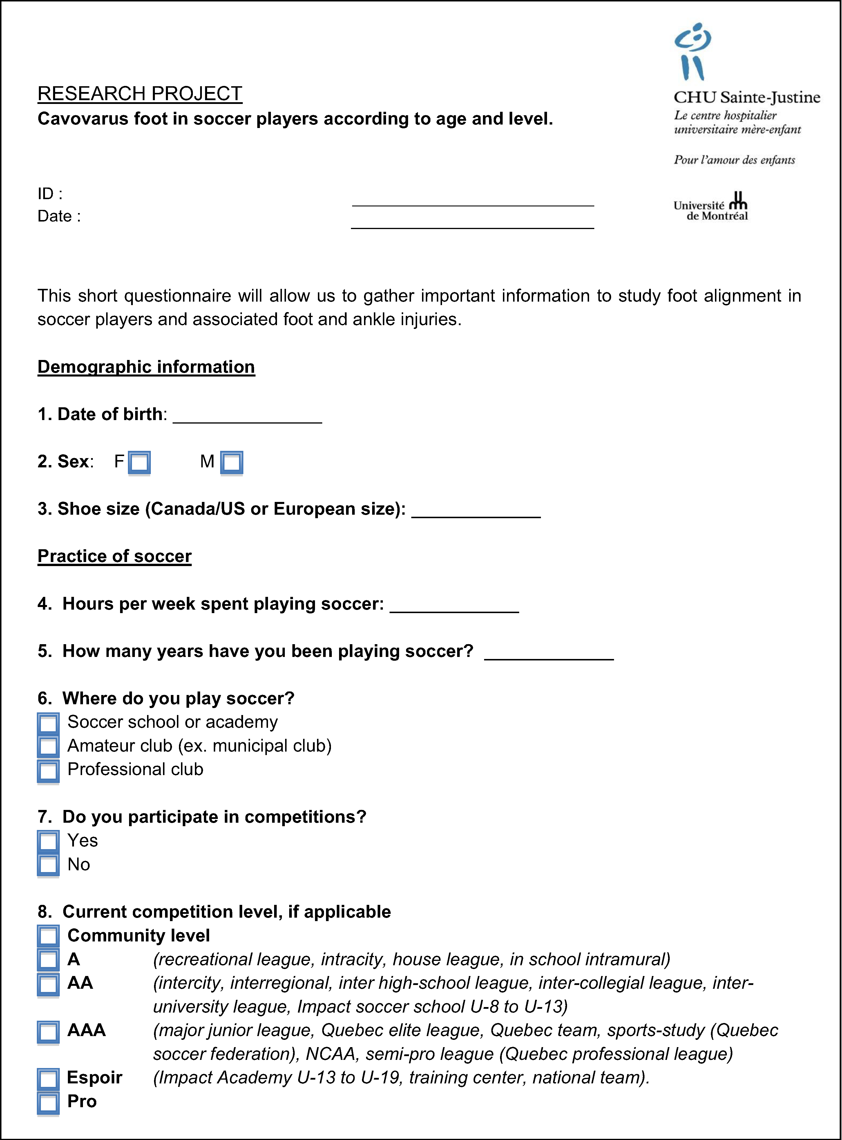
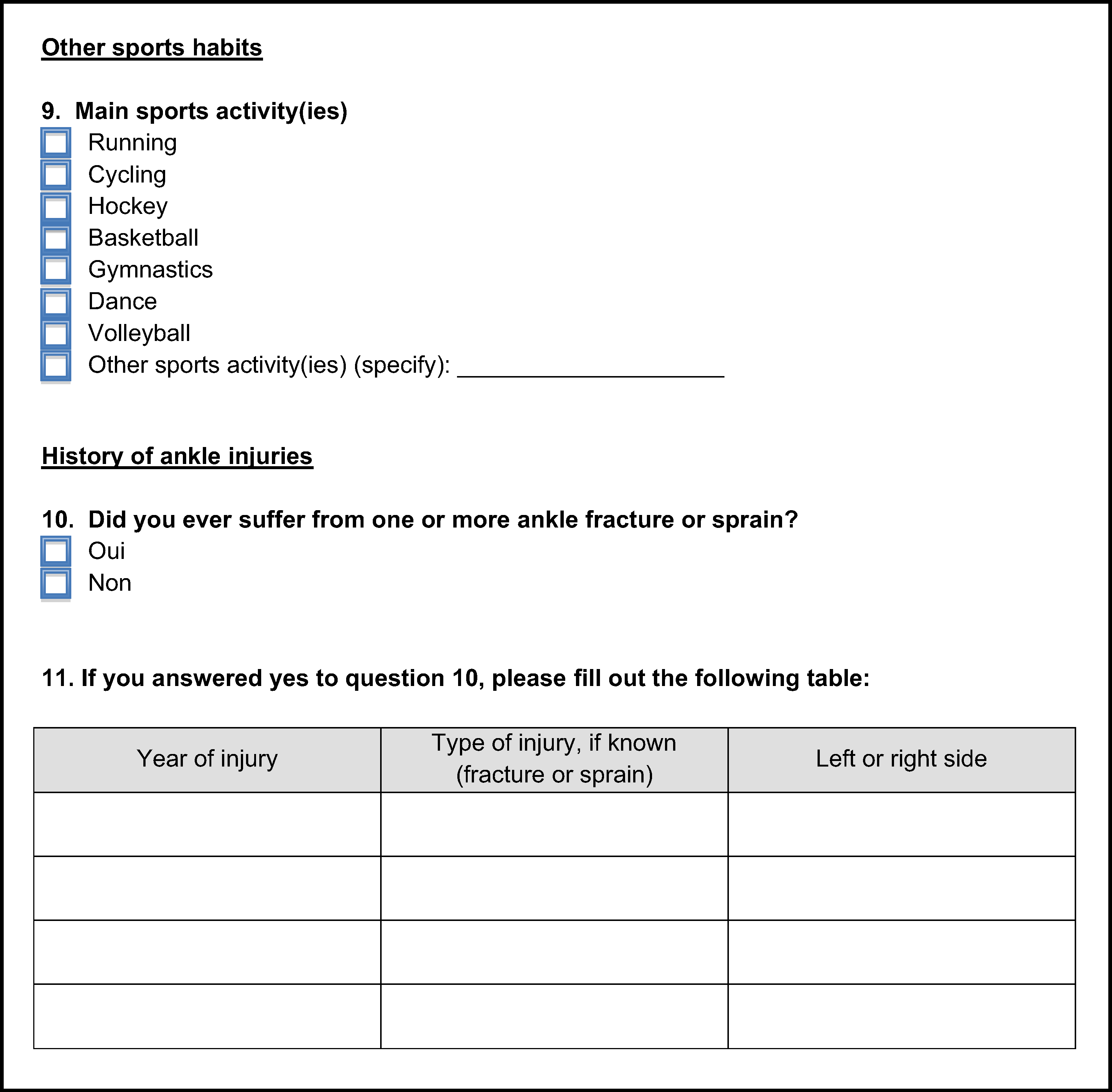
Players completed a soccer-related questionnaire that included injury and soccer histories (Appendix A). Based on their answers, players were classified into one of two groups: healthy or previously injured. More specifically, injury type, affected side, and time of injury were self-reported. Regarding soccer history, players were asked when (year) they started playing soccer, the number of active playing years, and the highest level they reached in their soccer career. Levels were classified into two categories: AA and below (regional level or below) and AAA and above (national level or above).
Foot Measurements
Foot measurements were performed on both feet, including foot length in the static full weightbearing condition using a Brannock device. A vertical digital caliper was used to place a marker on the top surface of each foot at midfoot length. Arch heights were then measured in two positions: 1) sitting with both feet on the ground (approximately 10% of body weight on each foot) and 2) standing on both feet (50% of body weight on each foot). The arch height flexibility was calculated as the difference between sitting and standing arch heights at midfoot normalized to body weight.[21,22] The simplified Foot Posture Index-6 (FPI-6) was used to provide a clinical characterization of each foot.[23,24] The simplified FPI-6 is an evaluation based on six criteria: talar head palpation, lateral malleolar curvature, calcaneal frontal plane position, talonavicular joint bulging, medial longitudinal arch evaluation, and abduction/adduction of the forefoot on the rearfoot. Each criterion was given a score from –2, indicating a more supinated position, to +2, indicating a more pronated position, for a total final FPI-6 score varying from –12 to +12. Normal-arched feet scored from 0 to 5, inclusively.[25] Based on the literature, feet with an FPI-6 score less than or equal to –2 were classified as cavus.[24,26] Two research students were trained to use the same measurement tools and the FPI-6.
Statistical Analyses
Although measurements were performed bilaterally, the right foot was systematically chosen for the analyses as recommended by Langley et al[25] for statistical considerations.[27] Variables included continuous variables (such as age, body mass, foot length, and arch height flexibility) and categorical variables (such as sex, highest level of play, presence of cavus foot type based on FPI-6 values, and presence of injury). The Shapiro-Wilk test showed that continuous variables did not follow a normal distribution, hence nonparametric Mann-Whitney U tests were conducted between the normal arch and cavus foot groups (based on their FPI-6 values), and effect sizes were estimated. To improve the characterization of the injured players group, categories were defined as follows: younger or older than 18 years and AA and below (regional leagues or below) or AAA and above (performance divisions or above) for highest level of play. These dichotomous variables were considered as independent variables when conducting statistical analyses. χ2 Tests were used to study associations between injuries and each previously mentioned variable (sex, age, level of play, and cavus foot). The threshold for statistical significance was set at P < .05. Cramer V values were reported to estimate effect sizes of χ2 tests. Values less than 0.2 indicated that the association was weak, between 0.2 and 0.6 moderate, and greater than 0.6 strong. Post hoc power calculations were also computed. Statistical analyses were performed using IBM SPSS Statistics for Windows, Version 25.0 (IBM Corp, Armonk, New York).
Results
Cohort Description
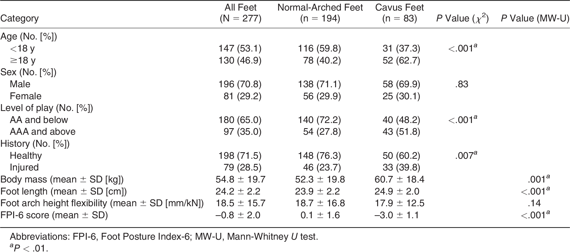
The cohort comprised 277 soccer players, including male (70.8%) and female (29.2%) players. The mean ± SD age of participants was 16.8 ± 7.5 years. Overall, mean ± SD foot length was 24.2 ± 2.2 cm and arch height flexibility was 18.5 ± 15.7 mm/kN. The mean ± SD FPI-6 value was –0.8 ± 2.0. The level of play was AA or below for 65.0% of the players, and 28.5% of the players sustained at least one foot and ankle injury (Table 1).

Table 1.
Descriptive Data of the Players Divided into Normal-Arched and Cavus Feet
Effect of Foot Type: Normal Arched versus Cavus
Players with normal-arched feet were significantly younger (mean ± SD age, 16.0 ± 7.5 years) compared with players with cavus feet (mean ± SD age, 18.8 ± 7.3 years) (P = .001). Results were similar when splitting the cohort into pediatric versus adult populations (Table 1). Along with age, body mass and foot length were also significantly lower in the normal-arched group versus the cavus group (Table 1). A higher level of play (AAA and above) was found more frequently in the cavus foot type group (P < .001) (Table 1), where players were older. All of the post hoc power calculations exceeded 90% except for injury history (76.6%). Sex and arch height flexibility were not significantly different between the normal-arched and cavus foot groups (P > .05).
Risk Factors Related to Foot and Ankle Injuries
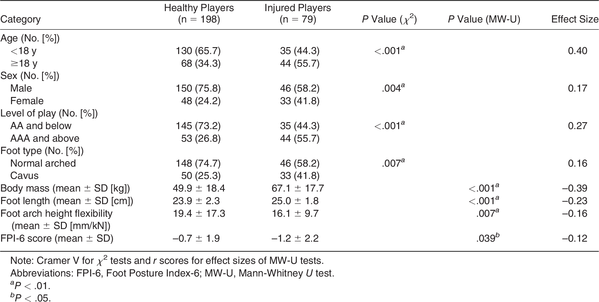
Healthy and injured players had a mean ± SD age of 14.7 ± 6.2 years and 22.1 ± 8.0 years, respectively (P < .001). Foot length, body mass, and highest level played (AA and below versus AAA and above) were all found to be significant (P < .001) regarding injury, with moderate effect sizes (–0.23, –0.39, and 0.27, respectively) (Table 2) and statistical power greater than 95%. Being female was also associated with injuries (P = .004), with 41.8% of females among injured players versus 24.2% in the healthy group (post hoc power of 81.8%). Foot type was significantly different between healthy and injured players, with 41.8% of cavus foot in injured players and 25.3% in the normal-arched group, with power of 76.3%. The effect size (Cramer V = 0.16) for foot type was weak (Table 2).
Table 2.
Comparison of Healthy versus Injured Players
Discussion
The aim of this study was to analyze the relationship between cavus foot type, and foot and ankle injuries in male and female soccer players. This study combined pediatric and adult soccer players to observe the evolution over time, although not in the same individuals. Two analyses were performed comparing 1) the effect of foot type (normal-arched versus cavus) and 2) characteristics of healthy (never injured) versus injured (at least once) players.
Cavus Foot Type Prevalent in Soccer Players
In the present soccer player cohort, the proportion of cavus foot type was 30%, which is two to three times more than in a similar ethnic population with a similar age range.[26,28] Gijon-Nogueron et al[26] tested 3,217 children and found 11% supinated feet. A similar percentage was also observed in a nonathletic population, with 14% cavus feet in individuals aged 10 to 20 years.[28] However, the criteria to define the normal and cavus foot types were not described, whereas the present study used a cutoff value of –2 for the cavus foot group. The mean ± SD FPI-6 score for this study (–0.8 ± 2.0) is similar to that of experienced handball players (–0.4 ± 6.9) with similar age (mean, 21.8 years) found in the literature.[29] Indeed, when the FPI-6 values across different sports were compared, runners and basketball players showed typical mean ± SD normal-arched foot values (2.9 ± 2.8 and 3.9 ± 4.1, respectively).[29] The authors compared adult cohorts and suggested that the FPI-6 difference could be explained by a tendency toward lateralization when jumping in handball, hence the supinated position.[29] A similar parallel could be drawn for soccer players, where ball control requires constant movement from the foot and ankle and a constant tibialis posterior muscle contraction. Specifically, the instep kick movement significantly increases pressure on the lateral part of the foot.[30]
Cavus Foot Type More Prevalent in Experienced Soccer Players
Interestingly, the present study also found an increased prevalence of cavus foot with higher-level players, in both older and younger players. This study is the first to identify an association between a higher level of play and the cavus foot type, although this association was quite weak and should be further investigated. One could wonder whether a cavus foot type provides a competitive advantage. As a result of a natural selection process, players with cavus feet continued practicing soccer over the years, and a higher concentration of players with cavus feet would be found among the professional ranks. In addition to soccer skills, the cavus foot type may also be related to footwear. Soccer shoes place the ankle in a dorsiflexion position that is 7° greater than regular running shoes, limiting the functional range of motion.[30] Future studies should look into the mechanical aspects of foot development, footwear, and injuries.
Higher Injury Risks in Soccer with the Cavus Foot Type
Injury risk factors found in this study were 1) being a female, 2) being older, 3) playing at a higher level, and 4) having a cavus foot. The female sex is already known to be associated with a higher risk of injury.[2,10] Being older and playing at a higher level have also been previously identified as risk factors in the literature,[5,31] and both factors are significantly associated with a Cramer V effect size of 0.58. From an injury prevention perspective, soccer players most at risk for injury can be identified based on their sex, age, and level of play. However, the cavus foot type is a risk factor for injury that can be acted on. Efforts can, therefore, be directed toward the development of orthoses or adapted footwear to correct foot alignment in soccer-specific shoes.
Limitations
Some limitations ought to be presented and discussed. First, this study was cross-sectional and compared young beginners with more experienced and skilled soccer players. A longitudinal follow-up of a young prospective cohort would provide a better understanding of injury and cavus occurrence. In the pediatric subgroup, note that they were analyzed as a single group and their skeletal maturity was not assessed and must be quite different. Second, we used the FPI-6 to characterize and classify feet because it is commonly used, robust, and repeatable. However, in the present cohort, most soccer players had feet bordering between the normal-arched and cavus categories. When studying foot types in a homogenous population, FPI-6 thresholds might not optimally reflect patterns in that population. Foot plantar pressure distribution could help improve foot type quantification to discriminate healthy and potentially pathological cavus feet.[32,33] Third, no details were collected regarding injury types or mechanisms. In future studies, injuries should be recorded and subdivided into two categories based on injury mechanism: player intrinsic (noncontact) and contact, which is defined as a collision with a player or an object.[1] There could be a correlation between noncontact injuries and foot type, which would also provide a basis to guide targeted prevention strategies for players at risk.
Conclusions
This study provided data on foot morphology and injury history in pediatric and adult soccer players. The prevalence of cavus feet in this soccer player cohort was 30%, and this rate increased significantly in more experienced soccer players, regardless of age. The risk factors for foot and ankle injuries identified in this cohort were female sex, older age (>18 years), more experienced (level of play), and a lower FPI-6 score (cavus foot type).
Acknowledgments
Éric Manfoumbi-Mouity for valuable help during data collection, Kathleen Beaumont for manuscript editing and revision, and Nathalie Jourdain for research assistance.
Financial Disclosure
This research was conducted as part of TransMedTech Institute’s activities and was funded in part by the Canada First Research Excellence Fund. We also want to thank the PREMIER program from University of Montreal, a research grant program for undergraduate students.
Conflicts of Interest
Marie-Lyne Nault reports departmental funding to CHU Sainte-Justine from Orthopaediatrics. This project did not receive any funding from this entity and they were not involved in any aspect of the submitted work. The other authors declare that they have no conflict of interest.
References
- FIFA: 270 million people active in football. Available at: https://www.yumpu.com/en/document/read/7283064/265-million-playing-football-fifacom.
- Wong P, Hong Y:Soccer injury in the lower extremities.Br J Sports Med 39:473, 2005.
- Inklaar H:Soccer injuries: I.Incidence and severity. Sports Med 18:55, 1994.
- Jansen P, Lehmann, Fellner B, et al:Relation of injuries and psychological symptoms in amateur soccer players.BMJ Open Sport Exerc Med 5:e000522, 2019.
- Nery C, Raduan F, Baumfeld D:Foot and ankle injuries in professional soccer players: diagnosis, treatment, and expectations.Foot Ankle Clin 21:391, 2016.
- Froholdt A, Olsen OE, Bahr R:Low risk of injuries among children playing organized soccer: a prospective cohort study.Am J Sports Med 37:1155, 2009.
- Silva DCF, Macedo R, Montes AM, et al:Does the cleat model interfere with ankle sprain risk factors in artificial grass? Clin Biomech 63:119, 2019.
- Calloway SP, Hardin DM, Crawford MD, et al:Injury surveillance in major league soccer: a 4-year comparison of injury on natural grass versus artificial turf field.Am J Sports Med 47:2279, 2019.
- Inklaar H:Soccer injuries: II.Aetiology and prevention. Sports Med 18:81, 1994.
- Vanlommel L, Vanlommel J, Bollars P, et al:Incidence and risk factors of lower leg fractures in Belgian soccer players.Injury 44:1847, 2013.
- Engebretsen AH, Myklebust G, Holme I, et al:Intrinsic risk factors for acute ankle injuries among male soccer players: a prospective cohort study.Scand J Med Sci Sports 20:403, 2010.
- Schwebel DC, Banaszek MM, McDaniel M:Brief report: behavioral risk factors for youth soccer (football) injury.J Pediatr Psychol 32:411, 2007.
- Aydog ST, Tetik O, Demirel HA, et al:Differences in sole arch indices in various sports.Br J Sports Med 39:e5, 2005.
- Barker HB, Beynnon BD, Renström PA:Ankle injury risk factors in sports.Sports Med 23:69, 1997.
- Dahle LK, Mueller MJ, Delitto A, et al:Visual assessment of foot type and relationship of foot type to lower extremity injury.J Orthop Sports Phys Ther 14:70, 1991.
- Schwend RM, Drennan JC:Cavus foot deformity in children.J Am Acad Orthop Surg 11:201, 2003.
- Abbasian A, Pomeroy G:The idiopathic cavus foot: not so subtle after all.Foot Ankle Clin 18:629, 2013.
- Tong JW, Kong PW:Association between foot type and lower extremity injuries: systematic literature review with meta-analysis.J Orthop Sports Phys Ther 43:700, 2013.
- Matsuda S, Fukubayashi T, Hirose N:Characteristics of the foot static alignment and the plantar pressure associated with fifth metatarsal stress fracture history in male soccer players: a case-control study.Sports Med Open 3:27, 2017.
- Rosenbaum D: “Assessing Pediatric Foot Deformities by Pedobarography,” in Handbook of Human Motion, ed by B Müller, S Wolf, G-P Brueggemann, et al, p 711, Springer, Cham, Switzerland, 2016.
- Zifchock RA, Theriot C, Hillstrom HJ, et al:The relationship between arch height and arch flexibility.JAPMA 107:119, 2017.
- Zifchock RA, Parker R, Willahelm W, et al:The relationship between foot arch flexibility and medial-lateral ground reaction force distribution.Gait Posture 69:46, 2019.
- Hillstrom HJ, Song J, Kraszewski AP, et al:Foot type biomechanics: part 1. Structure and function of the asymptomatic foot.Gait Posture 37:445, 2013.
- Redmond AC, Crane YZ, Menz HB:Normative values for the foot posture index.J Foot Ankle Res 31:6, 2008.
- Langley B, Cramp M, Morrison SC:Clinical measures of static foot posture do not agree.J Foot Ankle Res 9:1, 2016.
- Gijon-Nogueron G, Martinez-Nova A, Alfageme-Garcia P, et al:International normative data for paediatric foot posture assessment: a cross-sectional investigation.BMJ Open 9:1, 2019.
- Menz HB:Two feet, or one person? problems associated with statistical analysis of paired data in foot and ankle medicine.Foot 14:2, 2004.
- Troiano G, Nante N, Citarelli GL:Pes planus and pes cavus in Southern Italy: a 5 years study.Ann 1st Super Sanita 53:142, 2017.
- Martínez-Nova A, Gómez-Blázquez E, Escamilla-Martínez E, et al:The foot posture index in men practicing three sports different in their biomechanical gestures.JAPMA 104:154, 2014.
- Blanchard S, Palestri J, Guer JL, et al:Current soccer footwear, its role in injuries and potential for improvement.Sports Med Int Open 2:E52, 2018.
- Oztekin HH, Boya H, Ozcan O, et al:Foot and ankle injuries and time lost from play in professional soccer players.Foot 19:22, 2009.
- Azevedo RR, da Rocha ES, Franco PS, et al:Plantar pressure asymmetry and risk of stress injuries in the foot of young soccer players.Phys Ther Sport 24:39, 2017.
- Franklyn-Miller A, Bilzon J, Wilson C, et al:Can RSScan footscan® D3D™ software predict injury in military population following plantar pressure assessment? a prospective cohort study.Foot 24:6, 2014.
Appendix A. Questionnaire Submitted to Players
© 2023 American Podiatric Medical Association