


Abstract
Necrotizing fasciitis of the foot is a relatively rare diagnosis and has traditionally been treated with distal amputation. A 30-year-old diabetic man with Charcot-Marie-Tooth muscular atrophy developed necrotizing fasciitis of the dorsal foot and underwent surgical debridement resulting in a significant wound with exposed tendons. Serial debridements were performed, eventually followed by a staged free flap reconstruction using an anterolateral thigh fasciocutaneous flap. After allowing time for flap healing, subsequent staged equinovarus reconstruction was also performed successfully. There were no flap or postoperative complications, and the patient is progressing as expected. Flap refinement procedures have been used to enhance cosmetic and functional outcomes. This report not only showcases the success of a procedure high on the reconstructive ladder in a patient at high risk for complications but also highlights an approach in which functional recovery is also optimized successfully in a planned staged multidisciplinary manner.
The annual incidence of necrotizing fasciitis ranges from 0.3 to 5.0 cases per 100,000 population [1,2]. Approximately one-third of these infections are associated with diabetes mellitus [2–6], and 38% to 54% affect the lower extremities [2,3]. The prevalence of Charcot-Marie-Tooth disease (CMT) in the general population is 10% with variable phenotypic expression, and the overall prevalence in type 2 diabetes is even more rare, with only case studies noted in the literature [7,8]. Furthermore, the presentation of necrotizing fasciitis of the foot in a patient with type 2 diabetes with severe CMT deformity has not been formally described in the literature to date.
Excisional debridement is the primary treatment of necrotizing soft-tissue infections, and multiple debridements are often required to achieve appropriate source control [1,9]. Debridement often results in a large wound that may not be durably covered with local or synthetic reconstructive options. Particularly in the extremities, these defects pose a therapeutic dilemma, and the literature is scarce regarding indications for free flap reconstruction versus amputation. Necrotizing fasciitis of the foot in the context of diabetes, where there is an increased risk of peripheral vascular disease, has traditionally been treated with preemptive distal limb amputation [10,11]. However, studies have shown that salvage of the distal limb can be achieved using microvascular free tissue transfer [12–14]. The frequency of this treatment is low, and less than 10% of all necrotizing fasciitis cases receive free tissue transfer reconstruction [3,15]. Thus, we present a unique case in which a diabetic man with a significant dorsal foot wound secondary to necrotizing fasciitis undergoes reconstruction with a free flap followed by a staged reconstruction of a CMT equinovarus deformity.
Case Report
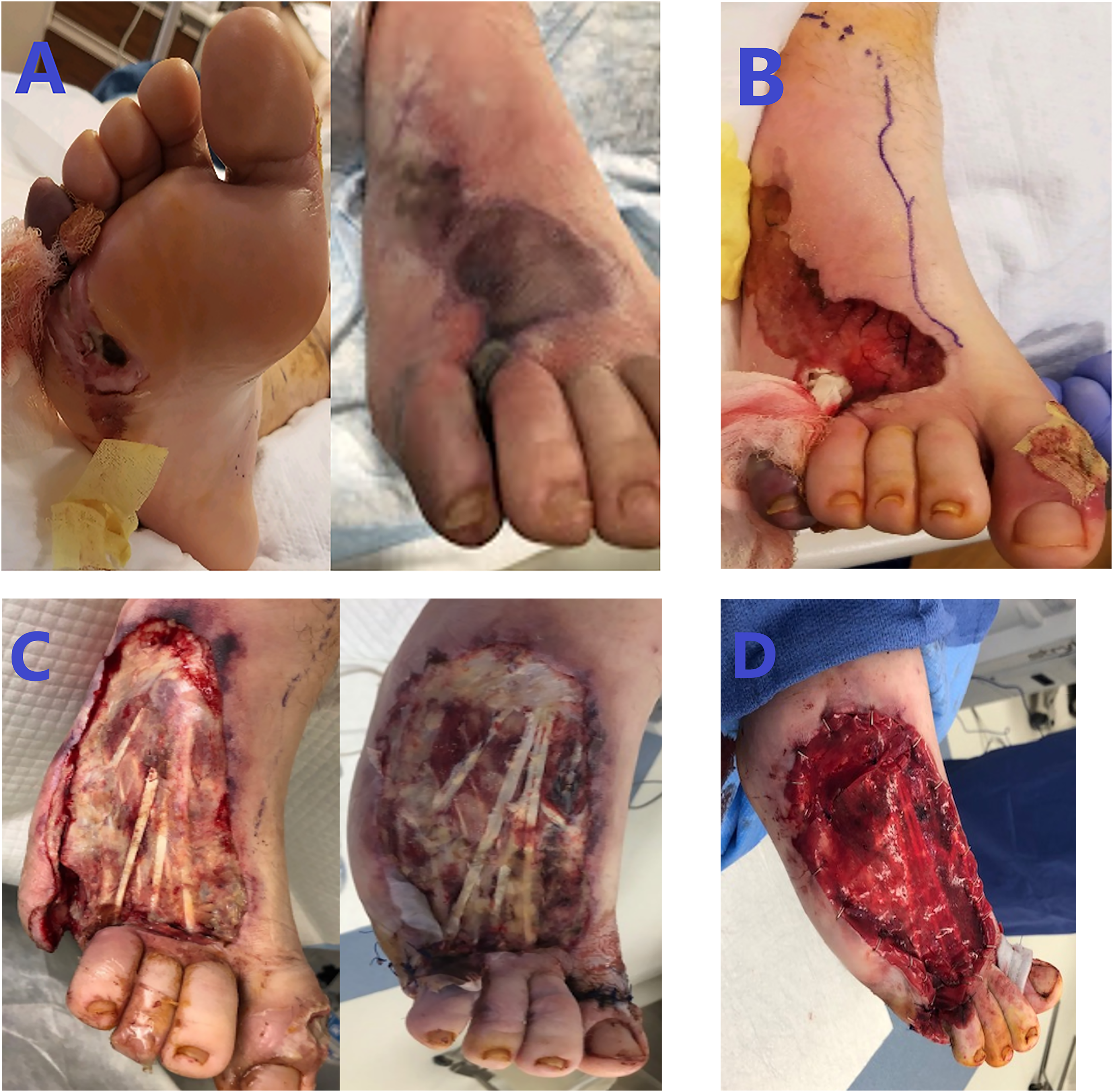
A 30-year-old man with a medical history of poorly controlled latent autoimmune diabetes of adulthood, hypertension, and CMT disease diagnosed by phenotypic expression and previous neurology evaluation presented in early March 2019 to an outside hospital with a large hemorrhagic blister between his right fourth and fifth toes extending toward the dorsum of the foot with peripheral bluish discoloration and increased swelling (Fig. 1A). He was tachycardic, with a white blood cell count of 27 × 109/L and a plain film of the right foot demonstrating gas in the dorsal soft tissues. He was started on intravenous vancomycin, clindamycin, and zosyn. He was taken immediately to the operating room by podiatric surgery and underwent a right foot debridement and partial fifth-ray amputation (Fig. 1B). Intraoperative wound cultures identified group B streptococcus (Streptococcus agalactiae). He was then transferred to our tertiary hospital for complex reconstruction evaluation.
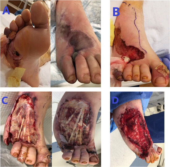
Figure 1.
Images of the right foot at presentation (A), status post initial outside hospital debridement (B), status post debridement after transfer (C), and status post additional debridement, proximal interphalangeal joint resection of the hallux, application of Integra wound graft (Integra LifeSciences, Cincinnati, Ohio) over the area, extensor hallucis longus lengthening, and metatarsophalangeal joint capsulotomy of digits 1 through 4 (D).
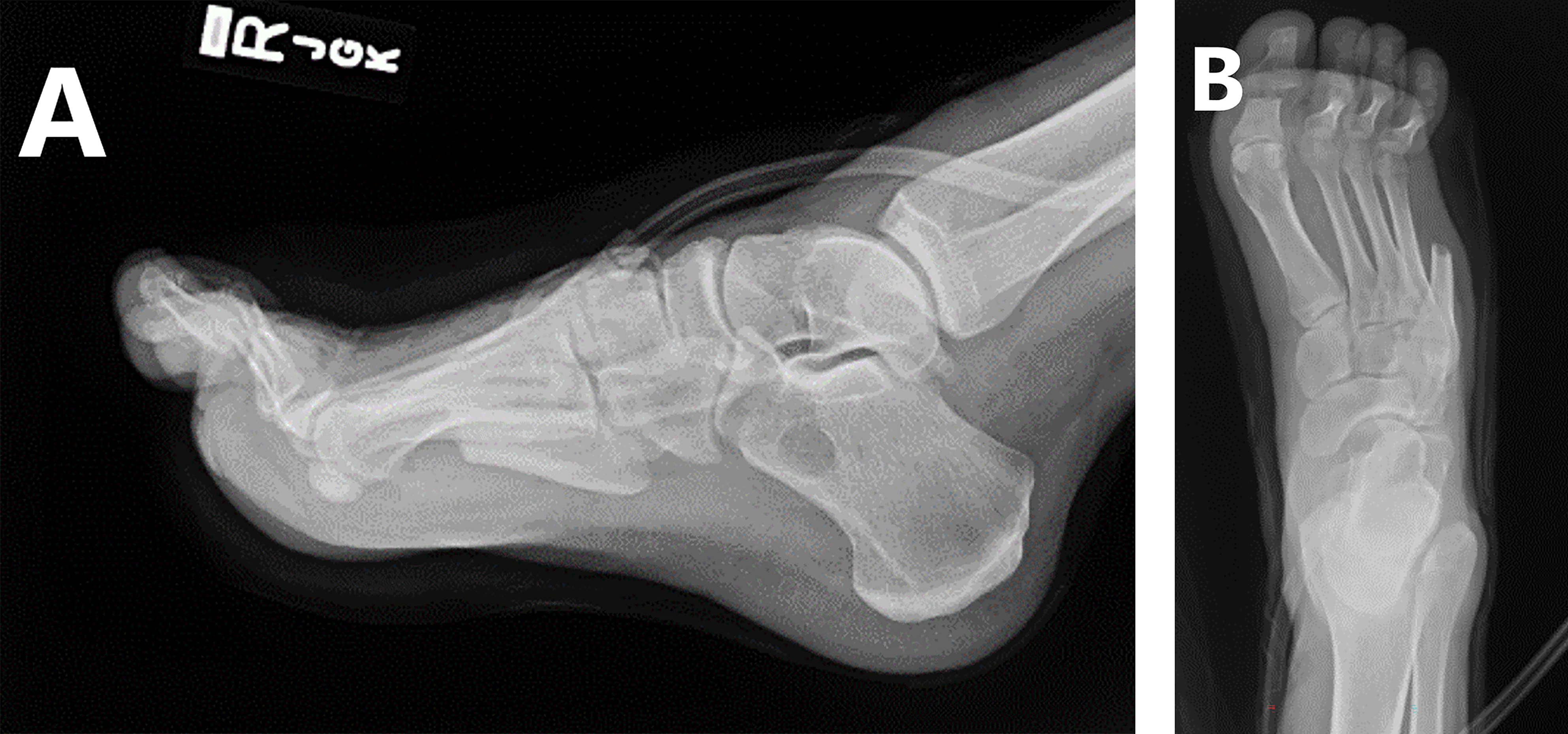
At our institution, physical examination showed an open wound involving most of the dorsum of the foot extending laterally and distally toward the fifth-ray amputation site with exposed tendon. Repeated plain films of the foot demonstrated the pes cavus deformity with gastrocnemius and soleus muscle equinus of 27° and an absence of soft-tissue gas (Fig. 2).
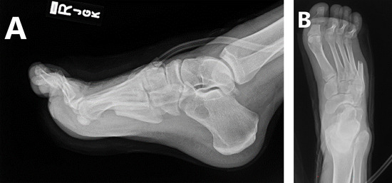
Figure 2.
Lateral (A) and anteroposterior (B) radiographs of the right foot after hospital transfer.
He was taken to the operating room several times for additional debridements, bony resections (right hallux’s proximal and distal phalanx, fourth digit at the metatarsophalangeal joint), extensor hallucis longus lengthening, and metatarsophalangeal joint capsulotomy of digits 1 through 4 before wound bed preparation for flap placement (Fig. 1D). The prepared wound bed measured 8 × 15 cm.
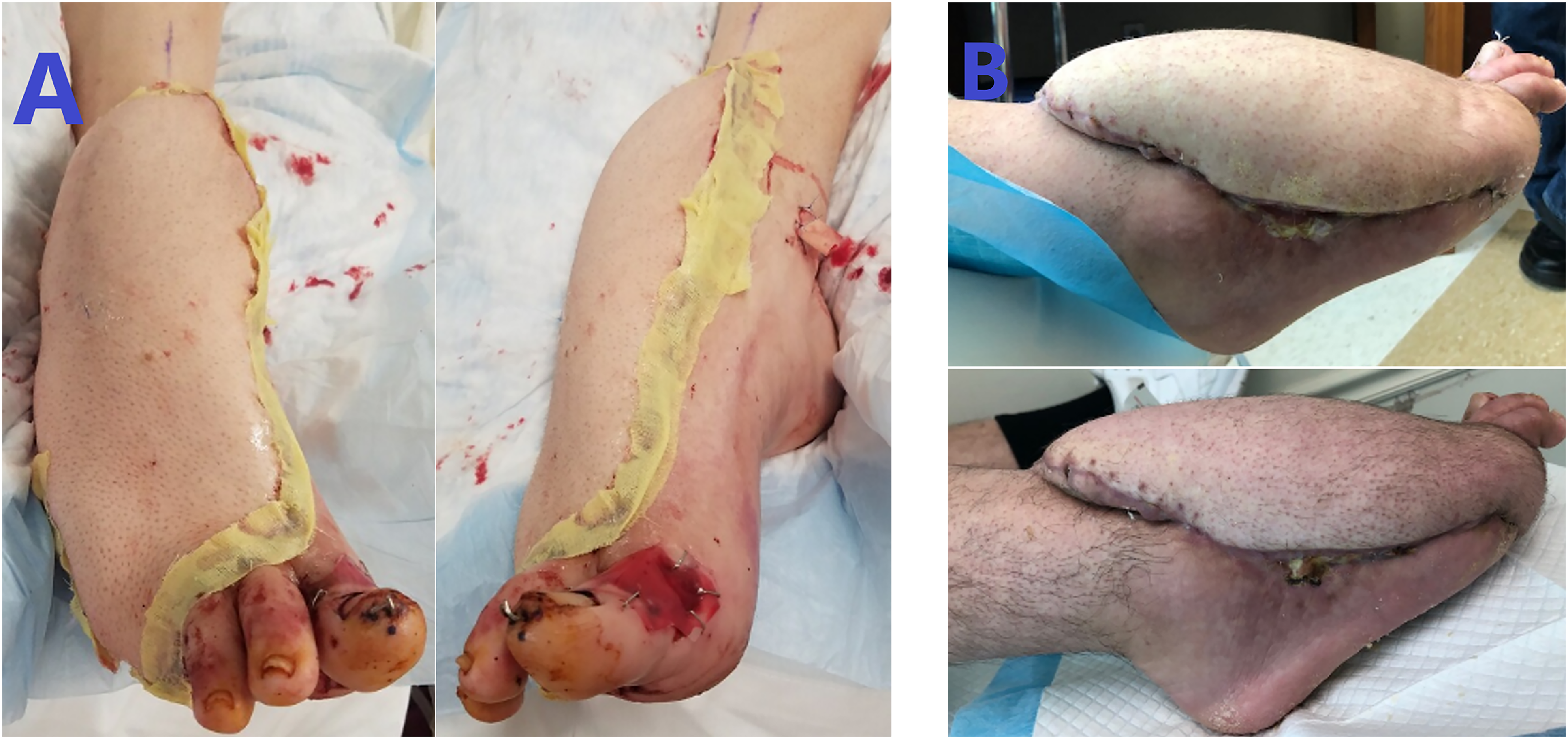
The patient underwent a left anterolateral thigh free flap to the right foot (Fig. 3A). The anterolateral thigh flap was raised in the standard fashion and measured 14 × 32 cm. The anterior tibial artery and the greater saphenous vein were exposed and prepared. The flap was then transferred successfully, with resultant cutaneous Doppler signals. Postoperatively, he progressed through the flap check and dangling protocol without issue. The right lower extremity remained elevated in a posterior splint with nonweightbearing restriction for 14 days. No postoperative complications were noted. The total length of stay was 50 days. The patient returned to the clinic monthly, during which the flap was noted to be healing appropriately (Fig. 3B).
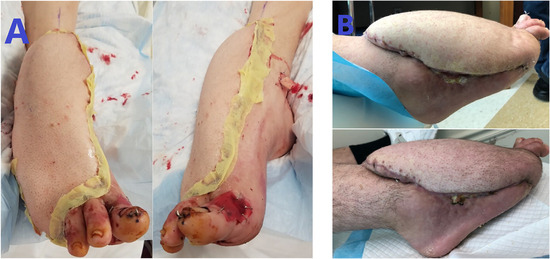
Figure 3.
Left anterolateral thigh fasciocutaneous free flap to the dorsum of the right foot immediately postoperatively (A) and on postoperative days 14 and 35 (B).
Equinocavovarus reconstruction was performed 4 months after the flap procedure by podiatric surgery with a Steindler stripping, open Achilles lengthening, dorsiflexory wedge osteotomy of the first metatarsal, interphalangeal joint arthrodesis, and Dwyer calcaneal osteotomy (Fig. 4). The patient was discharged postoperatively in a posterior splint and continued to be nonweightbearing for the right lower extremity. No complications were noted in his postoperative course, and outpatient plain films demonstrated optimal reconstruction. At 6-week follow-up he was weaned from narcotic pain medications and transitioned to a controlled ankle movement boot with advancement of weightbearing status to weightbearing as tolerated. The patient continued to improve as expected and then had his flap debulked approximately 1.5 years after flap placement. He was recently seen in the clinic after debulking and was healing as expected (Fig. 5). His care and rehabilitation have been completed, with plantigrade pedal alignment (Fig. 6).
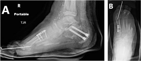
Figure 4.
Lateral (A) and anteroposterior (B) radiographs of the right foot immediately after cavus foot reconstruction.
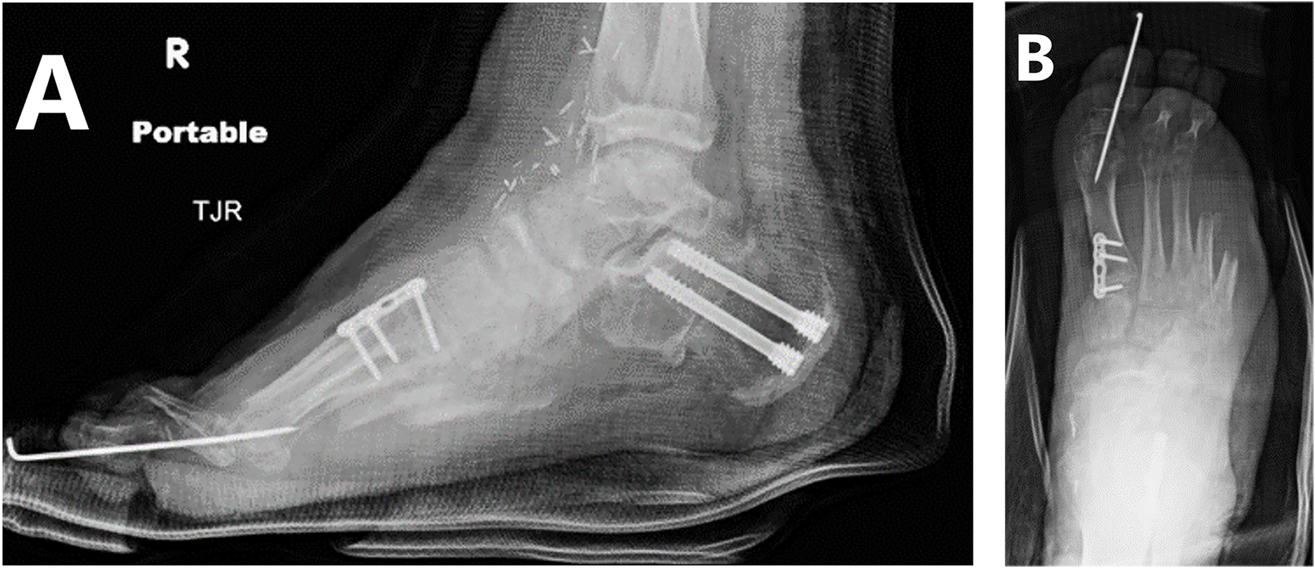
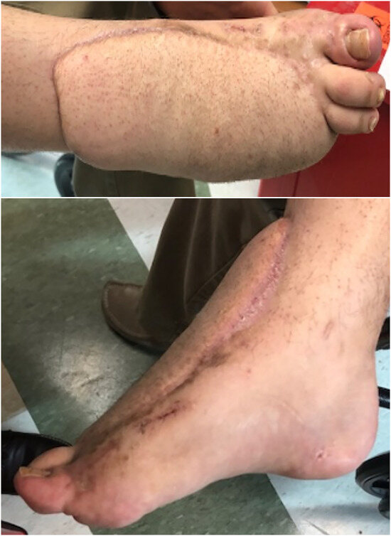
Figure 5.
Right foot at the 12-month clinic follow-up.
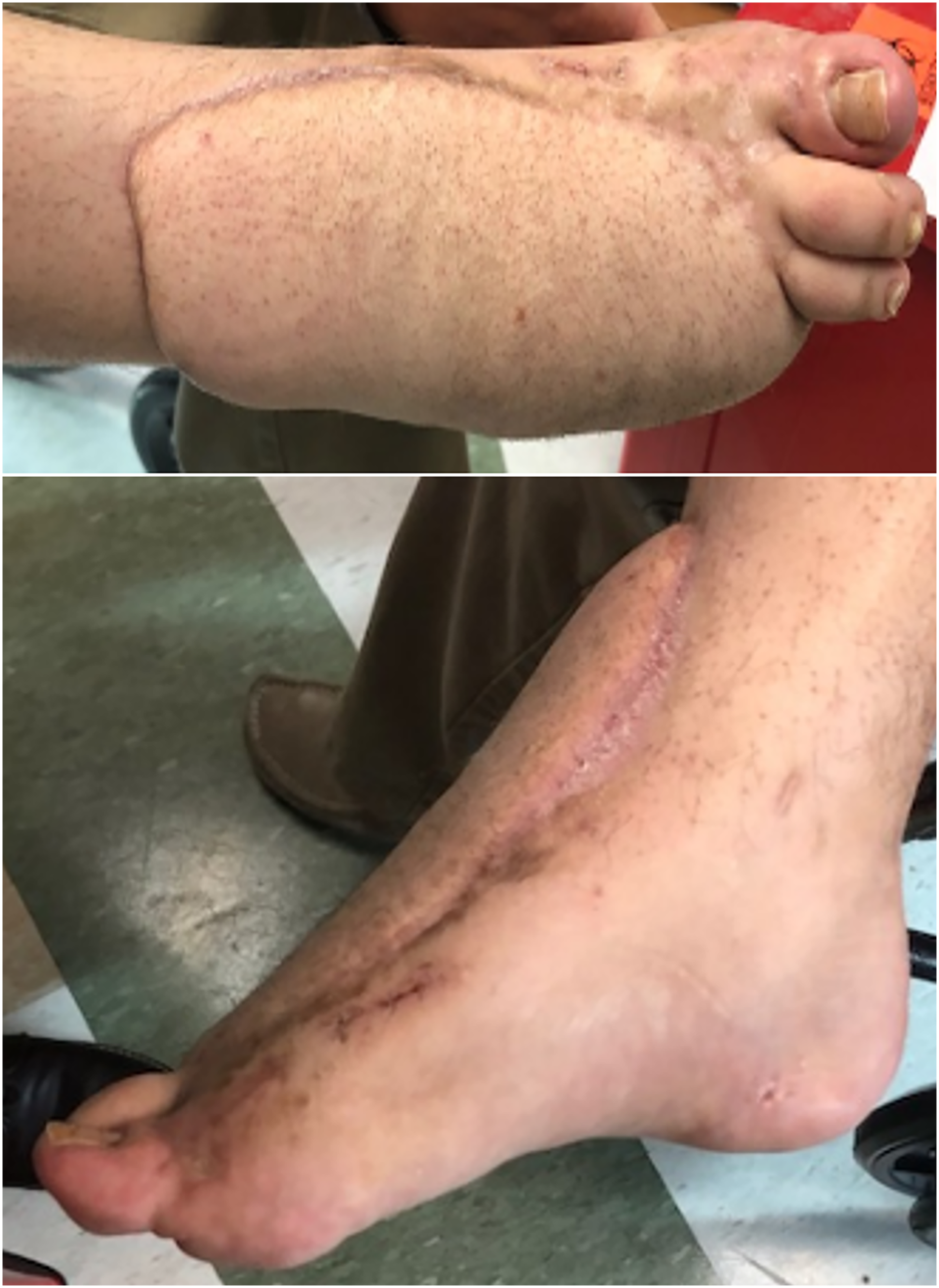
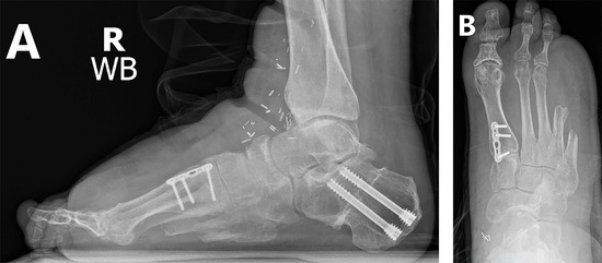
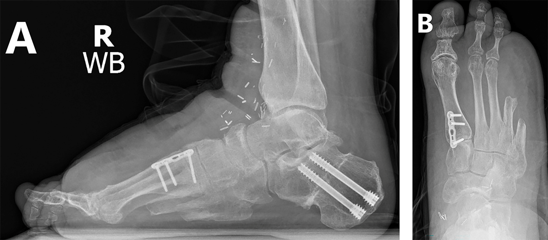
Figure 6.
Lateral (A) and anteroposterior (B) radiographs at 18-month follow-up demonstrating restoration of the plantigrade angle and talar first metatarsal axis.
Discussion
Although diabetic foot infections have excellent limb salvage rates, wound healing after debridement for necrotizing fasciitis is difficult due to underlying comorbidity-related factors, wound size, and the frequent presence of exposed musculoskeletal components [16]. This situation was particularly tenuous due to the presence of group B streptococcus, which is an exceedingly rare and aggressive cause of necrotizing fasciitis and has been reported in only case reports and series [17]. These wounds are not suitable for skin grafting or coverage with synthetic skin substitutes, and neither is there enough local skin for local flaps [4–10]. Thus, free flap reconstruction should be pursued in these patients, especially given that diabetic foot reconstructions have been shown to have a high rate of success, with a significant increase in 5-year survival [18].
Flap reconstruction should focus on adequate tissue coverage over initial cosmetic considerations. Pedicled flaps such as distally based dorsal metatarsal artery perforator flaps can be an adequate option; however, they are limited in wide or composite defects or when the vascular pedicle of the flap has been damaged [19–21]. In this instance, the wound defect was large, and no pedicled flaps were available. Thus, a free flap was the ideal option in this defect. The anterolateral thigh flap was chosen because it is comparable with other traditional free flaps and provides durable, supple coverage in lower-extremity limb salvage [21]. The anterolateral thigh flap is a safe, reliable, and aesthetically appealing option for resurfacing traumatic dorsal foot wounds [22–24].
Peripheral arterial disease and diabetes are well-established risk factors that increase the chance of flap loss [25,26]. However, with proper preoperative planning and optimization, foot and ankle free tissue transfers have generally been shown to be successful, with a low incidence of foot ischemia in diabetic patients [27,28].
The goal of pursuing reconstruction should ultimately be the restoration of function. The present patient had a CMT pes cavus deformity, which can be progressively debilitating and eventually lead to persistent wounds that could result in a major amputation, particularly when combined with type 2 diabetes [29]. Historically, CMT could be managed with custom ankle-foot orthoses, yet the progressive nature of this hereditary motor and sensory neuropathy could lead to a nonbraceable extremity in the context of an insensate foot susceptible to neuropathic ulceration. A myriad of surgical procedures has been proposed in the literature with the focus on multiplanar correction and prevention of recurrence and maintaining long-term functional outcomes. Good-to-excellent results have been demonstrated in the literature with extra-articular osteotomies. Faldini and colleagues [30] demonstrated favorable outcomes in 92% of patients treated with extra-articular procedures at mean follow-up of 6 years. Ward and colleagues [31] retrospectively reviewed 25 consecutive patients with CMT and performed a series of soft-tissue procedures and tendon transfers to rebalance the foot and ankle, with notable improvements in 36-Item Short-Form Health Survey and Foot Function Index scores as well as minimal progression to degenerative joint disease and clinical parameters with respect to gait at mean follow-up of 26.1 years. In this particular case, the authors opted for an Achilles tendon lengthening for the equinus component and obviated performing a posterior tibial tendon transfer given the lack of profound deforming influence at the midtarsal joint complex. In addition, a Steindler stripping was incorporated for the global cavus component of the deformity and use of the Dwyer calcaneal osteotomy for hindfoot varus correction based on the hindfoot axial alignment/varus resting calcaneal stance position.
We demonstrated successful wound reconstruction and structural deformity correction in a rare case of a diabetic patient with CMT after surgical management of necrotizing fasciitis. Even in the setting of a severe infection that frequently leads to major amputations, if there is limb viability with the chance for eventual independent function, then wound reconstruction can be pursued successfully with subsequent correction of any foot deformities to maximize functional restoration. More research will be needed to determine short- and long-term outcomes; however, we propose that free flap reconstruction with staged anatomical correction can be performed successfully on select diabetic patients with substantial pedal deformities and large wounds secondary to severe infections.
Financial Disclosure
None reported.
Conflicts of Interest
None reported.
References
- Stevens DL, Bryant AE: Necrotizing soft-tissue infections. N Engl J Med 377: 2253, 2017.
- Glass GE, Sheil F, Ruston JC, et al: Necrotising soft tissue infection in a UK metropolitan population. Ann R Coll Surg Engl 97: 46, 2015.
- Tsitsilonis S, Druschel C, Wichlas F, et al: Necrotizing fasciitis: is the bacterial spectrum changing? Langenbecks Arch Surg 398: 153, 2013.
- Miller LG, Perdreau-Remington F, Rieg G, et al: Necrotizing fasciitis caused by community-associated methicillin-resistant Staphylococcus aureus in Los Angeles. N Engl J Med 352: 1445, 2005.
- Bucca K, Spencer R, Orford N, et al: Early diagnosis and treatment of necrotizing fasciitis can improve survival: an observational intensive care unit cohort study. ANZ J Surg 83: 365, 2013.
- Bocking N, Matsumoto CL, Loewen K, et al: High incidence of invasive group A streptococcal infections in remote indigenous communities in northwestern Ontario, Canada. Open Forum Infect Dis 4: ofw243, 2017.
- Sachithanandam V, Joseph B: The influence of footwear on the prevalence of flat foot: a survey of 1846 skeletally mature persons. J Bone Joint Surg Br 77: 254, 1995.
- Win HH, Davenport C, Delaney S, et al: Charcot-Marie-Tooth disease complicating type 2 diabetes. JAPMA 101: 349, 2011.
- Caravaggi C, Cimmino M, Caruso S, et al: Intramedullary compressive nail fixation for the treatment of severe Charcot deformity of the ankle and rear foot. J Foot Ankle Surg 45: 20, 2006.
- Iacopi E, Coppelli A, Goretti C, et al: Necrotizing fasciitis and the diabetic foot. Int J Low Extrem Wounds 14: 316, 2015.
- Goh T, Goh LG, Ang CH, et al: Early diagnosis of necrotizing fasciitis. Br J Surg 101: e119, 2014.
- Randall KL, Booth BA, Miller AJ, et al: Use of an acellular regenerative tissue matrix in combination with vacuum-assisted closure therapy for treatment of a diabetic foot wound. J Foot Ankle Surg 47: 430, 2008.
- Fugitt JB, Puckett ML, Quigley MM, et al: Necrotizing fasciitis. Radiographics 24: 1472, 2004.
- Heller L, Levin LS: Lower extremity microsurgical reconstruction. Plast Reconstr Surg 108: 1029, 2001.
- Sunar H, Aygit CA, Afsar Y, et al: Arterial and venous reconstruction for free tissue transfer in diabetic ischemic foot ulcers. Eur J Vasc Endovasc Surg 27: 210, 2004.
- Xiong L, Gazyakan E, Kremer T, et al: Free flaps for reconstruction of soft tissue defects in lower extremity: a meta-analysis on microsurgical outcome and safety. Microsurgery 36: 511, 2016.
- Collis S, Hoskinson S: Fulminant group B streptococcal necrotizing fasciitis in a patient with undiagnosed malignancy: a case report. IDCases 19: e00655, 2019.
- Lee JY, Jung H, Kwon H, et al: Extended negative pressure wound therapy-assisted dermatotraction for the closure of large open fasciotomy wounds in necrotizing fasciitis patients. World J Emerg Surg 9: 29, 2014.
- Kim BS, Choi WJ, Baek MK, et al: Limb salvage in severe diabetic foot infection. Foot Ankle Int 32: 31, 2011.
- Cho EH, Garcia RM, Pien I, et al: Vascular considerations in foot and ankle free tissue transfer: analysis of 231 free flaps. Microsurgery 36: 276, 2016.
- van Alphen NA, Laungani AT, Christner JA, et al: The distally based dorsal metatarsal artery perforator flap: vascular study and clinical implications. J Reconstr Microsurg 32: 245, 2016.
- Pinsolle V, Reau AF, Pelissier P, et al: Soft-tissue reconstruction of the distal lower leg and foot: are free flaps the only choice? review of 215 cases. J Plast Reconstr Aesthet Surg 59: 912, 2006.
- Bibbo C, Nelson J, Fischer JP, et al: Lower extremity limb salvage after trauma: versatility of the anterolateral thigh free flap. J Orthop Trauma 29: 563, 2015.
- El-Gammal TA, El-Sayed A, Kotb MM, et al: Dorsal foot resurfacing using free anterolateral thigh (ALT) flap in children. Microsurgery 33: 259, 2013.
- Sato T, Yana Y, Ichioka S: Free flap reconstruction for diabetic foot limb salvage. J Plast Surg Hand Surg 51: 399, 2017.
- Myung Y, Yim S, Kim BK: A comparison of axial circumference between superficial circumflex iliac artery perforator flap and other workhorse flaps in dorsal foot reconstruction. J Plast Surg Hand Surg 51: 381, 2017.
- Althouse AD, Abbott JD, Forker AD, et al; BARI 2D Study Group: Risk factors for incident peripheral arterial disease in type 2 diabetes: results from the Bypass Angioplasty Revascularization Investigation in type 2 Diabetes (BARI 2D) Trial. Diabetes Care 37: 1346, 2014.
- Adler AI, Stevens RJ, Neil A, et al: UKPDS 59: hyperglycemia and other potentially modifiable risk factors for peripheral vascular disease in type 2 diabetes. Diabetes Care 25: 894, 2002.
- Sheth S, Francies K, Siskind CE, et al: Diabetes mellitus exacerbates motor and sensory impairment in CMT1A. J Peripher Nerv Syst 13: 299, 2008.
- Faldini C, Traina F, Nanni M, et al: Surgical treatment of cavus foot in Charcot-Marie-tooth disease: a review of twenty-four cases: AAOS exhibit selection. J Bone Joint Surg Am 97: e30, 2015.
- Ward CM, Dolan LA, Bennett DL, et al: Long-term results of reconstruction for treatment of a flexible cavovarus foot in Charcot-Marie-Tooth disease. J Bone Joint Surg Am 90: 2631, 2008.
© 2023 American Podiatric Medical Association