


Abstract
Herpetic whitlow is a viral infection of the fingers or toes caused by the herpes simplex virus. Herpes simplex virus is a common pathogen that causes infections in any cutaneous or mucocutaneous surface, most commonly gingivostomatitis or genital herpes. However, infection of the digits is also infrequently reported. Herpetic whitlow occurs when the virus infects the distal phalanx of the fingers or toes by means of direct inoculation, causing pain, swelling, erythema, and vesicle formation. The proper diagnosis is important because the condition can mimic various other podiatric abnormalities such as paronychia, bacterial cellulitis, or even embolic disease. Improper diagnosis often leads to unnecessary work-up, antibiotic therapy, or even surgical intervention. This case will help illuminate the clinical presentation of herpetic whitlow in an atypical location, and the patient’s subsequent treatment. We present an atypical case of right hallux herpetic whitlow with delayed diagnosis and associated cellulitis. The patient was admitted after seeing multiple providers for a progressive right hallux infection that presented as a mixture of vesicular lesions and apparent cellulitis. His history was positive for biting his fingernails and toenails, and the lesions were noted to be honeycomb-like, with minimal drainage. The lesions were then deroofed and viral cultures were obtained, which were positive for herpes simplex virus type 1, thus confirming a diagnosis of herpetic whitlow. Although he remained afebrile with negative wound cultures during admission, a secondary bacterial infection could not be excluded because of his nail avulsion and surrounding cellulitis. He was discharged on oral antibiotics, antivirals, and wound care recommendations. Herpetic whitlow should be included in the differential diagnosis of pedal digital lesions that appear as vesicular or cellulitic in the pediatric population.
Herpetic whitlow is the cutaneous infection of the digits caused by the herpes simplex virus (HSV), either type 1 or type 2 [1–3]. The primary infection is through direct exogenous or autogenous inoculation through broken skin, often through a torn cuticle or nail biting. Exposure to HSV results in viral invasion and replication within the epidermal and dermal cells; however, in some cases, it may progress into the sensory dorsal root ganglion, where latency can be established [4].
Signs of infection usually occur within 2 to 20 days following exposure [2]. Vesicles appear on the skin or around the nail fold, and generally increase in number until they coalesce into a large, distinct honeycomb-appearing bulla at roughly 5 to 6 days. The infection itself is usually self-limited and resolves in approximately 2 to 4 weeks [2,5].
Although herpetic whitlow can be found in any age group, it is found most commonly in children who suck or bite their thumb/toes, and in health-care providers exposed to a patient's oral mucosa [6]. The highest health-care incidences are recorded in dental hygienists and respiratory therapists with an incidence rate of approximately 2.4 cases per 100,000 people per year [7].
A diagnosis of herpetic infections may be identified by performing a microscope examination of the margin of the vesicles in a Tzanck test, and also by unroofing a vesicle, swabbing it for a sample, and staining it with Giemsa, which shows characteristic multinucleated giant cells. Negative tests can show the herpes virus by electron microscopy, whereas polymerase chain reaction (PCR) may confirm the diagnosis by immunofluorescence and will differentiate type 1 from type 2 HSV [8].
The treatment algorithm for HSV digital infection is a combination of medications, and wound care. Treatment drugs for HSV infection include acyclovir, valacyclovir, or famciclovir in addition to antibiotics and analgesics. This treatment algorithm aims at halting viral replication, relieving pain, and preventing a bacterial superinfection. The course of infection in a nonimmunocompromised patient is typically 2 to 4 weeks, with the possibility of recurrence in 30% to 50% [9]. Although treatment with acyclovir may be effective in preventing recurrences in the latent or prodromal phases, its utility in established infections is unclear because of the limited number of studies [10]. Some data show that antiviral medications started within the first 48 hours of symptoms can possibly shorten the duration of the patient’s symptoms [9]. Dressing the digital infection not only covers the lesion and protects it, but also assists with preventing viral shedding until the epidermal layer has healed [11,12].
Herpetic whitlow should be differentiated from various other infections of the digits, such as a bacterial felon, cellulitis, acute thrombosis, and paronychia. When misdiagnosed, occasional surgical interventions such as incision and drainage or debridement are performed, which provide no symptomatic relief and have the ability to cause viremia, viral encephalitis, or a bacterial infection [10,11]. Clinical examination, the patient’s history, and pathologic findings should all be used to come to the proper diagnosis. In this article, we present a case report of herpetic whitlow of the right hallux with delayed diagnosis and associated cellulitis. The patient’s delay in diagnosis likely stemmed from it being an uncommon infection, its rare citation in the podiatric literature, and its uncommon prevalence in toes in the pediatric, nonimmunocompromised population.
Case Report
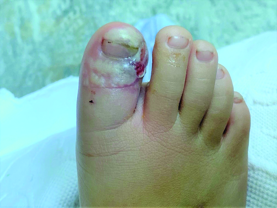
A 6-year-old, fully immunized boy with no significant medical history presented to our hospital after 10 days of a worsening right distal hallux infection. His symptoms started as right hallux pain, itching, and redness, and over the following 24 hours progressed into a pustular lesion. His mother attempted treatment by using antibiotic ointment and adhesive bandages at home, with no resolution. After 3 days, he was seen by an outside podiatrist, who diagnosed him with a paronychia, and the vesicle was drained (Fig. 1). The family was instructed to perform warm water soaks with Epsom salt and to perform bacitracin dressing changes, and given a 7-day course of cephalexin.
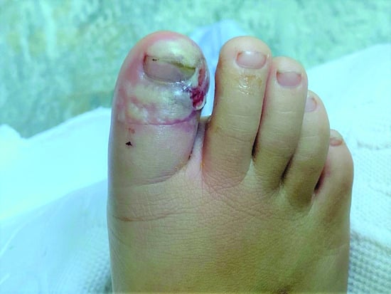
Figure 1.
Day 3.
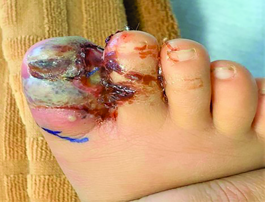
After 5 days of antibiotic therapy, the vesicles and erythema continued to spread, and he developed a one-time reported fever of 104.4°F and abdominal pain. He presented to yet another podiatrist for evaluation, where the right hallux nail plate was removed, and he was started on amoxicillin/clavulanic acid and told to take ibuprofen for pain (Fig. 2). His discomfort continued to progress until his mother decided to bring him to our hospital, where he was admitted for evaluation.
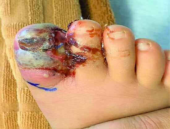
Figure 2.
Day 8.
On evaluation, his right hallux was edematous and erythematous, with mild pain on palpation. Multiple diffuse vesicles were present to the whole dorsal hallux, with a yellowish serous fluid on deroofing. His pain was localized to his distal hallux, with no pain over his extensor tendons, and no limitation to his range of motion. His vital signs were stable, with no recorded fevers, and his laboratory values showed a white blood cell count of 8,000/μl, a C-reactive protein level of 4.7 mg/L, and an erythrocyte sedimentation rate of 19 mm/h. There was no radiographic evidence of osseous changes, and a magnetic resonance imaging scan of the right foot was obtained, which showed acute osseous changes on T2-weighted imaging of both the distal and proximal phalanges of the hallux, which was believed to be reactive edema and not osteomyelitis.
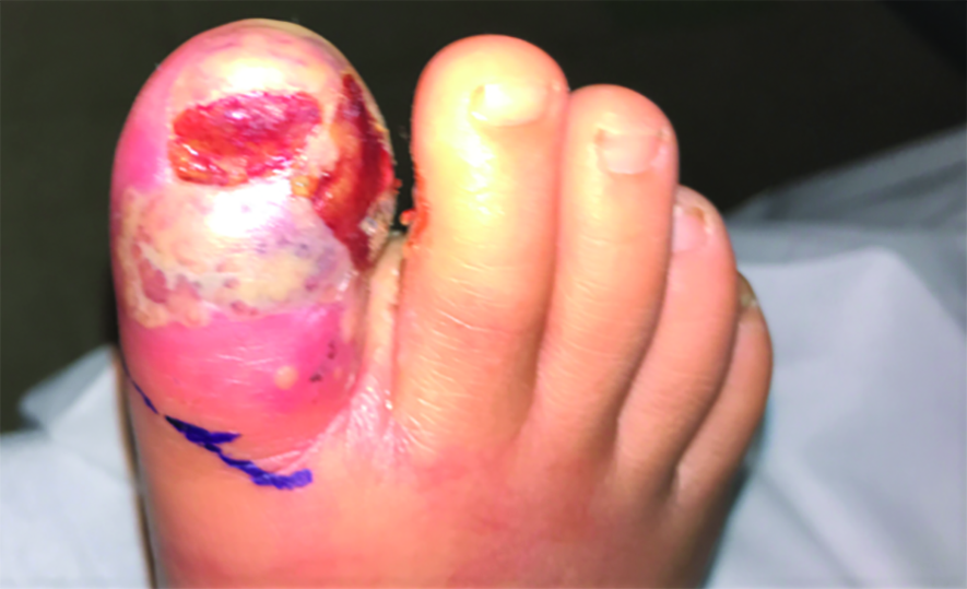
The pediatric team started him on intravenous vancomycin 330 mg one time, cefotaxime 1,100 mg one time, and acyclovir 110 mg one time. They were concerned based on his presentation that he had herpetic whitlow with a superinfection because, according to the mother, he would often bite and suck his toes. Once cultures were obtained of the vesicular fluid and PCR samples sent, it was determined that they were negative for any bacteria but positive for HSV type 1. Once his pain was controlled, he was discharged (5 days after admission) on a course of oral clindamycin and a 5-day course of acyclovir (Fig. 3). At his first follow-up appointment on January 22, all signs of edema had dissipated, no pain remained, and all lesions had fully crusted over. His final appointment was on February 12, at which time a fully healed hallux with complete skin healing and the beginning of new nail formation was observed.
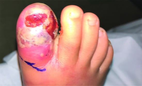
Figure 3.
Day 14.
Discussion
The term herpetic whitlow was used by Stern in 1959 in relation to neurosurgical units nursing staff with multiple finger infections [7]. The word whitlow comes from the Scandinavian word whichflaw, which means crack around the nail. At this time, all cases of digital herpetic infections are referred to as herpetic whitlow, including sparsely reported toe infections. Herpetic whitlow is caused by HSV types 1 and 2, which are members of the alpha-Herpesviridae family [13].
This is a presentation of a case of hallux herpetic whitlow that was initially mistaken for a paronychia and ingrown toenail. The patient was previously treated with oral antibiotics, incision and drainage of the blisters, and total nail avulsion. In classic cases, the herpes infection of the hands or toes is associated with finger and toe sucking in asymptomatic carriers. It was determined in the patient’s history that he had a history of sucking his fingers and toes, prodromal symptoms of itching and pain to the area before vesicle formation, and negative Gram’s stain.
Differential diagnoses for herpetic whitlow should be properly identified and ruled out, such as an ingrown toenail, cellulitis, or paronychia, before any intervention. Clinical examination, patient history, and pathologic findings should all be used to come to the proper diagnosis to prevent any excessive procedures. The patient had already undergone an incision and drainage of the vesicle/abscess, and a total nail avulsion. In cases of herpetic whitlow, this is contraindicated because of the risk of viremia, secondary bacterial superinfection, and encephalitis [10,11]. Diagnosis was confirmed by negative bacterial vesicle fluid cultures but a positive viral culture and PCR test for HSV type 1. It is possible that our bacterial culture results were confounded by the start of antibiotics before his hospitalization; however, based on his clinical picture, it is most likely that he had a secondary bacterial cellulitis associated with the previous incision and drainage and nail avulsion. This misdiagnosis is attributable to the rare nature of this disease, and it can often be forgotten and treated as a more common abnormality.
There are limited cases of herpetic whitlow of the feet mentioned in the literature, and its occurrence is considered atypical. In a case by Feder et al, [14] a 3-year-old girl developed an HSV type 1 infection of her toenails after her mother used her teeth to trim her daughter’s toenails. Mohler et al [15] found an HSV type 1 infection in a 13-month-old girl in her left fifth toe, which was believed to be caused by autoinoculation of unknown route. It is believed that in this case report, the patient most likely transmitted the infection by autoinoculation, biting his hallux toenail.
Herpetic whitlow of the hallux is an uncommon form of HSV infection and can be caused by varying forms of transmission. Clinicians should consider the diagnosis of herpetic whitlow when going over differential diagnoses for a possible bacterial toe infection. It can improve diagnostic accuracy and prevent potentially harmful surgical procedures or antibiotics.
Conclusions
Herpetic whitlow of the lower extremities is uncommon but a possible form of HSV infection. Clinicians should consider this diagnosis when considering any form of pediatric toe infection, and simple questions about nail biting/toe sucking can prevent misdiagnosis and unnecessary testing and debridement.
Funding
None reported.
Acknowledgments
This case report was supported by the Division of Vascular Surgery, New York Presbyterian Hospital, University Hospitals of Weill Cornell and Columbia.
Conflicts of Interest
None reported.
References
- Foti C, Romita P, Mascia P, et al: Atypical herpetic whitlow: a diagnosis to consider. Endocr Metab Immune Disord Drug Targets 17: 3, 2017.
- Adışen E, Önder M: Acral manifestations of viral infections. Clin Dermatol 35: 40, 2017.
- Camasmie HR, Léda SB, Lupi O, et al: Chronic herpetic whitlow as the first manifestation of HIV infection. AIDS 30: 2254, 2016.
- Walker LG, Simmons BP, Lovallo JL: Pediatric herpetic hand infections. J Hand Surg Am 15: 176, 1990.
- Patel R, Kumar H, More B, et al: Paediatric recurrent herpetic whitlow. BMJ Case Rep 2013: bcr2013010207, 2013.
- Avitzur Y, Amir J: Herpetic whitlow infection in a general pediatrician—an occupational hazard. Infection 30: 234, 2002.
- Gill MJ, Arlette J, Buchan KA: Herpes simplex virus infection of the hand. J Am Acad Dermatol 22: 111, 1990.
- Singh A, Preiksaitis J, Ferenczy A, et al: The laboratory diagnosis of herpes simplex virus infections. Can J Infect Dis Med Microbiol 16: 92, 2005.
- Rerucha CM, Ewing JT, Oppenlander KE, et al: Acute hand infections. Am Fam Physician 99: 228, 2019.
- Gill M, Arlette J, Buchan K, et al: Therapy for recurrent herpetic whitlow. Ann Intern Med 105: 631, 1986
- Wu IB, Schwartz RA: Herpetic whitlow. Pediatr Dermatol 79: 193, 2007.
- Wilson R, Truesdell AG, Villines TC: Inflammatory lesions on every finger. Am Fam Physician 72: 317, 2005.
- Stanberry LR: Pathogenesis of herpes simplex virus infection and animal models for its study. Curr Top Microbiol Immunol 179: 15, 1992.
- Feder HM Jr, Geller RW: Herpetic whitlow of the great toe. N Engl J Med 326: 1295, 1992.
- Mohler A: Herpetic whitlow of the toe. J Am Board Fam Pract 13: 213, 2000.
© 2022, Journal of the American Podiatric Medical Association