


Abstract
Osteomyelitis of the calcaneus combined with a pathologic fracture is a rare and difficult presentation for any practicing foot and ankle surgeon. Treatment for achieving an aseptic nonunion involves a variety of steps, including surgical debridement, antibiotic administration, and fracture stabilization. In this case series, we report a novel technique for the treatment of a tongue-type calcaneal fracture in the setting of chronic osteomyelitis using the Biomet JuggerLoc bone-to-bone system for fixation.
Calcaneal fractures can lead to a vast array of complications that can cause long-term pathology for the patient. Although fractures of the hindfoot are uncommon, the calcaneus is the most injured tarsus bone.[1] The majority of calcaneal fractures described in the literature are said to affect male patients more commonly than female patients, and generally occur secondary to a severe trauma, such as a fall from height.[2] There is a small subset to the incidence of calcaneal fractures that fall into the category of pathologic in the setting of osteomyelitis.[2] Pathologic fractures secondary to osteomyelitis add an additional complication to current treatment protocols. Not only does there need to be an attempt at eradicating the bacteria and dead space, but attempts at fracture stabilization must also be put forth in an effort for limb salvage. These types of fractures have seldom been reported in current literature. Brucato et al[3] discuss five patients with pathologic calcaneal fractures from underlying osteomyelitis. Treatment options were discussed, yet results were unsatisfactory.
Attempting to achieve aseptic bony union requires some form of surgical debridement, antibiotic administration, and fracture stabilization.[4] It is well studied that fixation is complicated in the setting of osteomyelitis because of the lack of good quality bone.[3] Implanted hardware can be a source of continued infection with the development of a biofilm that adheres to the implanted hardware.[5] Therefore, most surgeons will choose to use external fixators or refrain from operative intervention until appropriate antibiotic therapy has been completed. There is literature to suggest that internal fixation can be useful in an infected fracture that is displaced, with holding reduction and maintaining stability.[5] One of the principal factors in eradicating osteomyelitis surgically is to remove any potential nidus of infection. McNally et al[6] propose a single-stage treatment of osteomyelitis. They prospectively studied 100 patients with chronic osteomyelitis treated with an injectable composite of calcium sulphate and hydroxyapatite loaded with gentamicin, 175 mg gentamicin in 10 ml of calcium sulphate/hydroxyapatite, which they call Cerament-V (BoneSupport Holding AB, Lund, Sweden). They noted eradication of infection in 96 of 100 patients by a single-stage operation.[6]
The management of isolated calcaneal fractures is somewhat controversial. Historically, tongue-type calcaneal fractures have been treated successfully with percutaneous fixation. Tornetta[7] describes a modified percutaneous Essex-Lopresti fixation of the posterior facet. Twenty-six consecutive patients with tongue-type or Sanders type 2C calcaneal fractures that underwent percutaneous fixation of the posterior facet were included. They noted that 23 patients treated with percutaneous fixation went on to have acceptable reduction. Results from this study suggest that percutaneous fixation, for these types of fractures, is superior to open reduction and internal fixation.
Without any definitive treatment algorithms, and limited research, the pathologic calcaneal fracture in the setting of underlying osteomyelitis can present quite a challenge for any foot and ankle surgeon. Therefore, in the following report, we discuss a novel technique for the treatment of a tongue-type calcaneal fracture in the setting of chronic osteomyelitis.
Case Report
A 46-year-old man with a medical history consisting of diabetes complicated by peripheral neuropathy, chronic back pain, anemia, high cholesterol, hypertension, and pancreatitis presented with a nonhealing wound to his left heel. The patient had an extensive surgical history extending from July of 2018 through the present. Performed by an outside physician, the patient primarily underwent a left-side Achilles tendon lengthening procedure with wound debridement. Subsequently, 9 weeks after the primary procedure, while walking his dog, the patient tripped and suffered a left insertional Achilles tendon rupture, which was repaired using two bone anchors. Ten months after the repair, the patient developed blisters on the medial and lateral side of his left calcaneus. The aforementioned blisters turned into wounds that failed multiple bouts of conservative therapies including local wound care, offloading, padding, protective weightbearing, and changing shoe gear. A series of three operative bone biopsies and wound debridements with tissue-engineered graft applications were attempted over the course of 1 year.
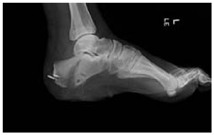
The patient subsequently was referred to our wound care center with a nonhealing wound to his left heel. Initial radiographs were unremarkable; thus, a white cell–labeled bone scan was ordered, revealing increased uptake at the plantar calcaneus in all three phases, indicative of osteomyelitis (Fig. 1). He was taken to the operating room and underwent wound debridement, calcaneal bone biopsy, drilling and injecting the calcaneus with flowable biphasic hydroxyapatite and calcium sulfate cement with vancomycin, application of a collagen antimicrobial wound matrix graft, and secondary closure using a tissue expander. Bone biopsy revealed methicillin-resistant Staphylococcus aureus. On the first postoperative visit, the patient noted a recent slip and fall with a pop and bruising to the back of his left foot and ankle. Radiographs revealed a closed, tongue-type calcaneal fracture (Fig. 2). Further operative intervention was required. Of relevance on physical examination specific to the left lower extremity, there was pain on palpation of the posterior aspect of the calcaneus at the insertion of the Achilles tendon and the lateral aspect of the left calcaneus. Moderate ecchymosis with minimal edema was noted to the left calcaneus. Dermatologically, the left foot revealed a 4 × 2 × 2-cm, chronic, nonhealing, Wagner grade 3 ulceration of the plantar calcaneus.
Figure 1.
White cell–labeled bone scan.
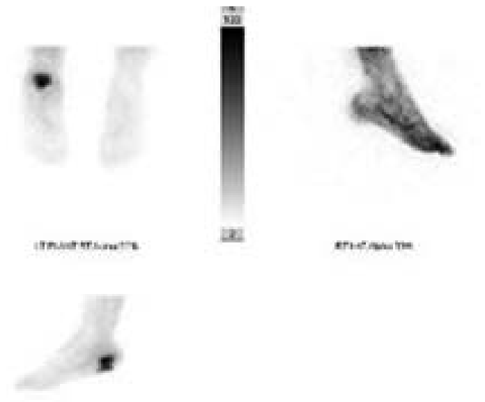
Figure 2.
Preoperative lateral view radiograph of pathologic tongue-type calcaneal fracture with underlying osteomyeltis.
Surgical Description
The patient was brought to the operating room and placed on the operating table in the prone position. A pneumatic calf tourniquet was placed about the left calf. Attention was directed to the posterosuperior aspect of the calcaneus at the insertion of the Achilles tendon, where a 4-cm midline linear incision was made down to the level of bone. A retained bone anchor was loose within the calcaneus, and was removed. Fluoroscopy visualized a second bone anchor, which was deep seated within the bone, and did not require extraction.
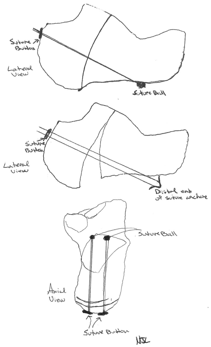
Manual closed reduction of the fracture was performed by means of a plantarflexory force applied to the distal forefoot, and a drill for the JuggerLoc anchor (Biomet Zimmer; Warsaw, Indiana) was inserted from the posterosuperior aspect of the calcaneus to the plantar inferior aspect of the calcaneus across the fracture fragment. A second drill point was entered just inferior to the first. Two JuggerLoc anchors were inserted within the previous drill holes, and the anchors were inserted according to the manufacturer and proper AO technique (Figs. 3 and 4). On tightening of the JuggerLoc system, fluoroscopy revealed that the calcaneus had restored its length, and the fracture fragment had been reduced into anatomical alignment (Figs. 5 and 6).
Figure 3.
JuggerLoc anchor insertion.
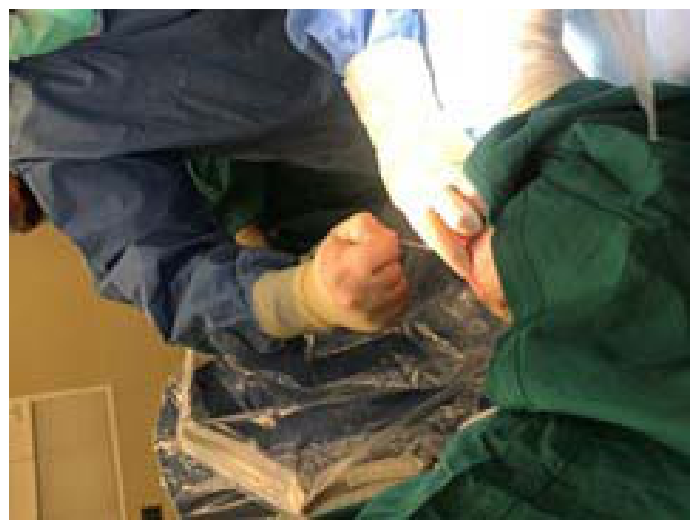
Figure 4.
Intraoperatively testing the integrity of the suture anchor as primary fixation of the calcaneal fracture.
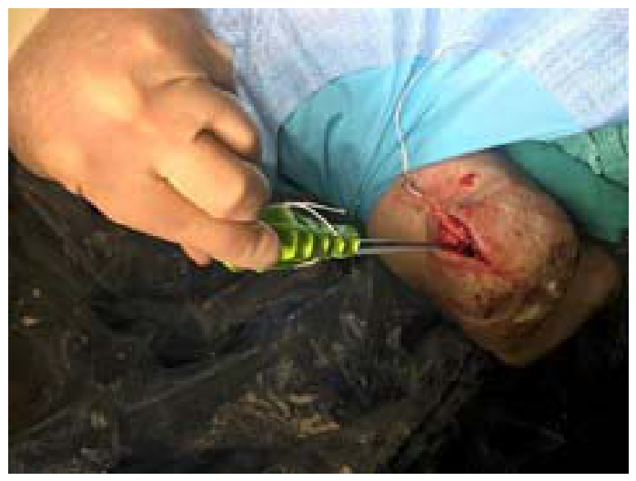
Figure 5.
Operating room spot film of two JuggerLoc anchors and fracture reduction with anatomical alignment achieved. Retained suture anchor from prior surgery inferior to the JuggerLoc anchors.
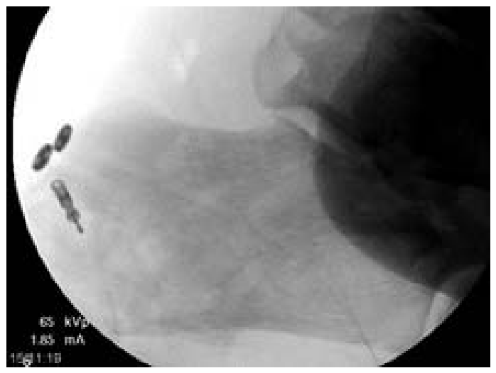
Figure 6.
Schematic drawing of the JuggerLoc anchor system holding fracture reduction for definitive fixation.
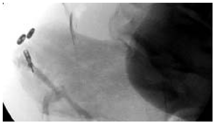
An inferior drill hole was made through the plantar portion of the calcaneus. Flowable biphasic hydroxyapatite and calcium sulfate cement was mixed with vancomycin on the back table according to the manufacturer's technique. Next, it was inserted within the drill hole, which had now tracked to the plantar wound (Fig. 7). The flowable cement with vancomycin (BoneSupport) was injected according to manufacturer technique without any complication or tracking into the wound (Figs. 8 and 9). The plantar calcaneal wound was sharply debrided using a No. 15 blade to allow for healthy bleeding margins. The incision site and the wound were flushed thoroughly with sterile saline. Primary closure of the plantar wound (4 × 2 × 2 cm) and the incision at the Achilles insertion was performed.
Figure 7.
Drilling a pilot hole in the calcaneus for the insertion of flowable biphasic hydroxyapatite and calcium sulfate cement with vancomycin.
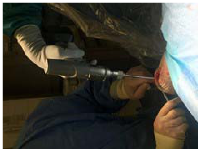
Figure 8.
Insertion of flowable biphasic hydroxyapatite and calcium sulfate cement with vancomycin with the calcaneus.
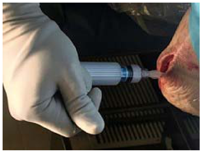
Figure 9.
Final operating room spot film after fixation and injection of the flowable biphasic hydroxyapatite and calcium sulfate cement with vancomycin.
After the procedure, a sterile compressive dressing was applied to the left foot. The left lower extremity was placed in a posterior splint in the operating room. The pneumatic calf tourniquet was deflated and prompt hyperemic response noted to all digits of the left foot. The patient tolerated the procedure and anesthesia well. The patient was transferred from the operating room to the recovery room with vital signs stable and neurovascular status to the left foot intact.
At 2-week follow-up, sutures were removed, and wound edges were well coapted. Because of the patient's extensive surgical history and multiple failed procedures, it was decided to place the patient in a total contact cast to the left lower extremity. The patient was seen weekly over the next 4 weeks for serial cast changes. At postoperative week 6, radiographs revealed anatomical fracture reduction with stability of the JuggerLoc anchors. The patient was transitioned to a short leg pneumatic walking boot and was protective weightbearing as tolerated. At 3 months postoperatively, the patient was ambulatory as tolerated without a limp.
Discussion
This case had two diagnoses to treat, and having them combined made the treatment options limited and difficult. The goal of our treatment was to provide the patient with an aseptic union of his calcaneal fracture secondary to osteomyelitis. Having several failed operative debridements and courses of antibiotics, we felt the need to incorporate a local antibiotic delivery system to treat the osteomyelitis. Based on current literature proposed by McNally et al,[6] we felt that this approach might mitigate the need for further surgery and eradicate osteomyelitis, all while providing the best chance at aseptic bony union. Fixation of the tongue-type calcaneal fracture presented another problem. Implanted hardware, as shown by Patzakis and Zalavras,[5] can be a source of continued infection with the development of a biofilm that adheres to the implanted hardware. To fixate and stabilize the fracture, two JuggerLoc anchors were used by means if the percutaneous technique described by Tornetta.[7] The anchors allowed anatomical reduction and restoration of height of the calcaneus, without introducing metallic hardware into the infected area of the calcaneus.
Limitations of the study include the lack of a preoperative American Orthopaedic Foot & Ankle Society score. Not having this preoperatively did not allow us to quantitatively assess how the patient rated his own pain level and functional outcome from the procedure. A second limitation was the inability to confirm whether deep bone infection was fully eradicated from the calcaneus. We did not feel it in the best interest of the patient to repeat a bone biopsy without any clinical signs of infection or open wounds in the postoperative course, although we do understand this is an area of weakness with our study. Lastly, follow-up was limited in this study to a 6-month time frame. We would have liked for at least a 1-year follow-up, but extenuating circumstances limited this from occurring. Although the limitations are there and need to be discussed, we consider this approach to be minimally invasive for the patient and one that is reproducible in the setting of pathologic tongue-type calcaneal fracture.
Conclusions
To our knowledge, this is the first described report of a stainless-steel suture anchor for the fixation of pathologic calcaneal fracture in the setting of chronic osteomyelitis. This novel technique provides inherent advantages to the traditional methods of fixating tongue-type calcaneal fractures. We hope this technique will allow for further research and discussion. We believe this is a rare fracture, and hope to provide reference should this occur in one's practice or facility.
Financial Disclosure
None reported.
Conflict of Interest
None reported.
References
- CrenshawA:“Fractures and Dislocations of the Foot,”inCampbell's Operative Orthopedics8th Ed,VolIVedited byCrenshaw,AHp2905St. Louis, MOMosby-Yearbook1992
- Mitchell, MJ; McKinley, JC; Robinson, CM:. The epidemiology of calcaneal fractures. Foot (Edinb) 2009, 19, 197. [Google Scholar]
- Brucato, MP; Wachtler, MF; Nasser, EM:. Osteomyelitis of the calcaneus with pathologic fracture. J Foot Ankle Surg 2019, 58, 591. [Google Scholar]
- Kim, Y; Inori, F; Yamanaka, K; et al. A case of osteomyelitis after calcaneal fracture treated by antibiotic-containing calcium phosphate cements. Case Rep Orthop 2018, 2018, 9321830. [Google Scholar]
- Patzakis, MJ; Zalavras, CG:. Chronic posttraumatic osteomyelitis and infected nonunion of the tibia: current management concepts. J Am Acad Orthop Surg 2005, 13, 417. [Google Scholar]
- McNally, MA; Ferguson, JY; Lau, AC; et al. Single-stage treatment of chronic osteomyelitis with a new absorbable, gentamicin-loaded, calcium sulphate/hydroxyapatite biocomposite: a prospective series of 100 cases. Bone Joint J Br 2016, 98, 1289. [Google Scholar]
- Tornetta, P, III. The Essex-Lopresti reduction for calcaneal fractures revisited. J Orthop Trauma 1998, 12, 469. [Google Scholar]
© 2021 American Podiatric Medical Association