


Abstract
The superficial fibular (peroneal) nerve traditionally courses through the anterolateral deep leg and pierces the deep crural fascia at the lower leg to divide into its terminal branches. Entrapment of the superficial fibular nerve is most commonly documented to occur at where it pierces the deep fascia, and numerous etiologies causing entrapment are described. In this case report, we describe an unusual cause of entrapment from a tertiary branch of the superficial fibular nerve taking a circumflex course and wrapping around the secondary branch of the main nerve. This was successfully treated by surgical excision. To the best of our knowledge, this cause of entrapment has not been described in the literature at the time of this publication.
Entrapment of the superficial fibular nerve (SFN) has been well described in the literature, with multiple etiologies such as systemic conditions, trauma, iatrogenic, and others [1,2]. In the lower leg, the SFN typically pierces the crural fascia approximately 12 cm proximal to the tip of the lateral malleolus and divides into the terminal branches consisting of the medial and intermediate dorsal cutaneous nerve shortly thereafter [3,4]. The site where the SFN pierces the fascia is the common site of entrapment [1,2,3]. Accessory branches of the SFN have been described, such as the accessory deep fibular nerve; in addition, entrapment of these accessory branches of the SFN has been described [5,6,7,8]. In this case report, we describe the successful surgical management of SFN entrapment caused by one of these accessory branches wrapping around the main nerve. At the time of this writing, this entity had not been described in the literature. It is the main objective of this article to describe the presentation and treatment of this unusual cause of SFN entrapment, as this can provide guidance to clinicians who might encounter this rare entity.
Case Report
The patient was a 65-year-old woman with an unremarkable health history, and she presented with a complaint of a painful mass in the lateral left leg. The pain was described as intermittent aching and tingling at the mass; in addition, physical contact to the mass would cause shooting pain down the leg into the foot. Physical examination was pertinent for a small mass at the lateral left leg near the peroneal tendons. Hypersensitivity on palpation of the mass was exhibited; in addition, the Tinel sign was elicited on percussion of the mass. Decreased light touch sensation was noted over the mass extending 2 cm distal to the mass; however, sensation was normal down the leg and into the foot. The rest of the lower extremity neurologic and musculoskeletal examinations were otherwise unremarkable, with no deficits noted. Radiographs were taken and were otherwise unremarkable.
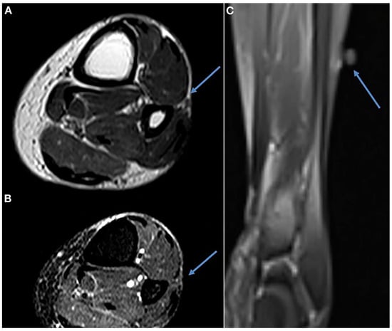
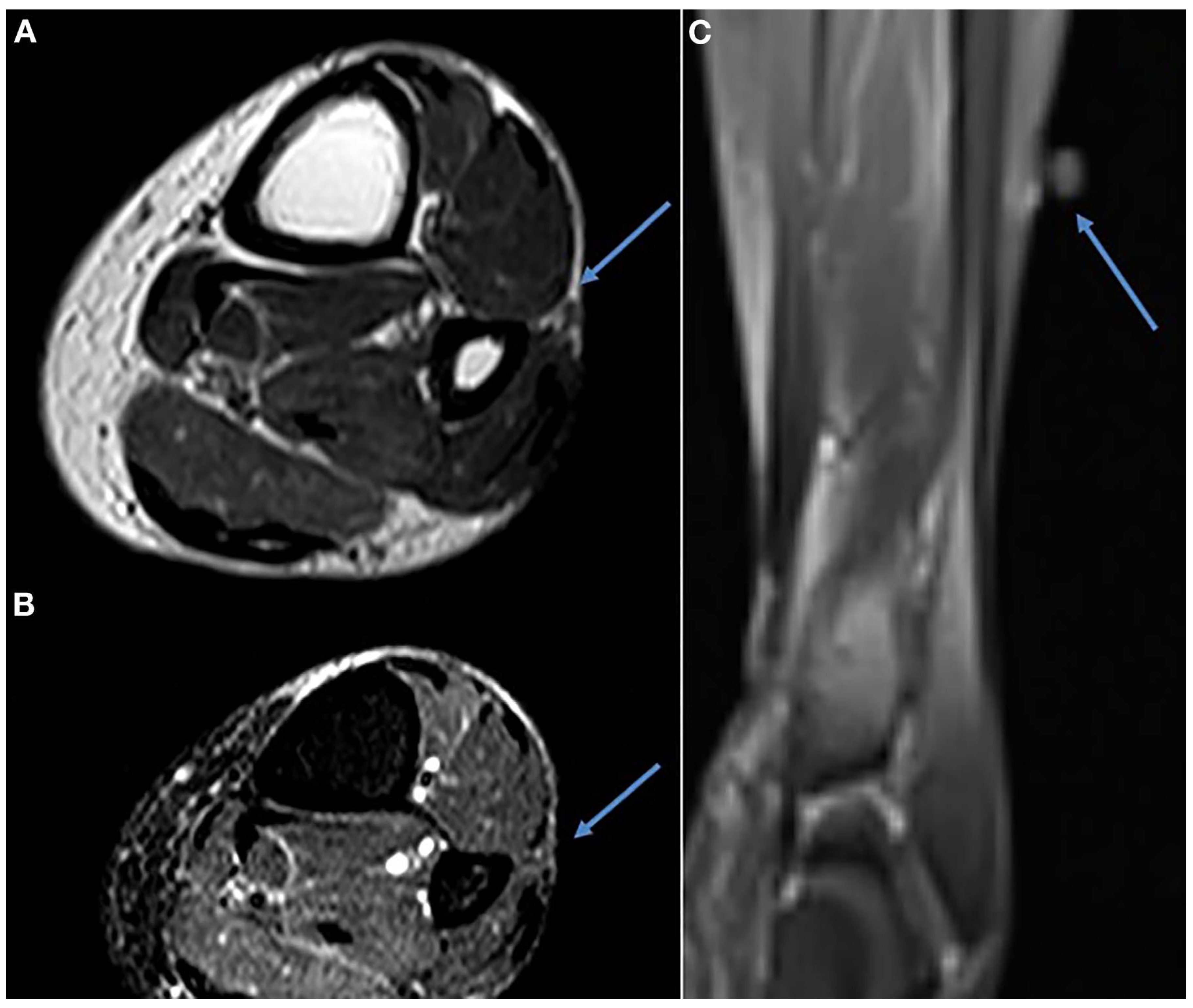
Lower extremity magnetic resonance imaging was performed (Figure 1), which demonstrated a focal soft-tissue mass approximately 3 mm in diameter, 11 cm proximal to the lateral malleolus. The mass had the radiographic appearance of a possible schwannoma or subcutaneous myxoma.
Figure 1.
Axial and frontal magnetic resonance imaging views displaying small mass in the area of the patient’s complaint.
Magnetic resonance imaging results were discussed with the patient, in addition to conservative and surgical treatment options. The patient chose surgical intervention and was scheduled for removal of the soft-tissue mass of the left leg.
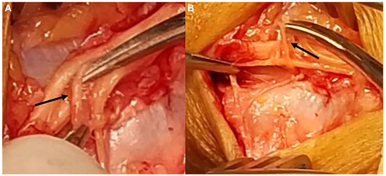
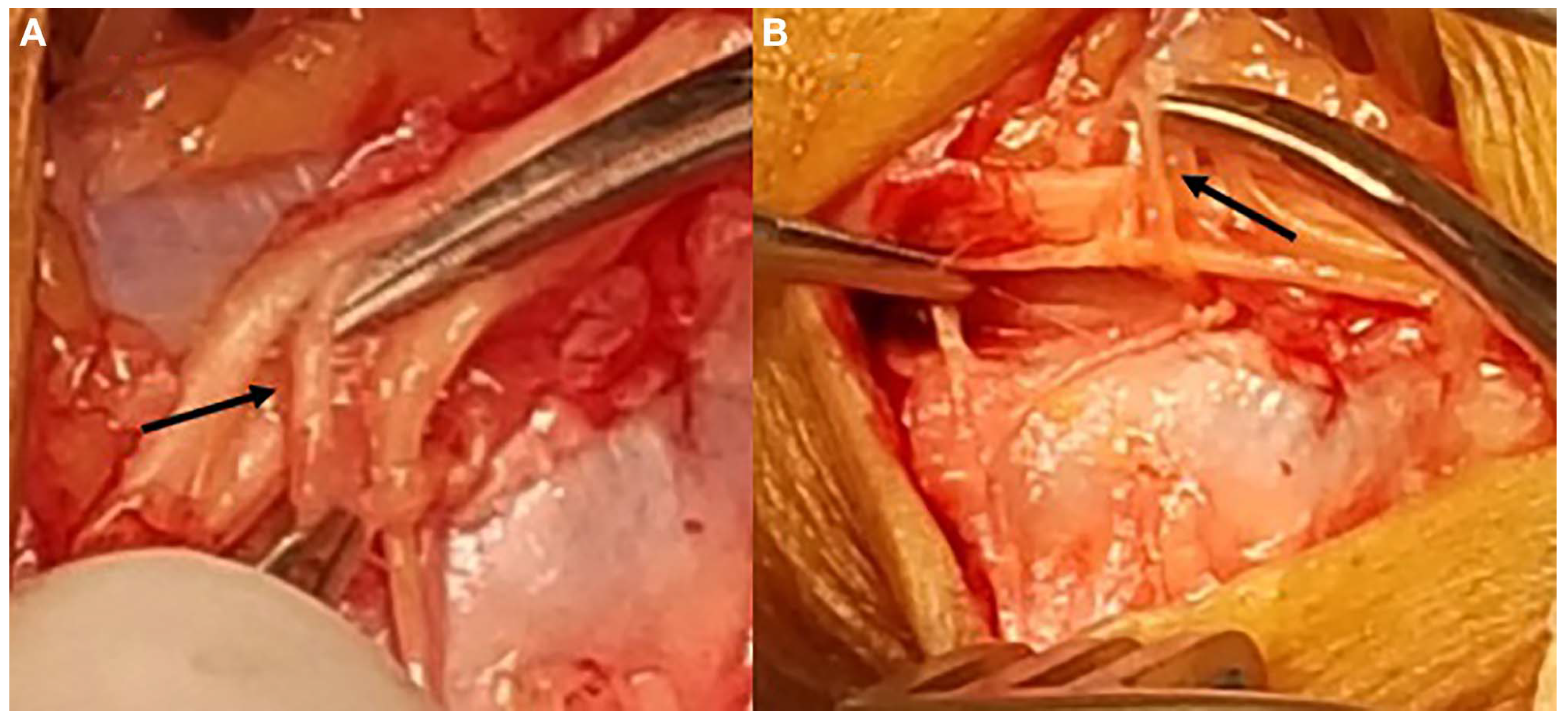
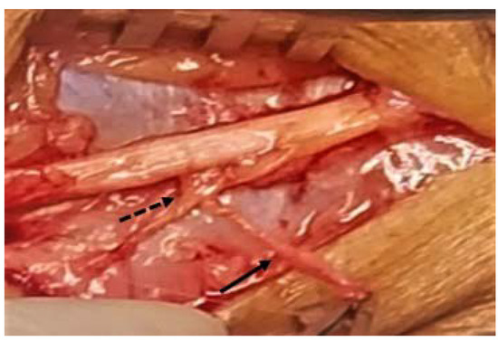
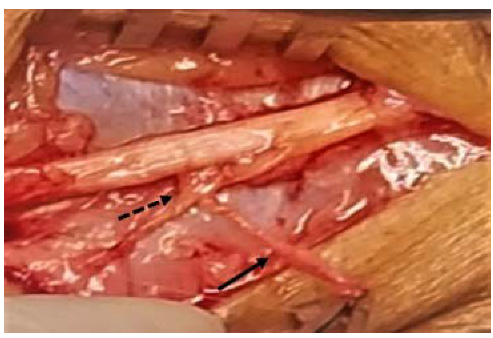
Surgical intervention performed consisted of a linear incision over the mass and dissection through subcutaneous tissue to identify the SFN. Once the SFN was identified, there were fibers appearing like nerve tissue wrapped around the SFN in a 360-degree manner, completely entangling the SFN (Figure 2). These fibers were dissected and traced to their origin, and these fibers were found to originate from an accessory branch of the SFN (Figure 3). This accessory SFN started distal to the entrapment and coursed posteriorly, and gave off a branch extending superiorly to the SFN. The circumflex course of this branch of the accessory SFN entangling the SFN is what gave the impression of a nerve tumor on magnetic resonance imaging.
Figure 2.
Circumflex branches (solid arrows) of the accessory superficial fibular nerve entangling the superficial fibular nerve, causing entrapment.
Figure 3.
Superficial fibular nerve after release. Circumflex branch of the accessory superficial fibular nerve (solid arrow) causing entrapment was traced back to an accessory superficial fibular nerve (dashed arrow).
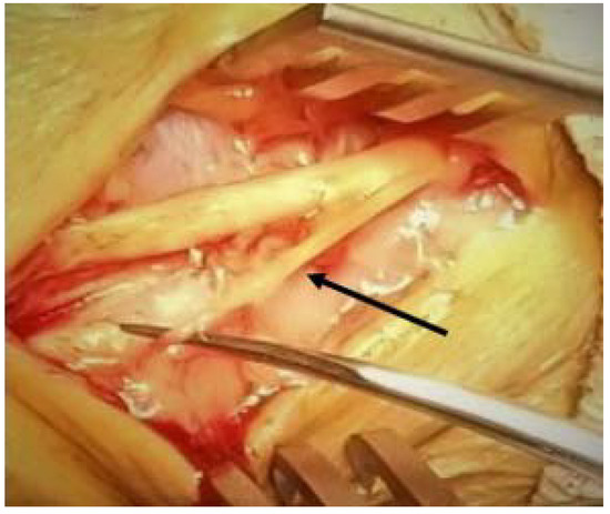
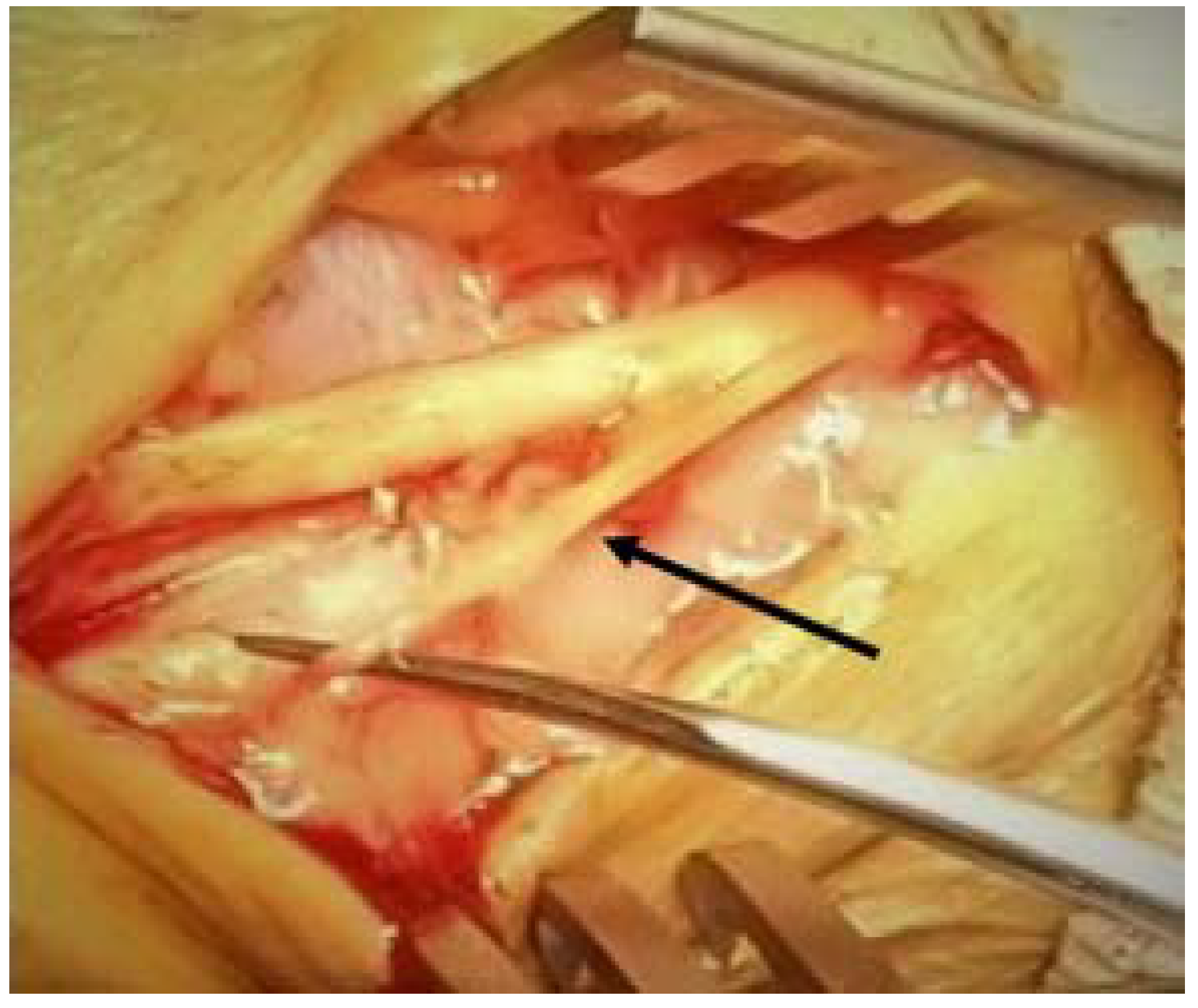
A complete neurectomy of the circumflex branch of the accessory SFN (Figure 4) was performed by transversely transecting this branch at its base. The transected branch was sent for pathologic identification. The accessory SFN and the stump of the circumflex accessory SFN branch were buried in muscle belly, and the surgical site was closed in standard layered manner with compression dressing applied.
Figure 4.
Superficial fibular nerve free of any entrapment. The accessory superficial fibular nerve is noted (solid arrow).
The patient had an uneventful recovery with no further complaints to the surgical site, and she was discharged from care after 4 months’ follow-up. Minimal sensory deficits were noted in the region of the excised accessory nerve; however, the patient did not report any pain or discomfort from this. Histopathologic studies of this excised nerve branch were noted to be regular nerve tissue, with no neoplasm or other disease noted. The patient was contacted by means of telephone 13 months after surgery as a final follow-up. As of the time of this writing, the patient remains pain-free and satisfied overall with the outcome of the intervention.
Discussion
Entrapment of the SFN is a well-documented condition in the literature with several possible etiologies. This case highlights an unusual cause of entrapment of the SFN by entanglement of a circumflex accessory branch of the SFN. At the time of this publication, there does not seem to be other cases that describe this cause of SFN entrapment.
Although this entrapment was detected by MRI, magnetic resonance imaging may not always detect nontraumatic etiologies of nerve pathology. According to Yablon et al. [9] MRI and ultrasound have a similar specificity for nerve pathologies; however, ultrasound has a much higher sensitivity versus MRI for detecting lower-extremity nerve pathology of 93% versus 67%. Clinicians of the foot and ankle should be reminded that ultrasound remains another option for work-up of lower-extremity nerve pathology.
The SFN is traditionally documented to divide into two terminal branches in the lower leg. Accessory branches of the SFN have been documented, with the most common described being the accessory deep fibular nerve [6,7,8]. Entrapment of these accessory bands have been documented; however, these entrapments are typically described as being caused by fascial bands around the lateral malleolus [5,7,8]. In contrast to cases in the literature, this entrapment is from the accessory nerve itself at a level proximal to what is typically documented. This could raise the question of whether the circumflex accessory nerve we encountered is an underrecognized entity, and whether this tertiary branch is a benign normal variant or results in a predisposition to nerve entrapments or other issues.
Conclusions
This case might provide guidance to clinicians who encounter this rare entity and should raise awareness of other potential congenital nerve anatomy variations that might explain a neuritic etiology of pain in an otherwise unremarkable work-up. It bears mentioning that although this nerve was large enough for detection on MRI, in cases of pain where MRI is negative, competent musculoskeletal ultrasonography is probably the best modality to identify rare cases of nontraumatic neurogenic pain from a local peripheral branch.
Financial Disclosure
None reported.
Conflicts of Interest
None reported.
References
- BREGMAN, PJ; SCHUENKE, M. Current diagnosis and treatment of superficial fibular nerve injuries and entrapment. Clin Podiatr Med Surg 2016, 33, 243. [Google Scholar] [CrossRef] [Scilit] [PubMed]
- MATSUMOTO, J; ISU, T; KIM, K. ET AL: Clinical features and surgical treatment of superficial peroneal nerve entrapment neuropathy. Neurol Med Chir (Tokyo) 2018, 58, 320. [Google Scholar] [CrossRef] [Scilit] [PubMed]
- TZIKA, M; PARASKEVAS, G; NATSIS, K. Entrapment of the superficial peroneal nerve: an anatomical insight. JAPMA 2015, 105, 150. [Google Scholar] [CrossRef] [Scilit] [PubMed]
- KELIKIAN, AS; SARRAFIAN, SK. Sarrafian’s Anatomy of the Foot and Ankle, Descriptive, Topographic, Functional; Lippincott Williams & Wilkins: Philadelphia, 2011. [Google Scholar]
- CUSHMAN, DM; WALKER, SE. Symptomatic accessory branch of the superficial peroneal nerve identified by ultrasound. Muscle Nerve 2020, 62, E46. [Google Scholar] [CrossRef] [Scilit] [PubMed]
- LAMBERT EH: The accessory deep peroneal nerve. A common variation in innervation of extensor digitorum brevis. Neurology 1969, 19, 1169. [CrossRef] [Scilit] [PubMed]
- TZIKA, M; PARASKEVAS, GK; KITSOULIS, P. The accessory deep peroneal nerve: a review of the literature. Foot (Edinb) 2012, 22, 232. [Google Scholar] [CrossRef] [Scilit] [PubMed]
- PARASKEVAS, GK; NATSIS, K; TZIKA, M; ET, AL. Potential entrapment of an accessory superficial peroneal sensory nerve at the lateral malleolus: a cadaveric case report and review of the literature. J Foot Ankle Surg 2014, 53, 92. [Google Scholar] [CrossRef] [Scilit] [PubMed]
- YABLON, CM; HAMMER, MR; MORAG, Y. ET AL: US of the peripheral nerves of the lower extremity: a landmark approach. Radiographics 2016, 36, 464. [Google Scholar] [CrossRef] [Scilit] [PubMed]
© 2021 American Podiatric Medical Association