


Abstract
A rare and unusual case of plasma cell dyscrasia of the calcaneus is presented. Clinically, the patient had a draining and painful ulcer that was treated with appropriate antibiotics and wound care but failed to show any signs of healing. Radiographic images showed cystic changes of the calcaneus in the vicinity of the ulcer. Blood work was negative for bone and soft-tissue infection, but uric acid and alkaline phosphatase levels were elevated. Nuclear bone scan showed increased uptake in the calcaneus suggestive of osteomyelitis. One possible differential diagnosis was an intraosseous gouty tophus deposit. Not convinced that this was either a bone infection or gout, the author performed a bone biopsy. Pathologic evaluation indicated plasma cell dyscrasia. Continued wound care healed the ulcer completely, with resolution of pain of his heel. Oncology/hematology was consulted, and 16 months after biopsy, he remains asymptomatic.
Tumor of the foot bones is considered rare. Bakotic and Huvos[1] published a series of 150 cases of tumors of the bones of the feet. Sixteen percent of the cases were found to be in the calcaneus.[1] In another series analyzing 196 cases of tumors of the foot and ankle, the calcaneus was found to be the most frequently involved bone (33% of the cases reviewed).[2] Both series further analyzed the data into percentages of benign versus malignant tumors, most common type of various categories of bone tumors, and others. Neither of these two series, and one other recently conducted series, specifically mentioned any finding of plasma cell dyscrasia in any of the foot bones cited.[3]
Plasma cell dyscrasia (PCD) is a heterogeneous group of bone disorders characterized by the expansion of monoclonal bone marrow plasma cells that produce a monoclonal immunoglobulin, which migrate to the gamma region of the electrophoretic strip, thus the term “monoclonal gammopathy.”[4] Each monoclonal protein consists of two heavy polypeptide chains of the same class (gamma, alpha, mu, delta, and epsilon) and two light polypeptide chains of the same type (kappa and lambda).[5] The International Myeloma Working Group has reviewed the criteria for diagnosis and advocated the classification of these disorders into four subgroups: 1) monoclonal gammopathy of undetermined significance (MGUS), 2) asymptomatic (smouldering) myeloma, 3) symptomatic multiple myeloma, and 4) solitary plasmacytoma of bone.[5,6] Although multiple myeloma is the one most readers would recognize, not only is MGUS the most prevalent plasma cell disorder, it has also been reported to be the precursor of multiple myeloma.[7,8] Occasionally, one may encounter this entity being reported in the literature as part of POEMS syndrome, a rare but unique multisystem disorder characterized by polyneuropathy, organomegaly, endocrinopathy, elevated M protein, and skin changes.[9]
Very few instances had been cited in the podiatric literature reporting PCD in the foot. Khoddami and Fernandes[10] cited four prior published case studies of solitary plasmacytoma, involving the middle cuneiform, calcaneus, cuboid, and talus. None of these prior reports were specific in terms of type of immunoglobulin being assayed from the bone. These authors presented the first reported case of solitary immunoglobulin (Ig) A plasmacytoma of the medial cuneiform.
Saviet et al[11] reported a case involving an individual who was first diagnosed with a plasmacytoma of the L3 vertebrae and was treated with surgery and radiation therapy. Two years later, new lytic lesions were found in the left femur, left proximal tibia, and right distal tibia. He underwent several cycles of biological therapy and autologous stem cell transplantation. Six years after the initial diagnosis, he was referred to see the authors because of swelling of his right foot. Radiographic images showed lytic lesions involving the third metatarsal bone at the base, and the head of the proximal phalanx of the second digit with pathologic fractures. The patient subsequently was found to have involvement of multiple bones and ultimately died as a result of this disease.
Solimando et al[12] reported a case involving an individual who had been treated for IgG-k multiple myeloma with chemotherapy and radiation therapy. Although rare, the monoclonal component in this case became a type I cryoglobulinemia. He developed persistent paresthesia and painful acrocyanosis of his fingers and toes. Because of the rapid biochemical response of the underlying disease, followed by bacterial infection of the wounds, the issue of his feet progressed to ischemic and necrotic ulcers that ultimately necessitated surgical amputation of both feet. After eight rounds of chemotherapy, the patient achieved a partial response of multiple myeloma, with disappearance of cryoglobulin.[12]
Case Report
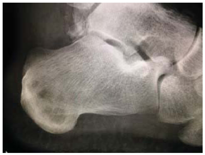
A 69-year-old man was referred to our clinic for a second opinion regarding a tender, nonhealing ulcer on his left heel. His medical history was significant for pulmonary emphysema, chronic systolic heart failure, hypertension, hyperlipidemia, asthma, and atrial fibrillation with implant defibrillator, currently on apixaban therapy. Approximately 9 weeks previously, he purchased and used an electric callus smoother that resulted in a “boil” on his posterior left heel. When he pulled off the scab, it became superficially infected. His primary care physician gave him an intramuscular ceftriaxone injection and prescribed oral doxycycline 100 mg twice daily for 10 days. Initial laboratory values were not indicative of an active infection, with a white blood cell count of 8.9 × 1000/mm3. Neither erythrocyte sedimentation rate nor C-reactive protein was ordered. Foot radiographs did not show any evidence of soft-tissue gas, air, or calcified pedal vessels. However, a radiologist did alert the patient's primary care physician to a “bubbly lytic lesion in the calcaneus,” which prompted a referral to podiatry for evaluation (Fig. 1).
Figure 1.
Lateral view of the left foot showing cluster of “bubbly lytic lesions” in the posterior calcaneus. Note that the cuboid appeared to be within normal limits in appearance.
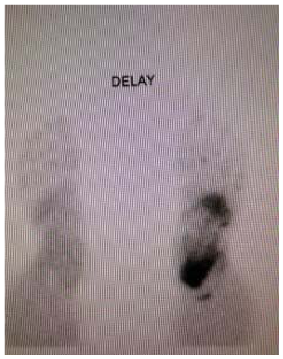
From the podiatrist's record, vascular examination showed nonpalpable pedal pulses, but signals were audible with Doppler examination. Dermatologic examination revealed “superficial excoriation in clusters of several 2- to 3-mm lesions flush with the surrounding skin, no gross abscess/pustule, on the posterior lateral left heel.” The area was very tender, and he had been taking tramadol and acetaminophen for pain control. The rest of his lower extremity examinations were noncontributory. A vascular consultation was placed, a refill for doxycycline was given, and wound care was ordered, which consisted of silver sulfadiazine cream covered with appropriate gauze and dressing. A heel offloading boot was also prescribed. The record from his second visit showed that the issue had progressed into a bleeding, pyogenic granuloma–like, partial thickness ulcer that was very painful. Wound culture was taken, and he was given amoxicillin 875 mg/clavulanate 125 mg to take twice daily instead, but with the same daily dressing order. Culture and Gram's stain were negative for any bacterial presence or growth. His oral antibiotic was discontinued because of reported nausea. Wound care was changed to hydrogen peroxide wet-to-dry dressing. A nuclear medicine bone scan was ordered, with the result indicating “perfusion, blood pool, and static image findings that were suggestive of osteomyelitis of the left calcaneus. A bone neoplasm was unlikely but was considered in the differential diagnosis” (Fig. 2). Interestingly, the scan also picked up a “mild increased uptake in the cuboid” even though radiographically the cuboid appeared to be without any cystic changes.
Figure 2.
Delay phase of nuclear bone scan showing increased uptake at the posterior calcaneus, left foot, with mild uptake in the cuboid, in comparison to the right foot.
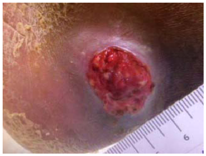
The patient was then referred to see the author. Clinically, the ulcer itself presented with few changes (Fig. 3). Noninvasive vascular study showed adequate perfusion, with ankle-brachial indices of 0.80 on the left posterior tibial artery and 1.16 on the left dorsalis pedis artery. Medical history was extensively reviewed. He denied any history of recent gouty attack or episode in the past. Interestingly, his uric acid level over the past 3 months was always above normal, ranging from 9.7 to 12.1 mg/dL (normal limits, 4.8–8.7 mg/dL), even though he had no symptoms. In the past 18 months, his alkaline phosphatase (ALP) level was consistently elevated, ranging from 135 to 363 IU/liter (normal limits, 36–109 IU/liter). His white blood cell count, erythrocyte sedimentation rate, and C-reactive protein values were always within normal limits. Because the author was not convinced that he had gout or osteomyelitis based on clinical presentation, history, laboratory evaluation, and imaging, a bone biopsy was recommended, as magnetic resonance imaging was not an option because of his implant defibrillator.
Figure 3.
Clinical presentation of the ulcer on the posterior lateral aspect of the left heel. Photograph taken nonweightbearing, with the ruler against the posterior heel, parallel to the Achilles tendon.
The patient consented to the procedure, which was performed using fluoroscopic imaging. Local anesthesia was achieved, and the foot was properly prepared and draped. Intraoperatively, there was no abscess, pus, or exudate observed on debridement of the ulcer bed. Specimen 1 was taken from the soft-tissue ulcer, and the pathology report showed “nonviable tissue, necrotic debris, and fragments of soft tissue with mixed inflammation and noncaseating granulomas, negative for tumor.” Culture and sensitivity study showed no bacterial or fungal growth, and no organism presented on Gram's stain.
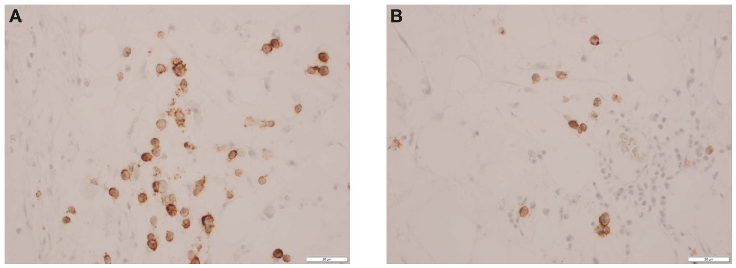
Three bone marrow biopsy needles were used, with care taken not to cross-contaminate any of the aspirates by using complete different sets of instruments during each biopsy. A small skin incision was made in each location with a blade, followed by blunt dissection using a hemostat to arrive at the level of the periosteum. Each location was verified by intraoperative fluoroscopy before obtaining bone marrow samples. Specimen 2 was taken from the cuboid (Fig. 4A) and specimen 3 was taken from the anterior calcaneus (Fig. 4B). Both specimens showed normal histopathologic findings. Specimen 4 was taken directly from the center of the cystic lesion in the posterior calcaneus (Fig. 4C). The pathology report read “atypical increased plasmacytoid proliferation, diffuse and in aggregates. Hypocellular bone marrow (5%) with trilineage hematopoiesis [Figs. 5 and 6]. Serology studies are recommended as well as a hematology/oncology consult to exclude an underlying plasma cell dyscrasia or plasmacytoma.” Each small incision was closed with nylon sutures.
Figure 4.
Intraoperative fluoroscopic image showing accurate needle biopsy placement with specimen being obtained from (A) cuboid, (B) anterior calcaneus, and (C) posterior calcaneus of the left foot.
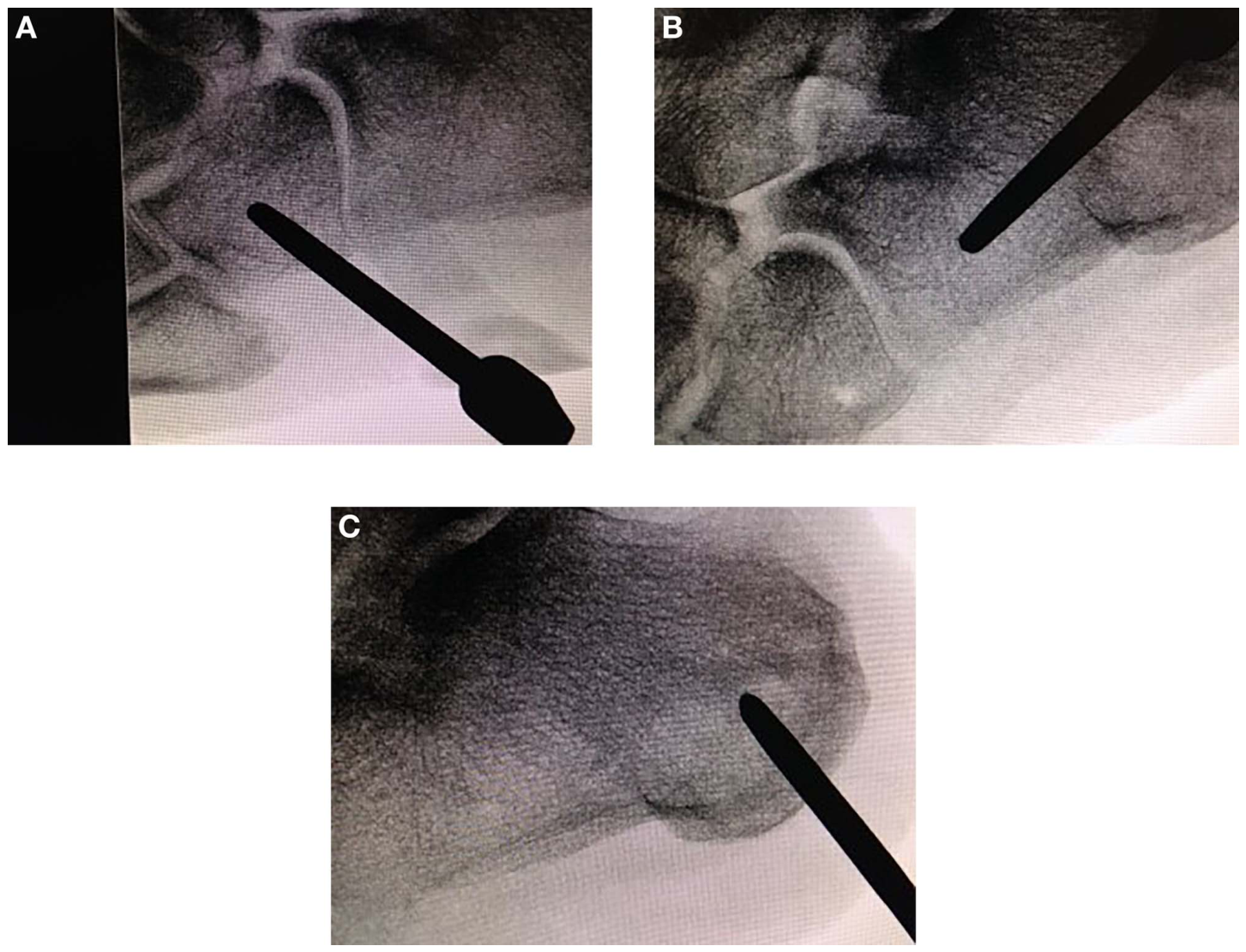
Figure 5.
Bone marrow staining in specimen obtained from the posterior calcaneus, showing abundance of plasma cells in (A) H&E stain and (B) immune peroxidase stain specifically for plasma cells (×40).
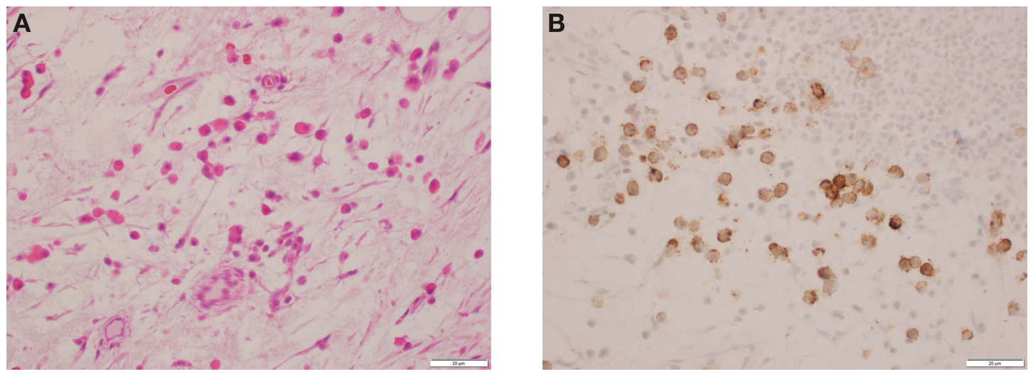
Figure 6.
Same cross-section as in Figure 5, using specific stain to pick up plasma cells with (A) kappa and (B) lambda chains (×40).
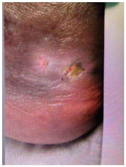
The patient had an uneventful postoperative course. No antibiotic was prescribed, and daily wound care consisted of applying calcium alginate dressing. By the first postoperative visit, he reported complete resolution of pain. By 8 weeks after biopsy, the ulcer was almost completely healed (Fig. 7). He was subsequently discharged from our clinic on complete healing of his ulcer and resolution of his symptoms, 14 weeks after biopsy. The patient was advised about the potential risk of pathologic fracture, and proper shoes and inserts were recommended for better support.
Figure 7.
Clinical presentation at 8 weeks after biopsy showing almost complete healing of the ulcer. Photograph taken with left foot weightbearing.
The patient had four visits with oncology/hematology for initial workup and follow-up evaluation. Initial test panels were all repeated 3 months later. One consistent finding was the presence of “one trace concentration band of free lambda monoclonal protein migrating in the gamma region” on his immunofixation studies. The other consistent findings were elevated serum kappa light chain and lambda light chain. His alpha-1 globulin, beta-2 globulin, IgG, IgA, and beta-2 microglobulin levels were elevated in the first blood drawn, but were within normal limits in the subsequent tests. At the time of submission of this article, the patient had been discharged from the oncology/hematology service because of lack of “evidence of myeloma on extensive workup.”
Discussion
This is an unusual case involving the presence of an ulcer over an area of the calcaneus that has underlying intraosseous cystic changes. Even though the bone lesion appears to be an incidental finding, the challenge in this case had always been to discern among the seemingly confusing clinical and laboratory findings to arrive at an accurate diagnosis.
Given the history and location of the heel ulcer, the etiology was traumatic and mechanical in nature, and not neuropathic or vascular in origin. The persistent bleeding and oozing of thin serosanguineous fluid from the ulcer might be explained by his apixaban therapy. However, his prothrombin time and international normalized ratio had always been within normal limits. Because there was no clinical, laboratory, or microbiologic evidence that the ulcer was infected, proper selection of a wound care product was the key to the successful healing of this type of ulcer. Calcium alginate was ideal in this case, as it is highly absorbent and hemostatic, yet maintains a physiologically moist environment that promotes wound healing.[13]
The hyperuricemia in this case remained a challenge in determining the exact underlying etiology. He had no gout symptoms and no history of gouty attack reported in the past. His renal function had always been within normal limits, with no evidence of acute or chronic kidney disease. His medical history did not indicate that he had been given diuretics for hypertension or xanthine oxidase inhibitors for uric acid control. A review of his diet history showed that it had not been in excess for red meat, seafood, or alcohol intake. The author's concern was whether or not the consistent hyperuricemia might have been attributable to an underlying lytic bone lesion, such as multiple myeloma, but this had been proven not to be the case from the biopsy result.
Another interesting finding in this case presentation was the chronically elevated ALP, which is commonly associated with liver disease or bone disorders, but can also be caused by heart disease, kidney disorders, or bacterial infection. Besides his elevated ALP, his alanine transaminase and aspartate transaminase had consistently been within normal limits. It would be unlikely that the elevation was from a liver disease, as 4 months before the onset of his heel issue, he had an abdominal computed tomography examination that showed no disease. Although it is possible to have a slight elevation of ALP from a bone infection such as acute osteomyelitis, it is unlikely, as it had remained elevated for at least 3 years before the onset of his heel ulcer. His serum calcium and phosphate levels had always been within normal limits; therefore, the presence of a kidney disorder was unlikely. Given his history of cardiac issues, it would most likely be the cause (or contributing factor) of his elevated ALP.
The incidental finding of the lytic bone lesion is the most pertinent aspect of this case study. Differential diagnosis based on radiographic appearance would include bone cyst, intraosseous lipoma or ganglion, and osteochondroma, to name a few. With his history of elevated uric acid and ALP, the author also included an intraosseous gouty tophus and myeloma as part of the differential diagnosis. Based on the bone biopsy and subsequent panel of tests ordered by oncology/hematology, it would appear that the calcaneal lesion is a form of PCD. The incidental finding of increased uptake in the cuboid was probably stress related rather than neoplastic or infectious in origin. The diagnostic criteria for each of the four subclasses of PCD had been well established and published in the literature.[11] Based on these criteria, the author felt that most likely it represented an asymptomatic variant of MGUS, as it fits the criteria of low serum monoclonal protein, low monoclonal bone marrow plasma cell, and no evidence of end-organ damage attributable to clonal plasma cell disorder.[11]
Conclusions
The author presented the case of a rare and unusual presentation of plasma cell dyscrasia of the calcaneus. This case study illustrates the importance of making an accurate diagnosis, as a delayed or missed diagnoses may result in severe consequences.
Acknowledgment
Dr. Dennis Mosely, chief of pathology, Dr. David M. Tasso, staff cytopathologist, and Dr. Paul J. Chamas, staff podiatrist, for their invaluable input and assistance.
Financial Disclosure
None reported.
Conflict of Interest
None reported.
References
- Bakotic, B; Huvos, A:. Tumors of the bones of the feet: the clinicopathologic features of 150 cases. J Foot Ankle Surg 2001, 40, 277. [Google Scholar]
- Ozdemir, HM; Yilmaz, C; Yildiz, Y; et al. Tumors of the foot and ankle: analysis of 196 cases. J Foot Ankle Surg 1997, 36, 403. [Google Scholar]
- Ruggieri, P; Angelini, A; Jorge, FD; et al. Review of foot tumors seen in a university tumor institute. J Foot Ankle Surg 2014, 53, 282. [Google Scholar]
- Boccadoro, M; Pileri, A:. Plasma cell dyscrasias: classification, clinical and laboratory characteristics, and differential diagnosis. Baillieres Clin Haematol 1995, 8, 705. [Google Scholar]
- Kyle, RA:. Criteria for the classification of monoclonal gammopathies, multiple myeloma and related disorders: a report of the International Myeloma Working Group. Br J Haematol 2003, 121, 749. [Google Scholar]
- Agarwal, A; Ghobrial, IM:. Monoclonal gammopathy of undetermined significance and smoldering multiple myeloma: a review of the current understanding of epidemiology, biology, risk stratification, and management of myeloma precursor disease. Clin Cancer Res 2013, 19, 985. [Google Scholar]
- Kyle, RA; Therneau, TM; Rajkumar, SV; et al. Prevalence of monoclonal gammopathy of undetermined significance. N Engl J Med 2006, 354, 1362. [Google Scholar]
- Landgren, O; Kyle, RA; Pfeiffer, RM; et al. Monoclonal gammopathy of undetermined significance (MGUS) consistently precedes multiple myeloma: a prospective study. Blood 2009, 113, 5412. [Google Scholar]
- Aggarwal, S; Goulatia, RK; Sood, A; et al. POEMS syndrome: a rare variety of plasma cell dyscrasia. AJR Am J Roentgenol 1990, 155, 339. [Google Scholar]
- Khoddami, M; Fernandes, BJ:. Solitary IgA myeloma of the foot. A case report and review of the literature. JAPMA 1997, 87, 233. [Google Scholar]
- Saviet, BM; Marcoux, JT; Narayanasami, U:. Rare pedal manifestation of diffuse multiple myeloma lesions. J Foot Ankle Surg 2017, 56, 357. [Google Scholar]
- Solimando, AG; Sportelli, A; Troiano, T; et al. A multiple myeloma that progressed as type I cryoglobulinemia with skin ulcers and foot necrosis. A case report. Medicine (Baltimore) 2018, 97, e12355. [Google Scholar]
- Wikipedia, Calcium alginate. Available at: https://en.wikipedia.org/w/index.php?title=Calcium_alginate&oldid=914213068. Accessed September 5.2019.
© 2021 American Podiatric Medical Association