


Abstract
Studies have been conducted to evaluate the efficacy of dehydrated human amnion chorion membrane (dHACM) in treating recalcitrant diabetic foot ulcers. A literature search was performed to review the data collected from the use of dHACM allografts. Two products were explicitly named in these publications, EpiFix and AmnioBand Membrane. Relevant results included the healing rate, number of wounds healed, and number of grafts used. Data had supported the potential of lowering the overall cost to manage a wound despite a relatively higher cost per dressing. However, discrepancy was observed in the rate of healing between several of the studies. Nonetheless, dHACM had demonstrated improvement in healing of recalcitrant diabetic foot ulcers compared to standard of care alone. These results provide grounds for more inclusive research on dHACM in the future.
Diabetes is a precipitously growing problem worldwide. In 2014, the World Health Organization gave a global estimate of 422 million adults living with diabetes, up 390% since 1980. In the United States alone, 29.1 million people, or 9.3% of the population, have diabetes [1]. Individuals diagnosed with diabetes have up to a 25% increased risk of developing foot ulcers (DFUs) compared with their healthy counterparts [2]. One major cause of DFUs is nerve dysfunction caused by hyperglycemia, which leads to weakness, numbness, and ultimately injury [3].
The indolent nature of DFUs lends itself to excess bioburden buildup, nonviable tissue accumulation, and increased risk of amputation [4]. The 5-year amputation rates for individuals with DFUs is 11%, with 45% of amputees facing mortality within another 5 years [5]. With the goal of limb salvage and mortality reduction, advanced therapies should be used in addition to standard of care (SOC) on recalcitrant wounds that have not shown a wound size reduction of 50% by 4 weeks [6,7].
Standard-of-care treatment of DFUs includes weekly sharp debridement, normal saline solution irrigation, silver dressing, antibacterial creams and ointments, methods of offloading, and instruction to patients on dressing changes. In an analysis of DFUs receiving SOC treatment, Margolis et al [4] reported complete healing rates of 24% at 12 weeks and 30% at 20 weeks. Advanced treatment of DFUs includes amnion/chorion membrane allografts, bioengineered living cellular constructs (BLCC), tissueengineered skin substitutes, and human fibroblastderived dermal substitutes (HFDS), among many others.
Physicians may choose from a variety of advanced treatments for the goal of healing recalcitrant DFUs. Included in this review are analyses of results of using dehydrated human amnion/chorion membrane allograft (dHACM) in the treatment of nonhealing DFUs. EpiFix (MiMedx Group Inc, Marietta, Georgia) and AmnioBand Membrane (Musculoskeletal Transplant Foundation, Edison, New Jersey) were two dHACM products that were extensively studied in the current literature. Dehydrated human amnion/chorion membrane allografts are composed of the amnion and chorion membrane derived from human placenta. The dehydrated placental membrane holds a variety of growth factors, granulocyte colony-stimulating factor, interleukins, and tissue inhibitors of metalloproteinases. The collective effects of dHACM include angiogenesis, extracellular matrix synthesis and regeneration, cell proliferation, antifibrosis, and regulation of inflammation [8,9,10]. The purpose of this review is to consolidate the current literature regarding the efficacy of dHACM use on DFUs.
Methods
A literature search of articles was performed using PubMed, the US National Library of Medicine and National Institutes of Health’s online database comprising biomedical literature from MEDLINE; life science journals; and online books. The initial search used PubMed’s advanced search builder to include the terms ‘‘diabetic foot ulcer’’ and ‘‘dehydrated amnion chorion membrane,’’ which resulted in 18 articles. A subsequent search used the same database to include the terms ‘‘diabetic foot ulcer’’ and ‘‘dHACM,’’ which resulted in 13 articles. A third search was conducted to include the terms ‘‘diabetic foot ulcer’’ and ‘‘EpiFix,’’ which resulted in eight articles. Finally, a search of ‘‘diabetic foot ulcer’’ and ‘‘AmnioBand’’ yielded three articles. The PubMed literature search yielded an aggregate of 42 articles, of which 19 remained after duplicate removal. The final selection of articles was confined by the following subject inclusion criteria: age 18 years or older, type 1 or type 2 diabetes, chronic ulcer size greater than or equal to 0.5 cm2, ulcer duration of greater than or equal to 4 weeks, and ulcer unresponsive to SOC. Exclusion criteria consisted of articles with the following: nondiabetic subjects, wounds of acute traumatic cause, use of non–sheet form of dHACM, and use of single-layer amniotic membrane. After evaluating for these inclusion and exclusion criteria, 15 were chosen from the PubMed database. In addition, manual searches through the reference lists of sources resulted in two additional articles. Included in this review are 14 relevant publications from 1980 through 2019. Relevant data included the healing rate, the number of wounds healed, and the number of grafts used.
Results
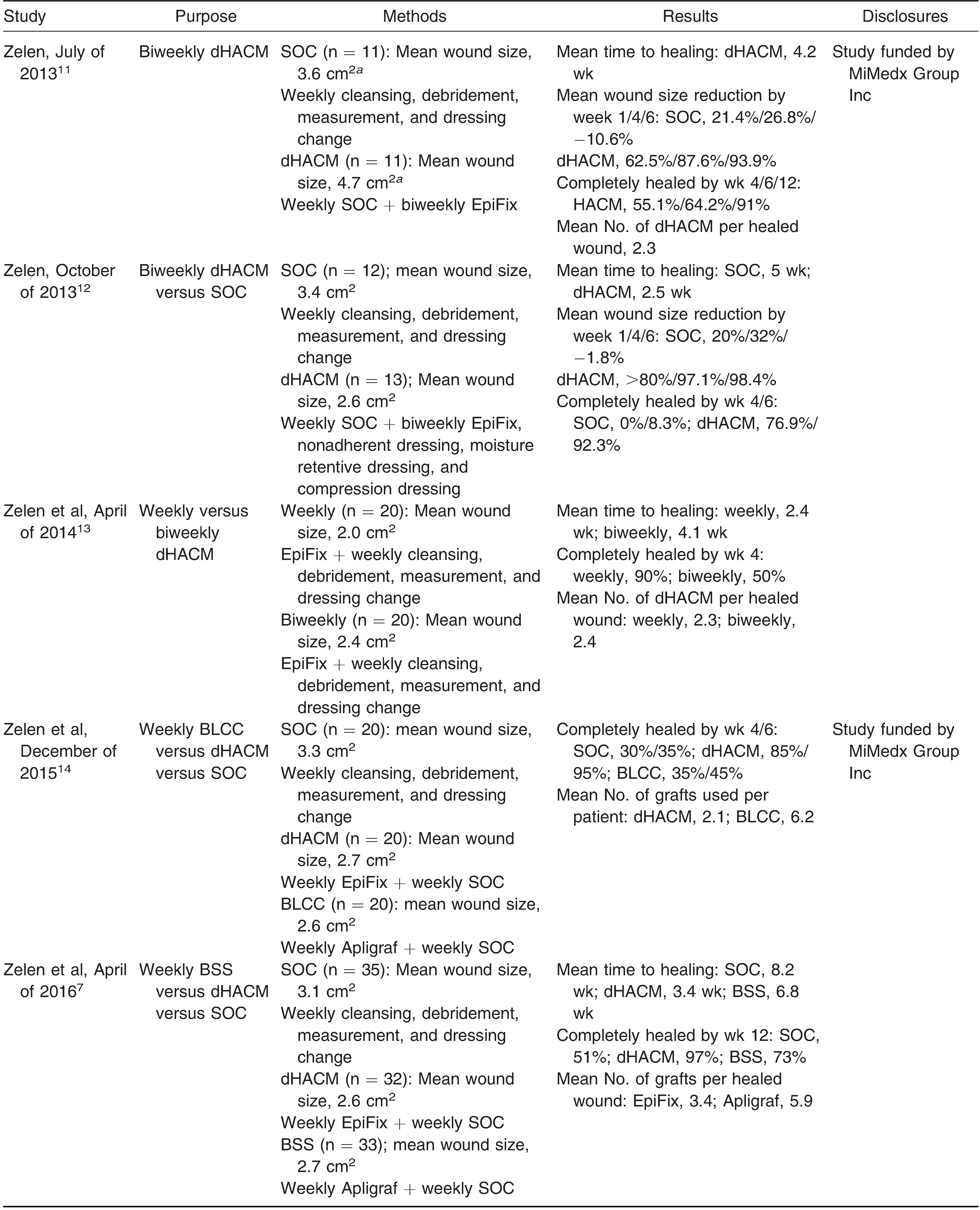
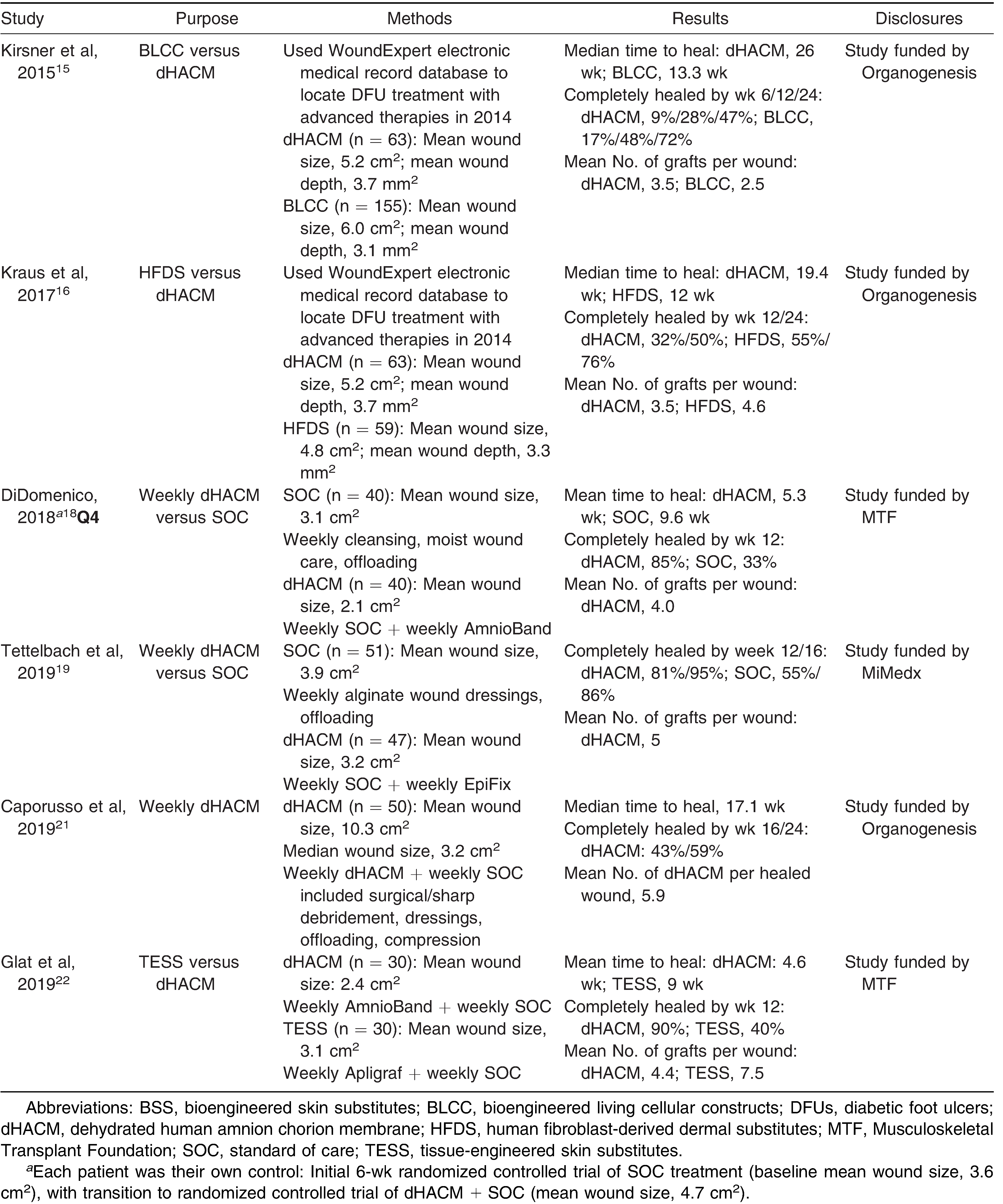
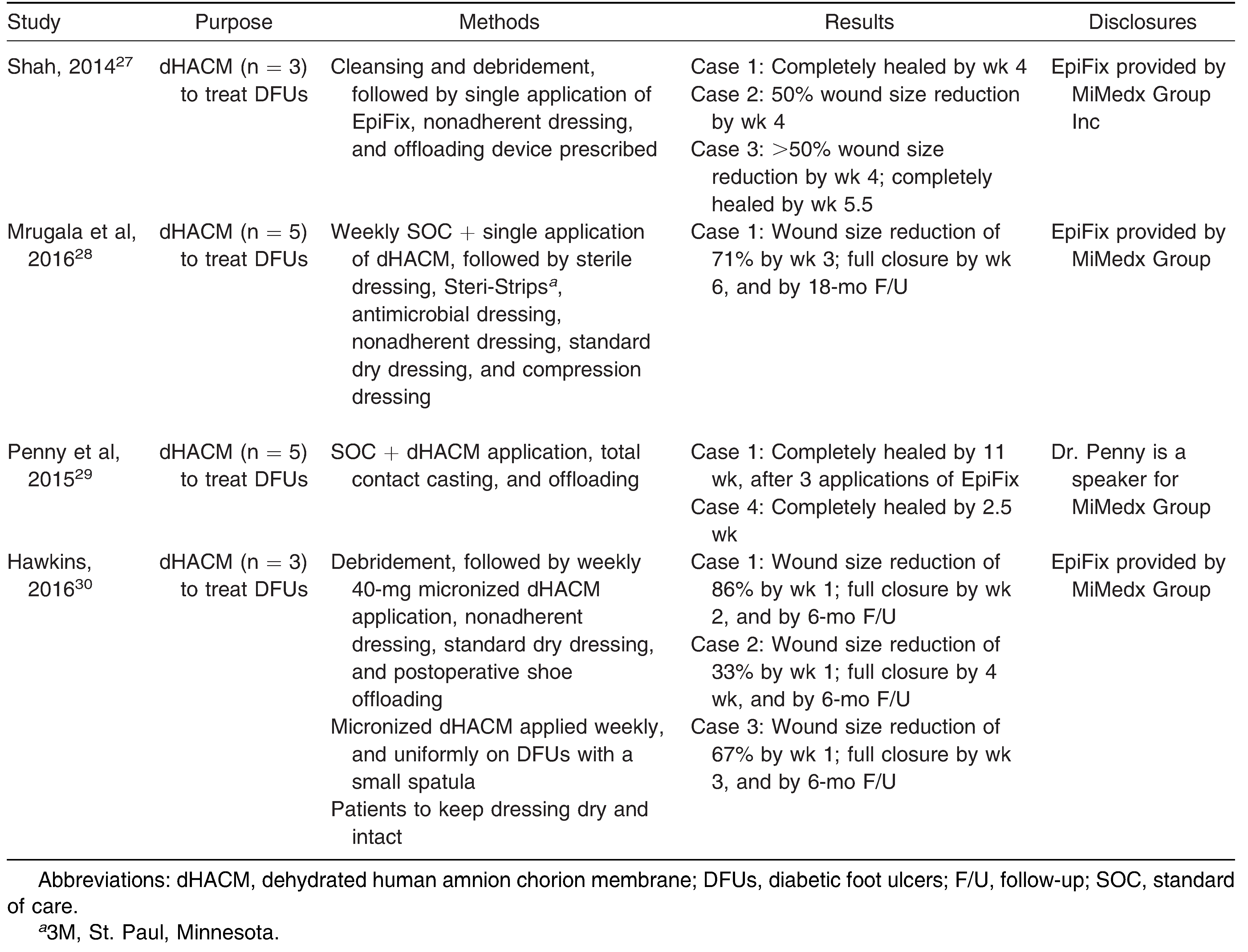
Eight peer-reviewed randomized controlled trial (RCT) articles and four peer-reviewed case studies examined the use of dHACM for the treatment of difficult-to-heal DFUs (Table 1). Three peer-reviewed comparative effectiveness articles used an electronic medical record (EMR) to retrospectively analyze DFU treatment with dHACM (Table 2). Zelen was the primary author in five of the RCTs, in July of 2013 and October of 2013, 2014, 2015, and 2016. The studies performed in July of 2013 and in 2015 were sponsored and funded by MiMedx Group Inc. The remaining studies by Zelen and colleagues provided no disclosure or conflict-of-interest statement.
Table 1.
Summary of Randomized Controlled Trials and Retrospective Studies Using dHACM in Treatment of Recalcitrant DFUs.
Table 2.
Summary of Case Studies Using dHACM in Treatment of Recalcitrant DFUs.
Zelen et al [11] first published a study of biweekly EpiFix use on 11 participants with DFUs in July of 2013. Complete ulcer healing occurred in 91% of participants by week 12, with 4.2 weeks as the mean time to complete healing. Average ulcer size reduction was stated to be 87.6% by week 4. In October of the same year, Zelen et al [12] conducted a similar RCT, of 6 weeks’ duration, to compare biweekly EpiFix treatment on DFUs (n = 13) to SOC treatment (n = 12). In this study, the average 4-week wound size reduction was 97% and 32% for EpiFix and SOC, respectively. Complete healing occurred in 77% of the EpiFix group and 0% of the SOC group. In 2014, Zelen et al [13] conducted a 12-week-long study (n = 40) on the effectiveness of weekly application as opposed to the previous biweekly application of EpiFix. By week 12, 92.5% of all treated ulcers showed complete healing, results that are in line with the July 2013 study. They reported that complete healing occurred by week 4 in 90% and 50% of participants in the weekly and biweekly groups, respectively.
In 2015, Zelen et al [14] conducted a 6-week-long study (n=60) to compare the efficacy of SOC versus EpiFix versus Apligraf, a BLCC, for DFU treatment. The 60 participants were randomized equally into three treatment groups. Eighty-five percent of EpiFix-treated wounds achieved complete healing by week 4, compared to 30% for the SOC group and 35% for the Apligraf-treated group. Within 6 weeks of treatment, these numbers climbed to 95%, 45%, and 35% for the EpiFix, Apligraf, and SOC groups, respectively. They further reported that the median time to healing was 13 days for the EpiFix-treated participants, compared to 49 days for both SOC- and Apligraf-treated participants.
In 2016, Zelen et al [7] extended their 2015 study to include a 12-week endpoint and increased the study enrollment to a total of 100 participants. The protocols for the two treatment groups remained the same; however, previous use of wet-to-dry dressings as the SOC was replaced by collagen alginate dressings. Complete healing was observed by 12 weeks in 97%, 73%, and 51% of participants treated with EpiFix, Apligraf, and SOC, respectively. The mean times to healing in respective order for the three groups were 3.4, 6.8, and 8.2 weeks, respectively.
The efficacy of dHACM was evaluated against BLCC in a 2015 study conducted by Kirsner et al [15]. In evaluating the effectiveness of both advanced therapies on the management of DFUs, the research team used similar inclusion criteria used by Zelen et al to search retrospectively through an EMR database. Data of 218 patients who were treated over a period of 24 weeks in 2014 were collected. They concluded that the median time to wound closure was 26 and 13.3 weeks for the dHACM- and BLCC-treated groups, respectively. Complete healing by 6 weeks was achieved in 9% and 17% of dHACM- and BLCC-treated patients, respectively. By 12 weeks, complete healing occurred in 28% and 48% of the respective treatment groups.
Kraus et al [16] in 2017 performed a similar study using EMR to analyze the efficacy of dHACM versus HFDS treatment under the same patient inclusion criteria. Kraus et al [16] concluded that the median time to wound closure was 19.4 and 12 weeks for the dHACM- and HFDS-treated groups, respectively. Complete healing rates by 12 weeks were reported to be 32% and 55% for dHACM- and HDFS-treated groups, respectively. At 24 weeks, complete healing had occurred in 50% and 76% of the respective treatment groups.
DiDomenico et al [17,18] completed an RCT comparing AmnioBand Membrane versus SOC of collagen alginate in 2016 (n = 40) and 2018 (n = 80). Dehydrated human amnion chorion membrane allografts were applied weekly for up to 12 weeks. By the end of the trial, healing had occurred for 85% in the dHACM group and 33% for the SOC group. The mean time to wound closure was 37 days for the dHACM group versus 67 days for the SOC group. The authors distinguished AmnioBand Membrane from EpiFix in the difference in cost after 12 weeks of weekly applications, $1,771 versus $2,798 per healed wound, respectively.
In 2019, Tettelbach et al [19] conducted a multicenter, RCT of 110 subjects in comparison of EpiFix and SOC of alginate dressings. They reported that 70% of the dHACM group and 50% in the SOC group healed at the completion of a 12-week trial.
Garoufalis et al [20] conducted a retrospective study of 101 subjects treated with micronized and/or sheet form of dHACM on lower-extremity wounds characterized not specifically to chronic DFUs. The subjects were included regardless of comorbidities or vascular status. Of the five subjects in the ischemic wound type group who had completed the study, complete wound healing was observed in only two after 12 weeks of weekly applications.
Caporusso et al [21] conducted a retrospective study in 2019 of 50 subjects treated with weekly applications of dHACM not limited to the foot and ankle. Participants with vascular insufficiency were included, but no ankle-brachial index was reported. The mean frequency of wound closure of the 24 DFUs was 42% at 16 weeks and 59% at 24 weeks, and the median time to healing was 120 days. The number of grafts used per wound over the study period was not specified, but the mean number of applications of dHACM for DFUs and venous leg ulcers was 5.9. The median wound area in the DFU group was 3.215 cm2, but with a mean of 10.387 cm2.
In 2019, Glat et al [22] published the results of an RCT (n = 60) comparing AmnioBand against Apligraf. Ninety percent of the patients in the AmnioBand group healed after 12 weeks of weekly graft applications compared to 40% in the Apligraf group. The mean number of dHACM grafts used per wound was 4.4, and the cost of wound closure by the end of 12 weeks was $2,200.
Adverse effects including infections and hospitalizations were reported, but none were related to dHACM use in five of the RCTs and four case studies. However, Tettelbach et al [19] reported three possible graft-related adverse reactions, including two positive wound cultures and one wound maceration. The RCT conducted by DiDomenico et al [17,18] reported one foot infection that progressed to osteomyelitis in the dHACM plus SOC group. They also noted development of osteomyelitis in three subjects in the SOC group. The four retrospective studies did not report serious dHACMrelated adverse effects.
Discussion
Patients who experience difficult-to-heal DFUs are at increased risk of infection, amputation, and mortality. Advanced therapy such as dHACM has been documented in the management of injuries ranging from elective surgical wounds, traumatic soft-tissue wounds, infected wounds, and burns since the late 1970s [23]. The field of wound care has recently introduced the use of human amnion chorion membrane for the treatment of diabetic foot ulcers, as the manufacturing of dehydrated product and ease of storage have shaped the graft into a treatment option that is more amenable for use [8]. An analysis of literature found by means of several PubMed database and manual reference list searches is thus provided to gain a clearer understanding of the effectiveness of dHACM use on recalcitrant DFUs.
Three of the 15 articles provided no conflict-ofinterest or disclosure statements; however, Zelen et al, the authors of these three articles, also authored two peer-reviewed RCTs that were funded by MiMedx Group Inc. Of the 13 articles that provided disclosure statements, three RCTs were funded by MiMedx Group Inc, two RCTs were funded by Organogenesis Inc, two RCTs were funded by the Musculoskeletal Transplant Foundation, one retrospective study was funded by Organogenesis Inc, three case studies were funded by the MiMedx Group Inc, and the lead author of one case study is a speaker for MiMedx Group Inc.
In an analysis of all five RCT articles authored by Zelen and colleagues, the initial and subsequent depth measurements were not reported even though each study indicated that ulcer depth was measured at each weekly follow-up. Ulcer depth, ulcer stage, and the presence of biofilm have been identified as healing outcome predictors of DFUs.[24] Identification of wound characteristics and locations are helpful in making better clinical assessments to individualize treatment plans. This information may help isolate the most appropriate advanced therapies for certain wounds and determine the wound characteristics that would benefit most from dHACM treatment.
Kirsner et al [15] and Kraus et al [16] reported results that were not in line with the data reported by Zelen et al. Zelen et al reported faster healing rates, and fewer graft applications per healed wound in favor of EpiFix over Apligraf and Dermagraft in a prospective and controlled setting. Kirsner et al [15] and Kraus et al, [16] in contrast, reported lower efficacy in both treatments, but with Apligraf having faster healing rates and fewer graft applications. Many factors may play a role in the dissimilarity of the results, including the use of the EMR in place of RCTs, a larger sample size, a longer study duration, assortment of clinical sites, differing treatment patterns, differing patient characteristics, and differing ulcer characteristics.
Adequate circulation is a controlled variable in all of the studies by Zelen et al in all mentioned RCTs except in July of 2013, in which the authors noted that only two of the 11 total participants were smokers. The inclusion criteria required normal ranges with either Doppler arterial waveforms, ankle-brachial index, or dorsum transcutaneous oxygen test. Kirsner et al [15] and Kraus et al [16] did not explicitly state the vascular status of the subjects that they included. However, they acknowledged the limitation of the EMR with the possibility of incomplete or unreliable medical history, surgical record, or concurrent medication. The retrospective study conducted by Garoufalis et al [20] discretely classified the subjects into a DFU group and an ischemic wound group. Although the sample size was small (n = 5), they reported a lower percentage of complete wound closure in the ischemic group (40%) compared to the DFU group (90.9%) at week 12. It was unclear how many of the subjects in the ischemic wound group were nondiabetic and how many of them received the micronized form of dHACM instead of the sheet form. Therefore, the data in this study do not meet our inclusion criteria. Proper circulation has been documented to be advantageous to wound healing. The uncertainty of vascular status of their selected subjects may be demonstrated as longer healing time compared to the results of the RCTs [25].
Kirsner et al [15] and Kraus et al [16] had a mean wound size that was larger than that reported by Zelen et al. The mean wound size studied by Kirsner et al and Kraus et al was 5.2 cm2, whereas the mean wound size studied by Zelen et al throughout all their RCTs was 2.7 cm2. Ince et al [26] noted that large baseline wound sizes predict slower healing of diabetic foot lesions. The difference in wound size may contribute to shorter healing time in the studies by Zelen et al. However, this does not necessarily indicate that EpiFix is superior to alternative advanced therapies such as Apligraf or Dermagraft in smaller wounds, as no specific range of wound size was studied.
Kirsner et al [15] and Kraus et al [16] used the same EMR with similar search criteria, and thus identified the same 63 patients with diabetic foot ulcers treated with dHACM. The Cox regression model estimated the median wound closure time with dHACM treatment to be 19.4 and 26 weeks for the two retrospective EMR studies, respectively. Complete healing results also differed by a few percentage points, with Kirsner et al [15] reporting 28% and 47% of dHACM-treated ulcers completely healed by 12 and 24 weeks, and Kraus et al [16] reporting 32% and 50% of dHACM-treated ulcers completely healed by 12 and 24 weeks, respectively. The disparity observed took into account the covariates, including ulcer size and duration, that influenced the predictive outcome of each treatment. Because the results from the real-world analyses are different from those of the RCTs, it can be inferred that dHACM is not necessarily the best option for all DFUs in the setting of complicated comorbidities. What can be appreciated from this is the need for individualized treatment options.
The subjects included in the retrospective study conducted by Caporusso et al [21] included patients with vascular insufficiency. However, their vascular status was not quantified with vascular study. The wound size was also not limited, with the largest being 62.50 cm2. The reported frequency of wound closure at week 24 was 59%, which is more consistent with the values reported by the studies that have less restrictive inclusion criteria.
So far, there is no literature that compared the efficacy of the two dHACM products, EpiFix and AmnioBand Membrane. The design of the RCTs that used either of these products yielded similar data except for the lower cost of AmnioBand Membrane.
The four presented case studies yielded results similar to those reported in the RCTs under similar patient selection criteria. The results obtained from EMR data analyses revealed lower efficacy of dHACM, possibly because of the difference in their subject inclusion and exclusion criteria. This treatment has been proven to substantially increase the healing rate with weekly application on vascularly intact patients with a wound size no greater than 2.5 cm2. There are variables including but not limited to ulcer depth, vascular status, and consistency in SOC that could cause the observed healing time to be vastly different in the retrospective EMR studies. Clinicians need to make a complete clinical assessment to optimize individual wound management and avoid prolongation of wound healing with less effective treatments.
Conclusions
Future multicenter RCTs without conflicts of interest are needed to further support the efficacy of dHACM treatment for DFUs. The study protocols should sample a wider range of wound dimensions and patients with multiple comorbidities to better represent the population that is commonly afflicted with DFUs. Proven indications for dHACM are defined in the inclusion criteria of the RCTs that were reviewed in the current article. One should be cautious when generalizing dHACM as the indicated treatment for all recalcitrant DFUs in the real-world setting. Dehydrated human amnion chorion membrane treatment has shown a significantly higher healing rate compared with SOC given that the patient is not vascularly compromised, does not have autoimmune disease or active infection, and the wound is not deep to tendon or bone. Similar results may not be reproducible if the patient is not appropriately selected.
A reduction in healing time with advanced wound treatment can considerably lower the cost of chronic wound management and costly complications such as amputation and hospitalization. In calculation of the cost-effectiveness, the overall long-term management of a wound in aspects other than the cost of an individual graft should be considered. The most economic plan is to heal a wound in the shortest amount of time without recurrence. Essentially, dHACM is a cost-effective advanced therapy option when used on the appropriate DFUs.
Financial Disclosure
None reported.
Conflicts of Interest
None reported.
References
- CENTERS FOR DISEASE CONTROL AND PREVENTION: National Diabetes Statistics Report, 2014: Estimates of Diabetes and Its Burden in the United States; US Department of Health and Human Services: Atlanta, GA, 2014.
- SINGH, N; ARMSTRONG, DG; LIPSKY, BA. Preventing foot ulcers in patients with diabetes. JAMA 2005, 293, 217. [Google Scholar] [CrossRef] [Scilit]
- EDWARDS, JL; VINCENT, AM; CHENG, HT; et al. Diabetic neuropathy: mechanisms to management. Pharmacol Ther 2008, 120, 1. [Google Scholar] [CrossRef] [Scilit]
- MARGOLIS, D; KANTOR, J; BERLIN, J. Healing of neuropathic ulcers: results of a meta-analysis. Diabetes Care 1999, 22, 692. [Google Scholar] [CrossRef] [Scilit]
- MOULIK, P; GILL, GV. Amputation and mortality in newonset diabetic foot ulcers stratified by etiology. Diabetes Care 2003, 26, 491. [Google Scholar] [CrossRef] [Scilit]
- WARRINER, RA; SNYDER, RJ. CARDINAL MH: Differentiating diabetic foot ulcers that are unlikely to heal by 12 weeks following achieving 50% percent area reduction at 4 weeks. Int Wound J 2011, 8, 632. [Google Scholar] [CrossRef] [Scilit] [PubMed]
- ZELEN, CM; SERENA, TE; GOULD, L; et al. Treatment of chronic diabetic lower extremity ulcers with advanced therapies: a prospective, randomised, controlled, multicentre comparative study examining clinical efficacy and cost. Int Wound J 2016, 13, 272. [Google Scholar] [CrossRef] [Scilit] [PubMed]
- KOOB, TJ; RENNERT, R; ZABEK, N; et al. Biological properties of dehydrated human amnion/chorion composite graft: implications for chronic wound healing. Int Wound J 2013, 10, 493. [Google Scholar] [CrossRef] [Scilit] [PubMed]
- KOOB, TJ; LIM, JJ; MASSEE, M; et al. Properties of dehydrated human amnion/chorion composite grafts: implications for wound repair and soft tissue regeneration. J Biomed Mater Res B Appl Biomater 2014, 102, 1353. [Google Scholar] [CrossRef] [Scilit]
- NIKNEJAD, H; HABIBOLLAH, P; MASOUMEH, J; et al. Properties of the amniotic membrane for potential use in tissue engineering. Eur Cell Mater 2008, 15, 88. [Google Scholar] [CrossRef] [Scilit]
- ZELEN CM: An evaluation of dehydrated human amniotic membrane allografts in patients with DFUs. J Wound Care 2013, 22, 347. [CrossRef] [Scilit]
- ZELEN, CM; SERENA, TE; DENOZIERE, G; et al. A prospective randomised comparative parallel study of amniotic membrane wound graft in the management of diabetic foot ulcers. Int Wound J 2013, 10, 502. [Google Scholar] [CrossRef] [Scilit]
- ZELEN, CM; SERENA, TE; SNYDER, RJ. A prospective, randomised comparative study of weekly versus biweekly application of dehydrated human amnion/ chorion membrane allograft in the management of diabetic foot ulcers. Int Wound J 2014, 11, 122. [Google Scholar] [CrossRef] [Scilit] [PubMed]
- ZELEN, CM; GOULD, L; SERENA, TE; et al. A prospective, randomised, controlled, multi-centre comparative effectiveness study of healing using dehydrated human amnion/chorion membrane allograft, bioengineered skin substitute or standard of care for treatment of chronic lower extremity diabetic ulcers. Int Wound J 2015, 12, 724. [Google Scholar]
- KIRSNER, RS; SABOLINSKI, ML; PARSONS, NB; et al. Comparative effectiveness of a bioengineered living cellular construct vs. a dehydrated human amniotic membrane allograft for the treatment of diabetic foot ulcers in a real-world setting. Wound Repair Regen 2015, 23, 737. [Google Scholar] [CrossRef] [Scilit] [PubMed]
- KRAUS, I; SABOLINSKI, ML; SKORNICKI, M; et al. The comparative effectiveness of a human fibroblast dermal substitute versus a dehydrated human amnion/chorion membrane allograft for the treatment of diabetic foot ulcers in a real-world setting. Wounds 2017, 29, 125. [Google Scholar]
- DIDOMENICO, LA; ORGILL, DP; GALIANO, RD; et al. Aseptically processed placental membrane improves healing of diabetic foot ulcerations: prospective, randomized clinical trial. Plast Reconstr Surg Glob Open 2016, 4, e1095. [Google Scholar] [CrossRef] [Scilit] [PubMed]
- DIDOMENICO, LA; ORGILL, DP; GALIANO, RD; et al. Use of an aseptically processed, dehydrated human amnion and chorion membrane improves likelihood and rate of healing in chronic diabetic foot ulcers: a prospective, randomised, multi-centre clinical trial in 80 patients. Int Wound J 2018, 15, 950. [Google Scholar] [CrossRef] [Scilit]
- TETTELBACH, W; CAZZELL, S; REYZELMAN, AM; et al. A confirmatory study on the efficacy of dehydrated human amnion/chorion membrane dHACM allograft in the management of diabetic foot ulcers: a prospective, multicentre, randomised, controlled study of 110 patients from 14 wound clinics. Int Wound J 2019, 16, 9. [Google Scholar]
- GAROUFALIS, M; NAGESH, D; SANCHEZ, PJ; et al. Use of dehydrated human amnion/chorion membrane allografts in more than 100 patients with six major types of refractory nonhealing wounds. JAPMA 2018, 108, 84. [Google Scholar] [CrossRef] [Scilit]
- CAPORUSSO, J; ABDO, R; KARR, J; et al. Clinical experience using a dehydrated amnion/chorion membrane construct for the management of wounds. Wounds 2019, 31, S19. [Google Scholar]
- GLAT, P; ORGILL, DP; GALIANO, R; et al. Placental membrane provides improved healing efficacy and lower cost versus a tissue-engineered human skin in the treatment of diabetic foot ulcerations. Plast Reconstr Surg Glob Open 2019, 7, e2371. [Google Scholar] [CrossRef] [Scilit] [PubMed]
- GRUSS, JS; JIRSCH, DW. Human amniotic membrane: a versatile wound dressing. Can Med Assoc J 1978, 118, 1237. [Google Scholar] [PubMed]
- VELLA L, FORMOSA C: Characteristics predicting the outcome in individuals with diabetic foot ulcerations. JAPMA 2017, 107, 180. [CrossRef] [Scilit]
- WHITNEY JD: The influence of tissue oxygen and perfusion on wound healing. AACN Clin Issues Crit Care Nurs 1990, 1, 578.
- INCE, P; KENDRICK, D; GAME, F; et al. The association between baseline characteristics and the outcome of foot lesions in a UK population with diabetes. Diabet Med 2007, 24, 977. [Google Scholar] [CrossRef] [Scilit]
- SHAH AP: Using amniotic membrane allografts in the treatment of neuropathic foot ulcers. JAPMA 2014, 104, 198. [CrossRef] [Scilit]
- MRUGALA, A; SUI, A; PLUMMER, M; et al. Amniotic membrane is a potential regenerative option for chronic nonhealing wounds: a report of five cases receiving dehydrated human amnion/chorion membrane allograft. Int Wound J 2016, 13, 485. [Google Scholar] [CrossRef] [Scilit]
- PENNY, H; RIFKAH, M; WEAVER, A; et al. Dehydrated human amnion/chorion tissue in difficult- to-heal DFUs: a case series. J Wound Care 2015, 24, 104. [Google Scholar] [CrossRef] [Scilit] [PubMed]
- HAWKINS B: The use of micronized dehydrated human amnion/chorion membrane allograft for the treatment of diabetic foot ulcers: a case series. Wounds 2016, 28, 152.

© 2021 American Podiatric Medical Association