


Abstract
A 34-year-old female recreational badminton player presented with left ankle pain 1 week after a recreational badminton game. She reported experiencing a similar pattern of pain in her right ankle 4 months before that had persisted for 3 months. On plain radiography, callus formation was evident on the right distal fibula, and a subtle lesion was observed on the left side. Ultrasound was performed with the clinical suspicion of bilateral, nonsimultaneous, distal fibular stress fracture. Focal hyperechoic thickening of the periosteum with irregularity and hypoechoic periosteal edema over the left distal fibula were identified. These findings were consistent with stress fracture, and an early phase of distal fibular stress fracture was diagnosed. This case report highlights that ultrasound can be an alternative modality to magnetic resonance imaging or bone scan scintigraphy for the early diagnosis of stress fracture.
Recently, the incidence of stress fracture has increased because more people are participating in recreational and competitive sports,[1,2] making early diagnosis and prompt treatment important. However, early diagnosis is often challenging because of ambiguous symptoms during the early phase of stress fracture and the low sensitivity of plain radiography.[3] As the criterion standard of stress fracture diagnosis, magnetic resonance imaging or bone scan scintigraphy can be considered[4]; however, routine investigation is not justified because these techniques are costly and time consuming. In contrast, ultrasound provides noninvasive, inexpensive, real-time dynamic images, and it is becoming more accepted as a first-line imaging modality in the orthopedic field.[5,6] This case report emphasizes the utility of ultrasound in early diagnosis of distal fibular stress fracture. The patient was informed that data concerning the case would be submitted for publication, and she provided consent.
Case Report
The patient was an otherwise healthy 34-year-old woman (height, 156 cm; weight, 53 kg; body mass index, 21.8 kg/m2) who presented with pain on the posterolateral side of the left ankle (visual analog scale score of 5). One week before, she had noticed left ankle pain while playing badminton, but this pain did not stop her from playing. After completion of the game, her pain increased and did not subside with rest and ice bag application. Interestingly, 4 months previously, she had experienced gradual pain in her right ankle while playing badminton, and the pain had persisted until 1 month previously. Initially, she did not visit a medical facility because the right ankle pain gradually diminished with activity restriction. However, resuming the activity resulted in a similar pattern of pain in her left ankle, so she decided to get a medical examination.
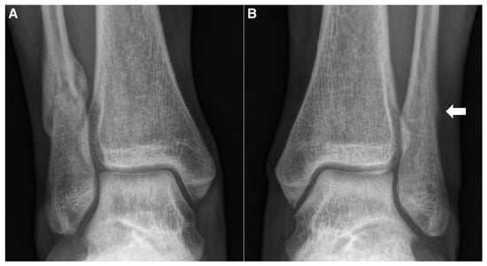
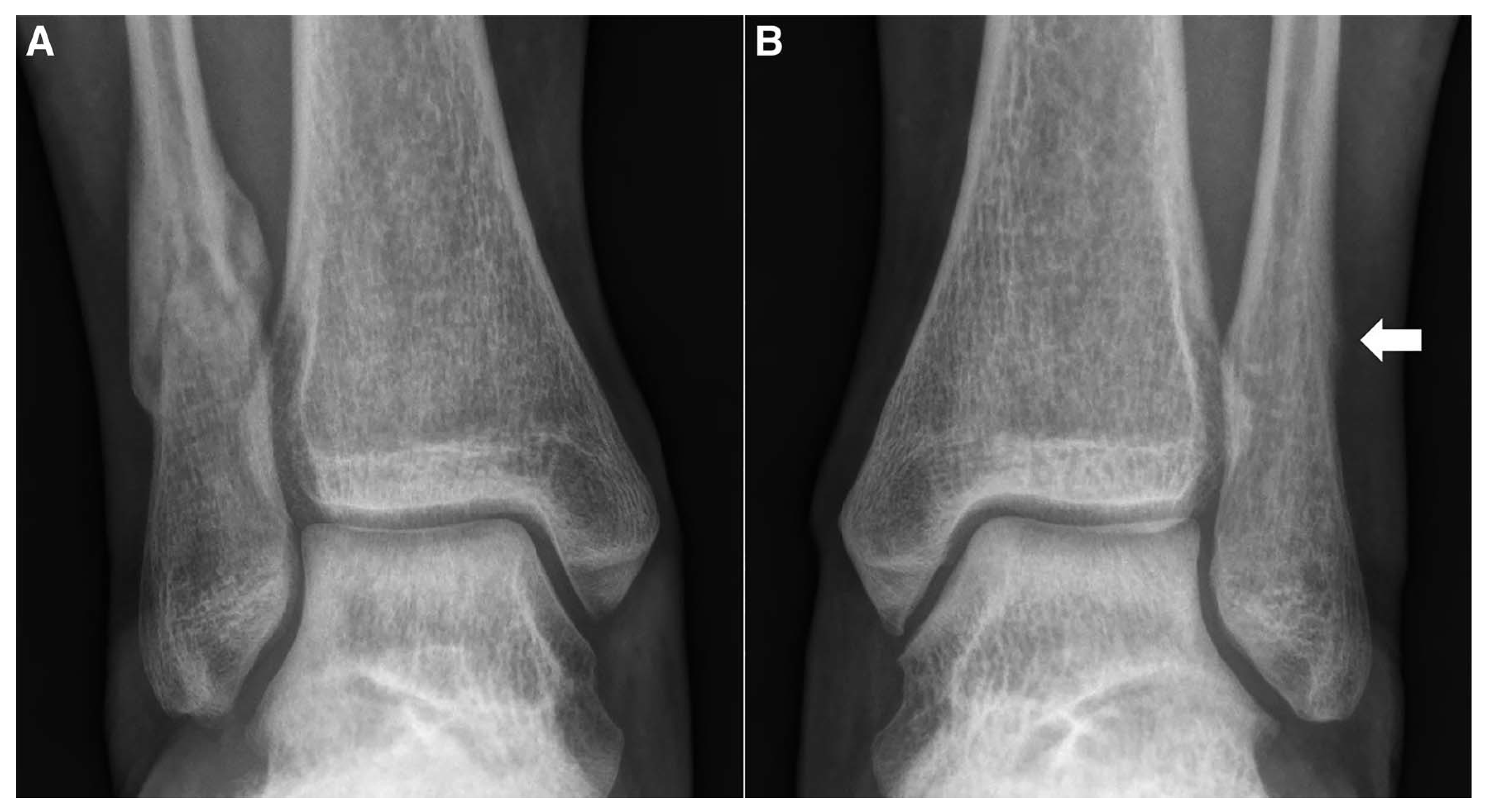
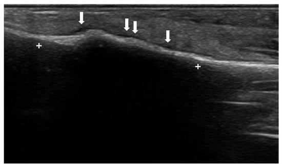
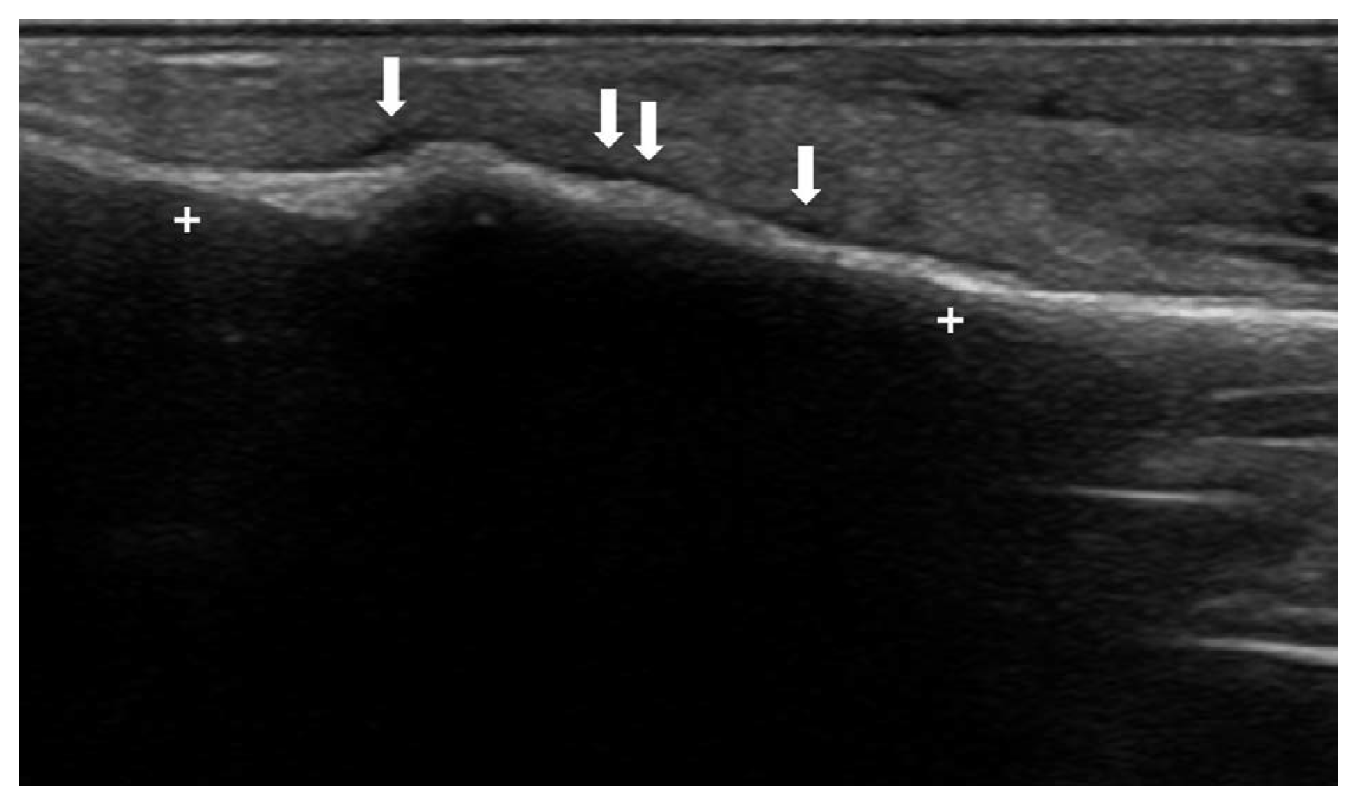
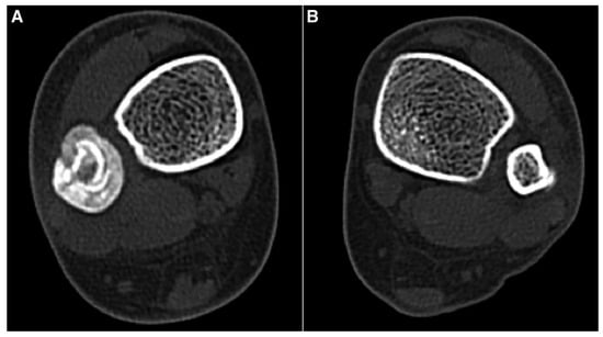
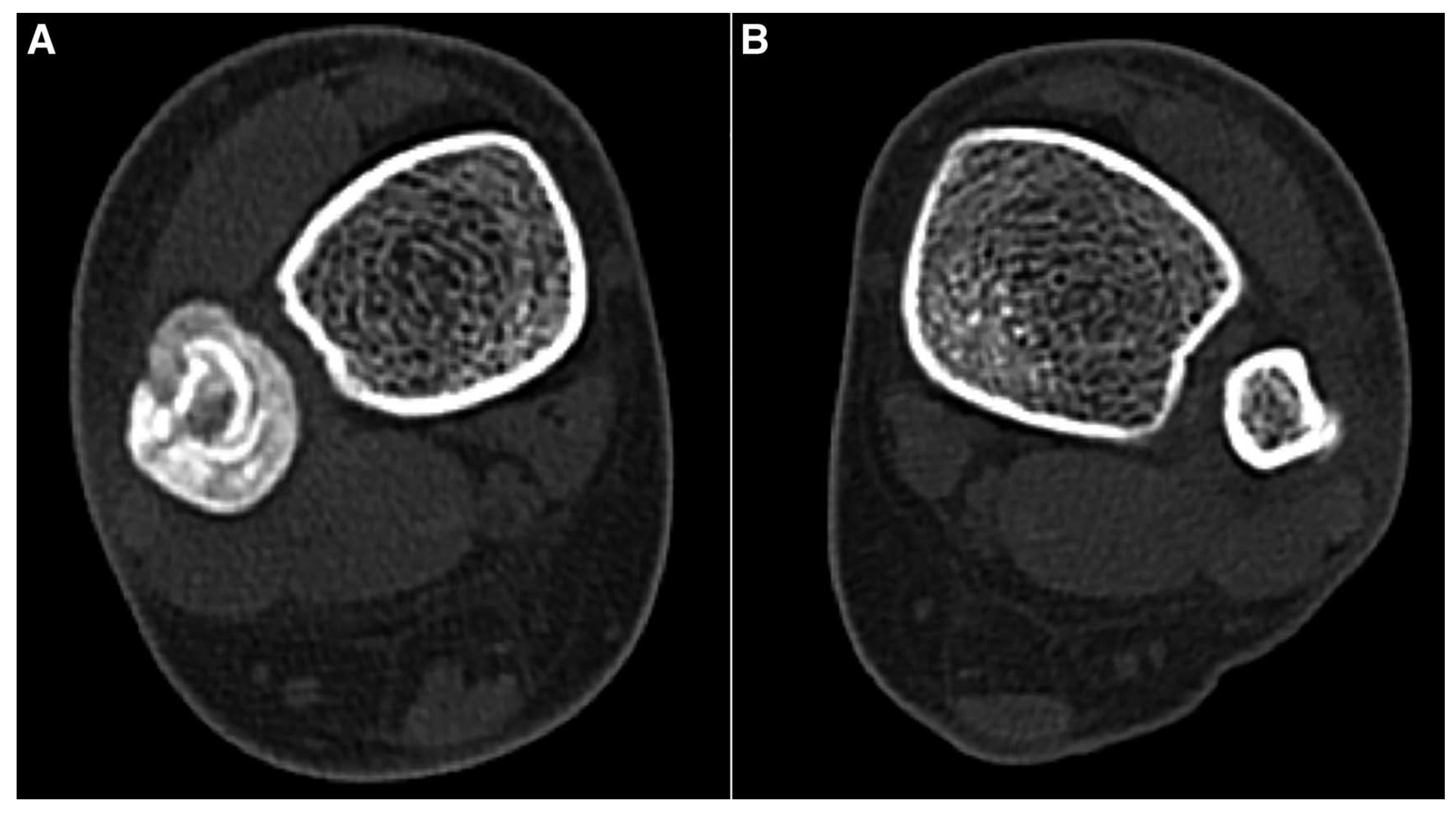
The examination revealed tenderness on the posterolateral border of the left distal fibula. Neurologic examination was within normal limits. Plain radiography of the lower extremity was performed. Abundant callus formation was observed on the right distal fibula, and slight bulging with a faint cortical line was observed on the lateral border of the left distal fibula just above the syndesmosis (Figure 1). However, because the radiologic findings of the left side were inconclusive, we could not confidently rule out pathologic findings. Therefore, ultrasound was performed by an experienced foot and ankle surgeon using a 9- to 15-MHz linear-array transducer (LOGIQ; GE Healthcare, Chalfont St Giles, United Kingdom) to obtain additional information. Ultrasound of the left distal tibia showed focal hyperechoic thickening of the periosteum with irregularity and hypoechoic periosteal edema over the left distal fibula that measured approximately 1 cm (Figure 2). On the basis of these findings, bilateral, nonsimultaneous, distal fibular stress fracture was diagnosed. Because the ultrasound findings were clear enough to diagnose stress fracture, magnetic resonance imaging or bone scan scintigraphy was not used to confirm the stress fracture. However, computed tomography was performed to evaluate whether the right distal fibular stress fracture had achieved union. The bilateral fibular computed tomographic scan showed complete union with circumferential callus formation on the right distal fibula and focal cortical disruption with slight callus deposition on the left distal fibula (Figure 3). These results were consistent with the findings of fibular stress fracture.
Figure 1.
Anteroposterior radiographs of both ankles. A, Abundant callus formation of the right distal fibula. B, Slight bulging of the lateral cortex of the left distal fibula (arrow).
Figure 2.
Longitudinal ultrasound image of the left distal fibula, taken 1 week after symptom onset. Focal hyperechoic thickening of the periosteum with irregularity (between the crosses) and hypoechoic periosteal edema (arrows) are shown.
Figure 3.
Axial plane computed tomographic scan showing (A) circumferential callus formation on the right distal fibula and (B) focal cortical disruption with slight callus deposition on the left distal fibula.
The patient was instructed to stop sport activity and given a rebound walking boot to wear that provided leg support. After using the walking boot, she did not report severe pain, so medication was not used. At 1-year follow-up, the patient was doing well, without any significant symptoms.
Discussion
We report novel ultrasound findings of an earlyphase distal fibular stress fracture when plain radiographs did not show definite pathologic signs. In this case, the early phase of the stress fracture showed focal hyperechoic thickening of the periosteum with irregularity and hypoechoic periosteal edema, whereas cortical disruption and increased periosteal color Doppler flow were not definite on ultrasound imaging of the stress fracture. Although these findings are also reported in a previous case of stress fracture and the sensitivity of ultrasound for diagnosis of stress fracture was 81.8%,[7,8] we believe that this report can be helpful for the diagnosis of early stress fractures by verifying ultrasound findings in the early phase of a fibular stress fracture.
Conclusions
The use of ultrasound in the diagnosis of stress fracture allows for noninvasive early diagnosis at a low cost.
Financial Disclosure
None reported.
Conflicts of Interest
None reported.
References
- GAETA, M; MILETO, A; ASCENTI, G. ET AL: Bone stress injuries of the leg in athletes. Radiol Med 2013, 118, 1034. [Google Scholar] [CrossRef] [Scilit] [PubMed]
- SNYDER, RA; KOESTER, MC; DUNN, WR. Epidemiology of stress fractures. Clin Sports Med 2006, 25, 37. [Google Scholar] [CrossRef] [Scilit] [PubMed]
- ROMANI, WA; PERRIN, DH; DUSSAULT, RG; et al. Identification of tibial stress fractures using therapeutic continuous ultrasound. J Orthop Sports Phys Ther 2000, 30, 444. [Google Scholar] [CrossRef] [Scilit] [PubMed]
- ISHIBASHI, Y; OKAMURA, Y; OTSUKA, H; et al. Comparison of scintigraphy and magnetic resonance imaging for stress injuries of bone. Clin J Sport Med 2002, 12, 79. [Google Scholar] [CrossRef] [Scilit] [PubMed]
- BACKHAUS, M; BURMESTER, GR; GERBER, T. ET AL: Guidelines for musculoskeletal ultrasound in rheumatology. Ann Rheum Dis 2001, 60, 641. [Google Scholar] [CrossRef] [Scilit] [PubMed]
- BLANKSTEIN, A. Ultrasound in the diagnosis of clinical orthopedics: the orthopedic stethoscope. World J Orthop 2011, 2, 13. [Google Scholar] [CrossRef] [Scilit] [PubMed]
- HOGLUND, LT; SILBERNAGEL, KG; TAWEEL, NR. Distal fibular stress fracture in a female recreational runner: a case report with musculoskeletal ultrasound imaging tindings. Int J Sports Phys Ther 2015, 10, 1050. [Google Scholar] [PubMed]
- PAPALADA, A; MALLIAROPOULOS, N; TSITAS, K. ET AL: Ultrasound as a primary evaluation tool of bone stress injuries in elite track and field athletes. Am J Sports Med 2012, 40, 915. [Google Scholar] [CrossRef] [Scilit] [PubMed]
© 2021 American Podiatric Medical Association