


Abstract
The Lauge-Hansen classification does not cover all types of ankle injuries. The present report details three cases of exceptional fragment of the medial tibia that differed from the traditional Lauge-Hansen supination–external rotation and pronation–external rotation fracture patterns. The information obtained from this study will be helpful for conducting basic research of this condition and determining appropriate surgical approaches.
Lauge-Hansen developed a commonly used classification system to describe the mechanism of injury of adult ankle fractures. This system classifies ankle fractures into four categories according to the foot position and direction of the force applied at the time of injury: supination-adduction, supination–external rotation (SER), pronation-abduction, and pronation–external rotation (PER) [1]. Lauge-Hansen demonstrated that the sequential application of morbid loading with a particular direction of force on a particular foot position will produce a predictable sequence of injuries [2]. However, the Lauge-Hansen classification does not cover all types of injuries. Atypical fracture patterns with a similar mechanism of injury may occur that cannot be classified accordingly [3]. The fractures presented in this article are extremely unusual and differ from traditional Lauge-Hansen patterns. All three patients provided written informed consent for the publication of the present report and the accompanying images.
Case Reports
Case 1
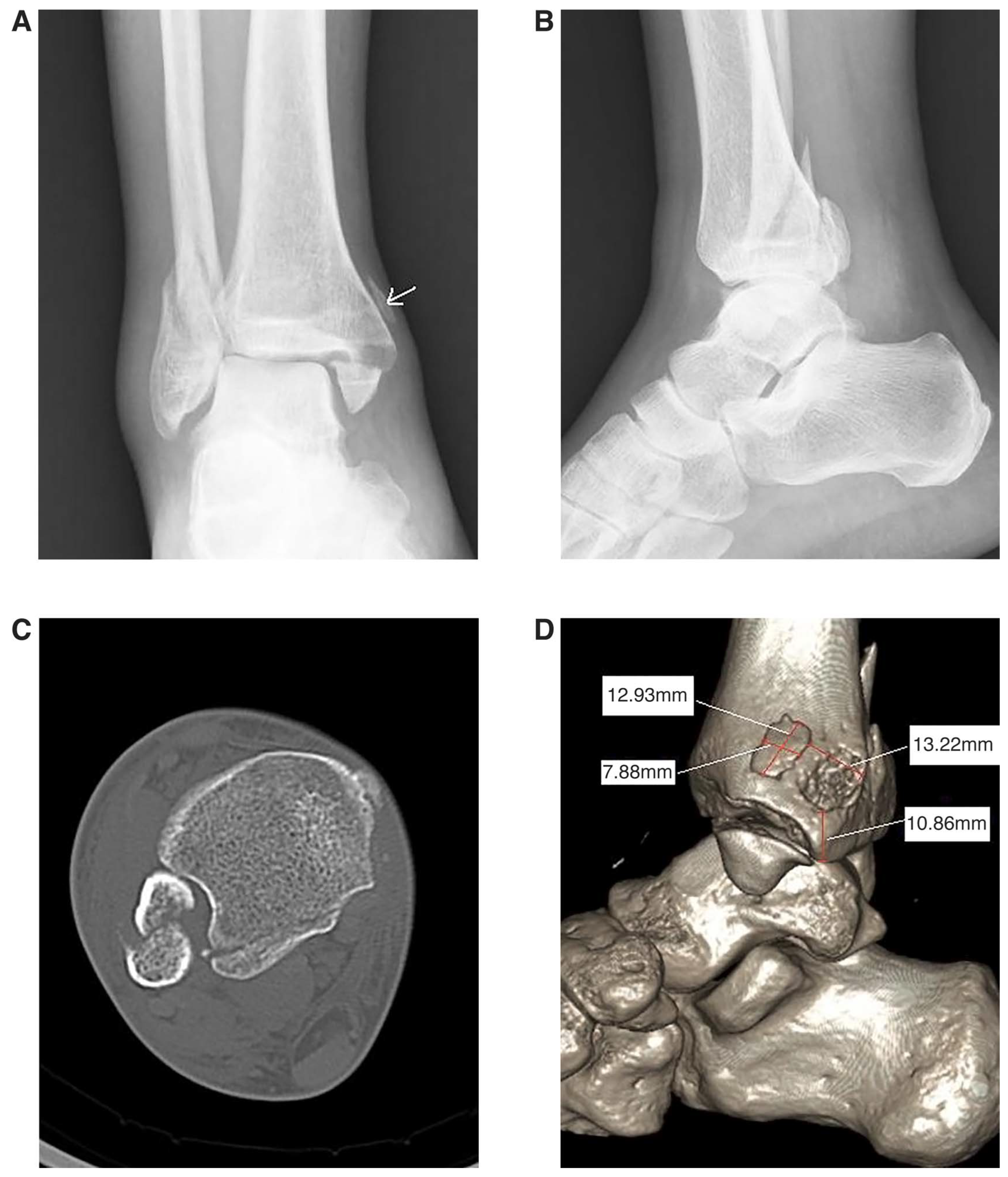
A 64-year-old man who was walking on grass tripped and sustained a twisting injury to his right ankle. He initially attended the emergency department and presented with pain, swelling, and bruising around the ankle joint. Clinical examination revealed the right ankle with swelling and painful restriction of ankle movement. Ecchymosis was present along the medial border of his ankle. Radiographs revealed an oblique fracture of the fibula and a medial malleolar fracture. Furthermore, an avulsion fracture of the posterolateral tibia and an avulsion fracture of the medial tibial margin were doubtful (Fig. 1A and B). Computed tomographic (CT) scans confirmed the presence of a Volkmann fracture and an unusual avulsion fragment of the medial tibial margin. We used image-analyzing software (ImageJ; Broken Symmetry Software, Bethesda, Maryland) to measure the exact size and location of the avulsion fracture. The fragment was 12.93 × 7.88 mm, with a forward displacement of 13.22 mm (Fig. 1C and D). The fragment was displaced obviously; thus, the decision was made to perform surgical fixation. The patient underwent fixation of the fracture with a single compression screw. The postoperative period was uneventful, and the patient was discharged on the first postoperative day. At the final follow-up visit, at 12 months after surgery, the patient was free of pain, and his ankle movements were normal.
Figure 1.
Computed tomographic imaging characteristics of Case 1. A, Anteroposterior view of the ankle demonstrating an exceptional avulsion fracture above the medial malleolus (arrow). B, Lateral view of the ankle revealing a posterior malleolar fracture. C, Axial and three-dimensional (D) examination of the ankle showing the posterior malleolar fractures and periosteal avulsion fracture.
Case 2
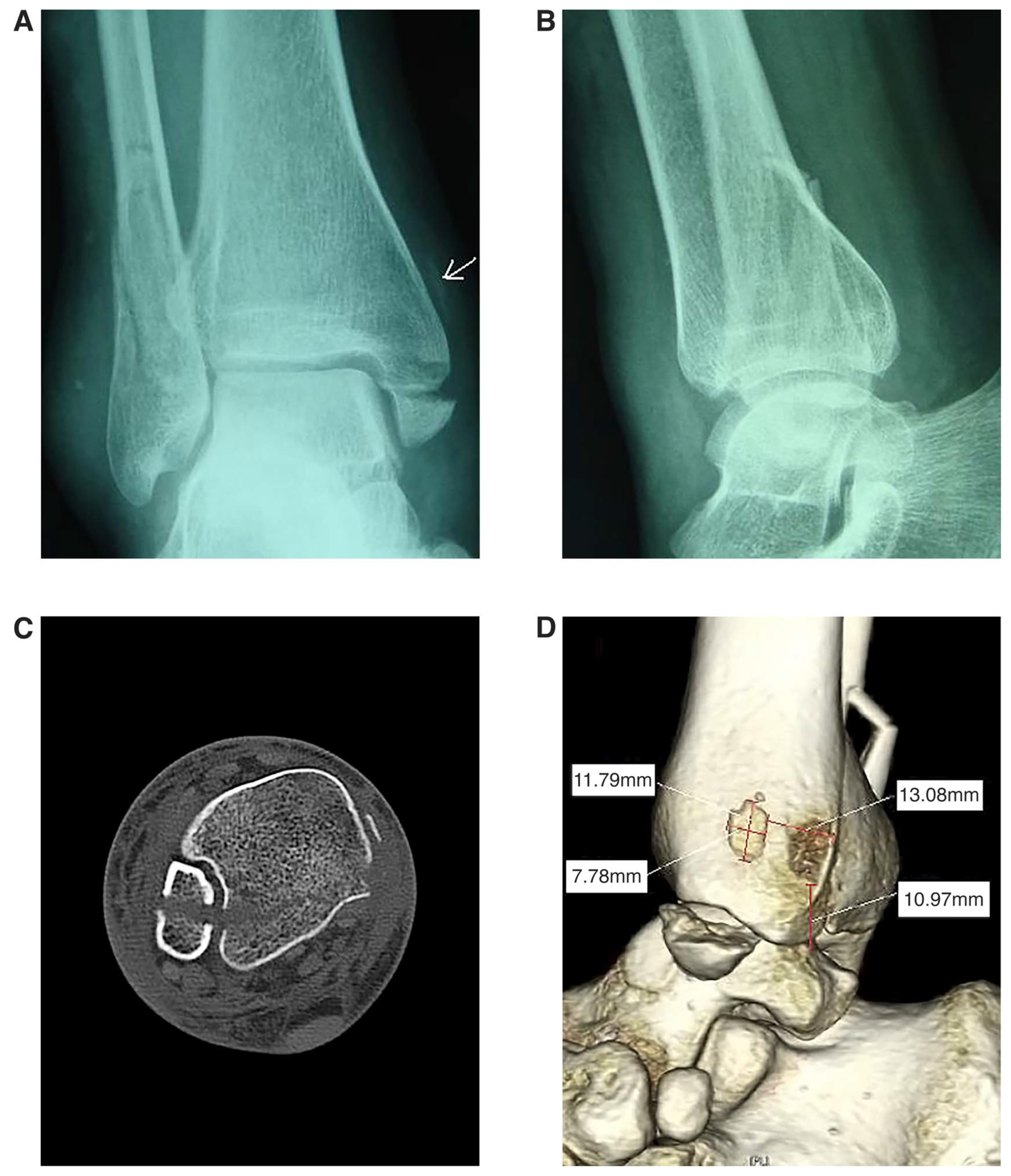
A 56-year-old woman sustained a twisting injury of her right ankle after a fall from a height of approximately 8 to 10 feet. On physical examination, swelling was present over both the lateral and medial aspects of her ankle. Passive ankle movements were restricted and painful. Significant tenderness was present over the medial aspect of the tibial margin. A review of the initial ankle radiographs showed a suspicious avulsion fracture of the medial tibia on the anteroposterior view (Fig. 2A) and a displaced posterior malleolar fracture on the lateral view (Fig. 2B). An additional CT examination was conducted to assess the extent of the fracture. The CT scan clearly revealed a displaced Volkmann fracture associated with a lateral and medial malleolar fracture and an exceptional avulsion fracture of the medial tibia displaced approximately 13.08 mm that was 11.79 × 7.78 mm (Fig. 2C and D).
Figure 2.
Computed tomographic imaging characteristics of Case 2. A, Anteroposterior view of the ankle showing a suspicious avulsion fracture above the medial malleolus (arrow). B, Lateral view of the ankle revealing a posterior malleolar fracture. C, Axial and three-dimensional (D) scans of the ankle showing the posterior malleolar fracture and periosteal avulsion fracture.
Case 3
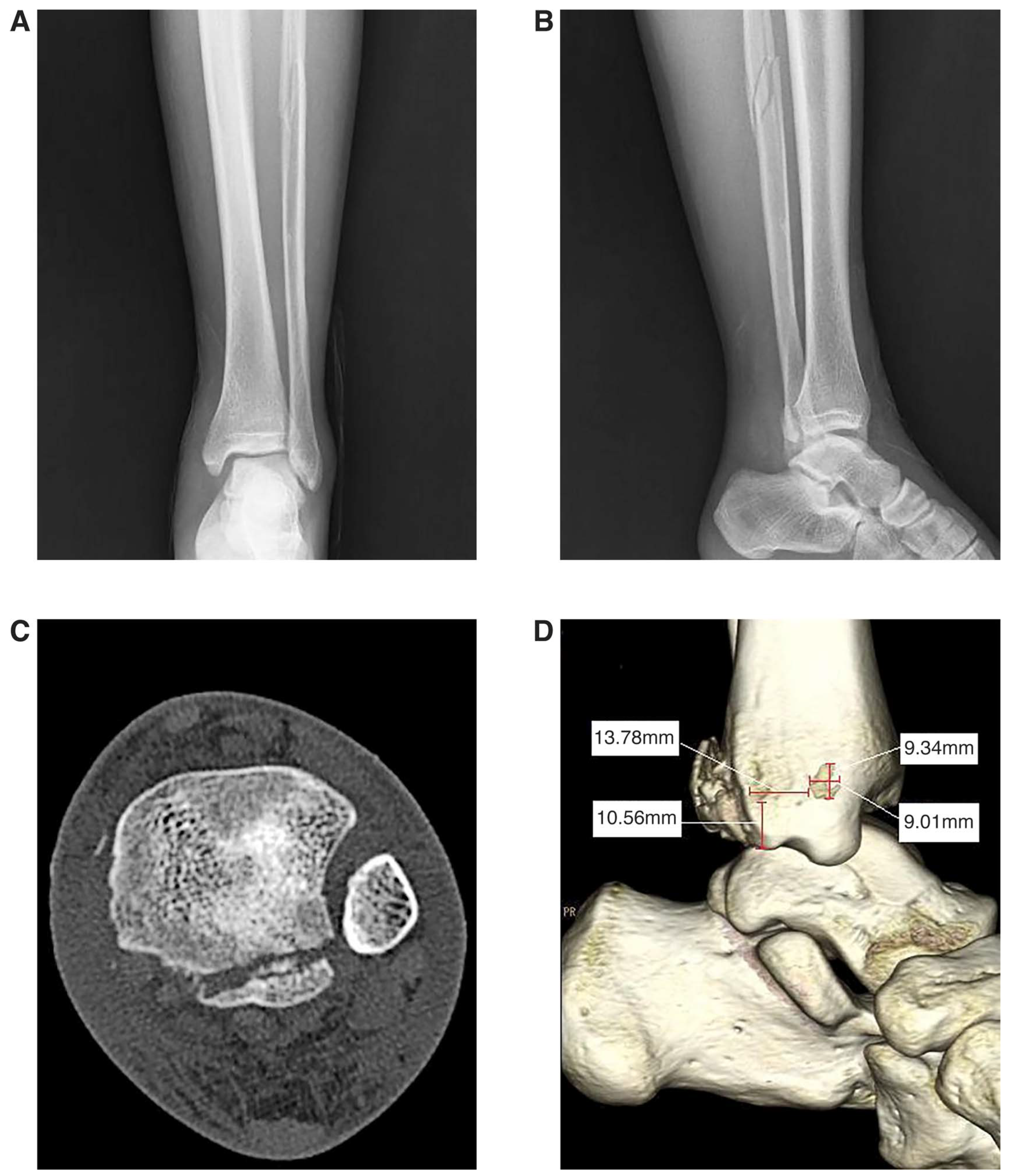
A 21-year-old man presented with injury to his left ankle after a fall from a height. Clinical examination revealed edema at the left ankle joint and painful restriction of ankle movement. Radiographs of the left ankle revealed a Volkmann fracture and an oblique high fibular fracture above the level of the syndesmosis (Fig. 3A and B). Computed tomographic scans were obtained to exclude the presence of a medial malleolar fracture. The scans inadvertently discovered the presence of an avulsion fracture of the medial tibia, with a displacement of approximately 13.78 mm (Fig. 3C and D). Because the fragment appeared cracked, no screw fixation of the fragment was performed. Postoperative management consisted of a short leg nonweightbearing cast for 4 weeks. After immobilization, examination revealed that the range of motion was equal to that of his right ankle. He was permitted to return to full activity at 9 weeks. A 12-month evaluation revealed that the patient had remained asymptomatic with no residual complaints.
Figure 3.
Computed tomographic imaging characteristics of Case 3. A, Anteroposterior view of the ankle demonstrating the high spiral fibula fracture. B, Lateral view of the ankle revealing a posterior malleolar fracture. C, Axial and three-dimensional (D) examination of the ankle showing the posterior malleolar fractures and avulsion fracture.
Discussion
The SER category of injury is divided into four stages [1]. In the first stage, supination of the foot with forced external rotation causes tension on the anteroinferior tibiofibular ligament (AITFL). Three types of injuries exist: 1) rupture of the AITFL, 2) avulsion fracture of the distal tibial attachment of the AITFL (also called a Tillaux fracture), and 3) avulsion fracture from the fibular attachment of the AITFL (also called a Wagstaffe–Le Fort fracture). In the second stage, as the rotation forces continue, a short oblique fracture of the fibula will occur. If the external rotational force is sufficient to continue, the talus will rotate farther laterally and the posteroinferior tibiofibular ligament (PITFL) will stretch. In the third stage, two types of injury exist: either the PITFL will rupture or an avulsion fracture from the posterolateral tibia at the attachment of the PITFL (Volkmann fracture or triangle) will occur. Further lateral rotation of the talus will ultimately create either a deltoid ligament injury or medial malleolar fracture at the fourth stage [4,5].
The PER category of injury is also divided into four stages. In the first stage, injury initiates in tension with either a deltoid ligament rupture or an avulsion fracture of the medial malleolus. The second stage of injury is a rupture of the distal tibiofibular syndesmosis. The third stage is unique in that it produces a spiral or oblique high fibular fracture well above the level of the syndesmosis. The fourth stage of the injury is rupture or avulsion of the PITFL [6].
As a medial malleolar fracture was present in the patients in cases 1 and 2, the sequence of events ended at stage 4 of the SER category. The patient in case 3 also was at the fourth stage of the PER category, as a Volkmann fracture was present. In contrast to the basic rule of sequential events, there was an unusual fragment of the medial tibia. These fractures tend to occur when there is an external rotation injury leading to an avulsion fracture of the posterolateral tibia. We conclude that the fragment that occurred in the present cases resulted from the pull of the periosteum of the posteromedial tibia. This view is based on the following considerations.
First, the fragments possess a similar anterior displacement in all three cases (generally, 13 mm). Moreover, the fragments had a similar shape when the fracture pattern was classified as SER as well in cases 1 and 2. These facts indicated that the possibility of coincidence was eliminated.
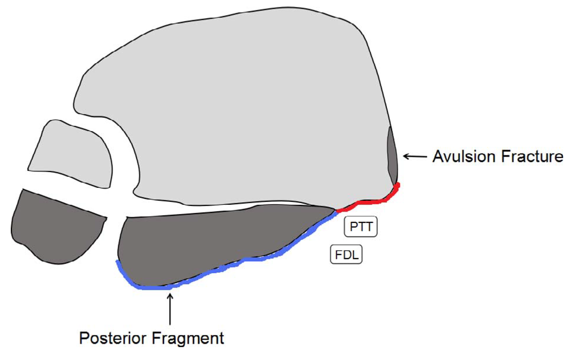
Furthermore, CT images of all three cases revealed that the posterior malleolar fractures had similar characteristics. The fracture line extended from the lateral rim of the posterior tibia to the malleolar groove for the posterior tibial tendon (Fig. 1C, Fig. 2C, and Fig. 3C). There might be a link between the greater avulsion fracture of the posterior tibia and the fragment of the medial tibia. Abducting or rotating the foot laterally under general anesthesia cannot increase the displacement of the fragment, which means that the fragment has little relation with the deltoid ligament injury or the medial malleolar fracture. Moreover, that the fragment with periosteum of the posteromedial tibia was connected with the periosteum of the fracture of the posterolateral tibia had been observed intraoperatively in cases 1 and 2. Magnetic resonance imaging demonstrated a periosteal lesion with high signal intensity on T2-weighted sequences in the region of the posteromedial tibia (Fig. 4). Thus, we can conclude that the mechanism of this injury is remnant external rotation force applied to the periosteum of the posteromedial tibia, resulting in tearing of the periosteum and an avulsion fragment of the medial surface of the distal tibia. As a result of losing tension of the posterior periosteum, the fragment most commonly moves forward (Fig. 5).
Figure 4.
Preoperative magnetic resonance images of the patient in case 3 showing a periosteal lesion with high signal intensity on T2-weighted sequences in the region of the posteromedial tibia on the axial views (red arrow).
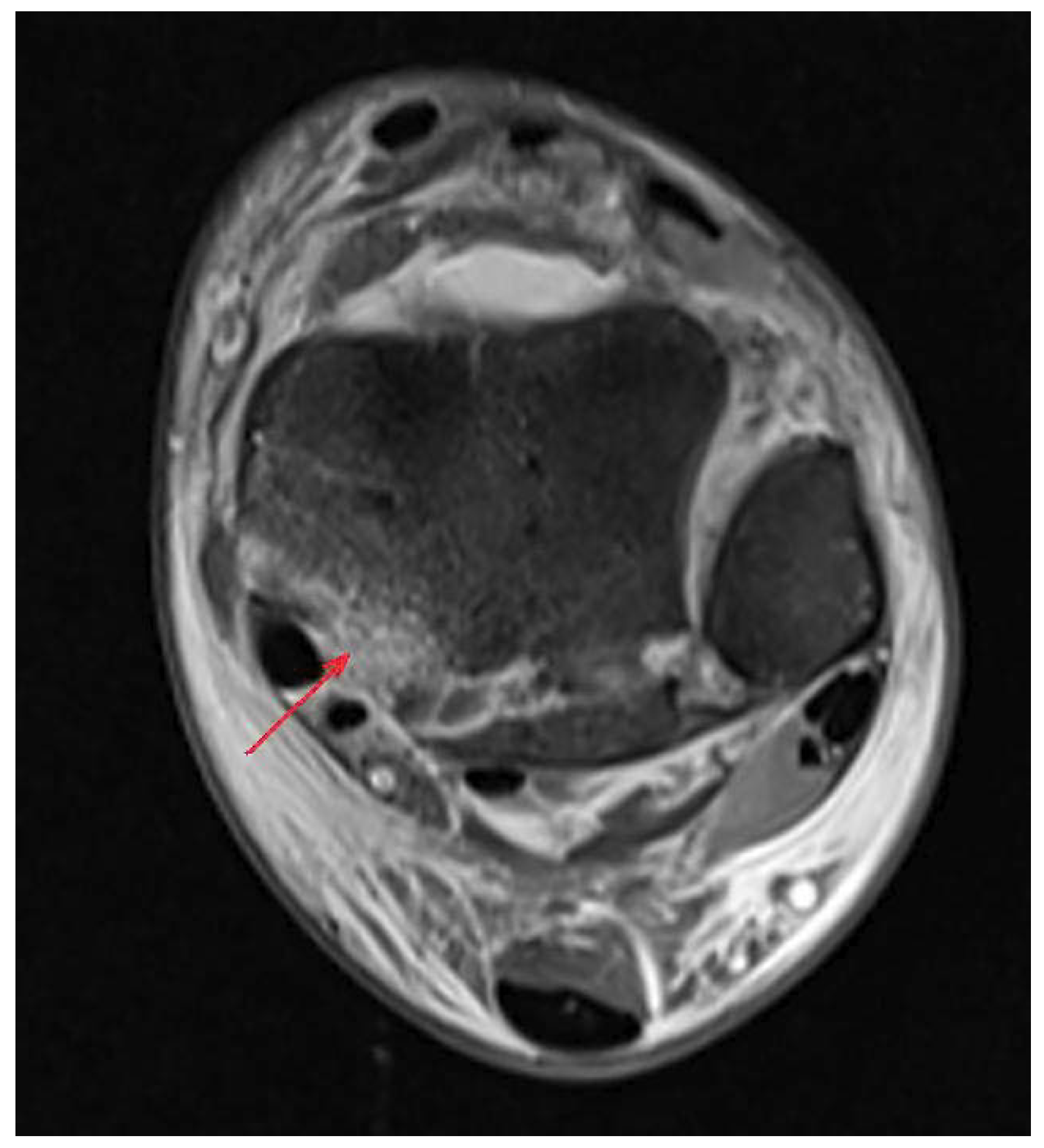
Figure 5.
Schematic diagram illustrating the fracture line of the posterolateral tibia progressing from lateral to medial, extending toward the medial tibia, and finally leading to the avulsion fracture of the cortex (the blue strip represents the periosteum attached to the fracture; the red strip represents the stripping of the periosteum). FDL, flexor digitorum longus; PTT, posterior tibial tendon.
Haraguchi et al [7] confirmed that a posterior malleolar fracture can be classified as one of three types: type I, the posterolateral-oblique type (67%); type II, the medial-extension type (19%); and type III, the small-shell type (14%). Their findings also showed that the fracture lines of posterior malleolar fractures vary greatly, and a medial-extension (type II) fracture usually has two fragments, a larger posteromedial and a smaller posterolateral fragment, and that some fragments involve almost the entire medial malleolus [7]. In the majority of cases, the pull of the periosteum will usually give way before damaging the posteromedial tibia, leading to most avulsion fracture injuries [8,9]. The fragments that we reported differ from the typical medial-extension type classified by Haraguchi et al [7].
The injury can occasionally be missed by a cursory examination of the radiographs. Computed tomography can be useful to confirm the diagnosis, clearly define the extent of the fracture, and rule out any associated injuries involving the tibia [10]. There is insufficient evidence to decide whether an operation is necessary and how to conduct it. After all, the fragment is sometimes too thin to be fixed, and no evidence of instability of the ankle is present following the restoration of other fractures. Unusual avulsion fractures, although uncommon, can be indicative of a type of external rotation force and a greater avulsion fracture of the posterior tibia occurring. It is important to recognize these distinct injuries and appropriately treat the pathologic features to prevent further instability and arthritis [11,12].
Conclusions
The fragment of the medial tibia, as a part of the traditional Lauge-Hansen SER and PER fracture patterns at the fourth stage, has not been reported previously [13,14]. The mechanism of this injury may be a remnant external rotation force applied to the periosteum of the posteromedial tibia. It may not matter so much whether the fragment is restored or not; however, it can act as a warning that Tillaux fracture might have occurred. A limitation of the study is that it included only three patients, and this injury pattern remains to be demonstrated in more patients.
Acknowledgments: This work was supported by the Scientific Research Project of Wuhan Health and Family Planning Commission (WX18Q01).
Financial Disclosure: None reported.
Conflict of Interest: None reported.
References
- Lauge-HansenN:Fractures of the ankle. II. Combined experimental-surgical and experimental-roentgenologic investigations. Arch Surg60: 957, 1950.
- Lauge-HansenN:Fractures of the ankle. III. Genetic roentgenologic diagnosis of fractures of the ankle. Am J Roentgenol Radium Ther Nucl Med71: 456, 1954.
- ThomsenNOOvergaardSOlsenLH: Observer variation in the radiographic classification of ankle fractures. J Bone Joint Surg Br73: 676, 1991.
- PaiVS:Medial malleolar fracture associated with deltoid ligament rupture. J Foot Ankle Surg38: 420, 1999.
- RasmussenSMadsenPVBennickeK:Observer variation in the Lauge-Hansen classification of ankle fractures. Precision improved by instruction. Acta Orthop Scand64: 693, 1993.
- LesicABumbasirevicM:Ankle fractures. Curr Orthop18: 232, 2004.
- HaraguchiNHaruyamaHTogaH: Pathoanatomy of posterior malleolar fractures of the ankle. J Bone Joint Surg Am88: 1085, 2006.
- MichelsonJD:Ankle fractures resulting from rotational injuries. J Am Acad Orthop Surg11: 403, 2003.
- ScheidtKBStiehlJBSkradeDA: Posterior malleolar ankle fractures: an in vitro biomechanical analysis of stability in the loaded and unloaded states. J Orthop Trauma6: 96, 1992.
- DonkenCCGoordenAJVerhofstadMH: The outcome at 20 years of conservatively treated “isolated” posterior malleolar fractures of the ankle: a case series. J Bone Joint Surg Br93: 1621, 2011.
- WeberM:Trimalleolar fractures with impaction of the posteromedial tibial plafond: implications for talar stability. Foot Ankle Int25: 716, 2004.
- McConnellTCreevyWTornettaP III:Stress examination of supination external rotation-type fibular fractures. J Bone Joint Surg Am86: 2171, 2004.
- KwonJYGitajnILWaltonP: A cadaver study revisiting the original methodology of Lauge-Hansen and a commentary on modern usage. J Bone Joint Surg Am97: 604, 2015.
- MichelsonJSolocoffDWaldmanB: Ankle fractures. The Lauge-Hansen classification revisited. Clin Orthop Relat Res345: 198, 1997.
© 2020 American Podiatric Medical Association