


Abstract
We report a unique case of the total loss of the intermediate cuneiform by posttraumatic avascular necrosis resulting from a left foot open fracture and dislocation of the navicular bone and medial and intermediate cuneiforms at the Chopart and Lisfranc joints in a 64-year-old woman. The injury was managed with open reduction and internal fixation with Kirschner wires and cannulated screws. During postoperative follow-up, we observed avascular necrosis of the intermediate cuneiform and the total loss of the bone. An extensive English literature search revealed only one case report published on this topic. Thus, we provide this case study to help guide clinical decision making in the future.
Injuries with a combined Chopart and Lisfranc fracture dislocation are often part of so-called crush injuries of the foot [1,2]. Although being less common, these injuries can be devastating, with associated severe soft-tissue injury, skin necrosis, redislocation, compartment syndrome, ligamentous instability, neurovascular injury and/or compromise, and high rates of deep infection [3].
We report a unique case of the total loss of the intermediate cuneiform by posttraumatic avascular necrosis (AVN) resulting from a left foot open fracture and dislocation of the navicular bone and medial and intermediate cuneiforms at the Chopart and Lisfranc joints in a 64-year-old woman. The injury was managed with open reduction and internal fixation with Kirschner wires and cannulated screws. During postoperative follow-up, we observed AVN of the intermediate cuneiform and total loss of the bone.
An extensive English literature search revealed only one case report published on this topic [4]. Thus, we provide this case study to help guide clinical decision making in the future.
Case Report
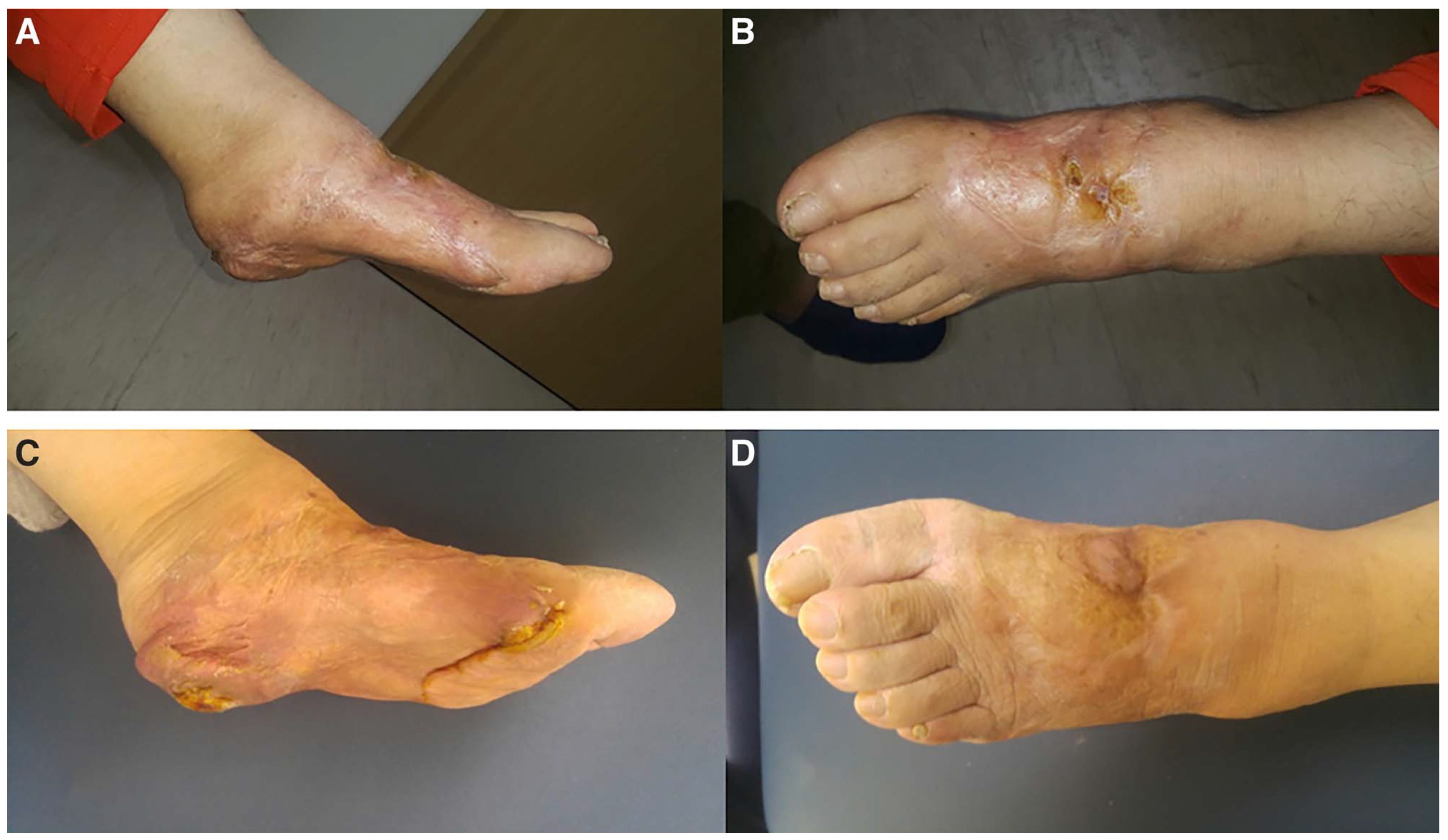
A 64-year-old woman presented to our emergency center after her left foot was run over by a car. The patient complained of severe left foot pain, with range-of-motion limitations. We found a U-shaped degloving injury of the plantar area, dorsalis pedis artery rupture, and extrusion of the medial cuneiform (Fig. 1). Plain radiographs (Fig. 2) and a computed tomographic scan (Fig. 3) of the left foot revealed navicular dislocation, extensive medial migration of the dislocated medial cuneiform, and plantar dislocation of the intermediate cuneiform. We performed massive irrigation, debridement, and open reduction and internal fixation with multiple Kirschner wires and cannulated screws and arteriorrhaphy (end-to-end anastomosis) of the dorsalis pedis artery. Immediate postoperative radiographs (Fig. 4) of the left foot demonstrated a concentrically reduced anatomical midfoot joint, and reduction of the medial and intermediate cuneiforms. Seven days postoperatively, we found skin necrosis on the initial soft-tissue degloving injury and performed debridement of the necrotic tissue and a split-thickness skin graft (Fig. 5). Postoperatively, we applied a short leg splint for 4 weeks and started gentle range-of-motion exercises of the ankle joint. Unfortunately, 6-month postoperative plain radiographs showed partial loss of the intermediate cuneiform, which progressed by 12 months and was completely lost 18 months postoperatively (Fig. 6). Plain radiographs taken 3 years postoperatively showed maintenance of anatomical alignment of the Lisfranc and Chopart joints and the intercuneiform joint, with a vacant space created by total loss of the intermediate cuneiform (Fig. 7). At the last follow-up, the patient could walk with limping (visual analogue scale score of 3) and had a relatively healthy foot, and completely healed soft tissue of the foot (Fig. 8).
Figure 1.
Initial wound photographs showing a U-shaped degloving injury of the medial and plantar area, dorsalis pedis artery rupture, and extrusion and migration of the medial cuneiform.
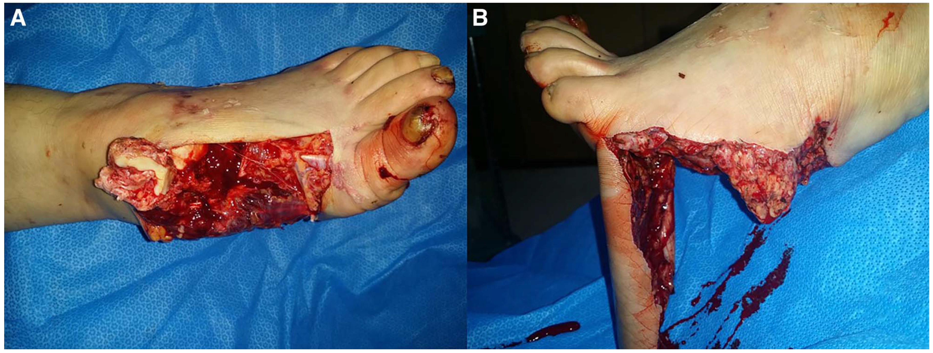
Figure 2.
Initial plain radiographs show Lisfranc and Chopart dislocation, extensive medial dislocation of the medial cuneiform, and plantar dislocation of the intermediate cuneiform. (A) Anteroposterior and (B) oblique views.
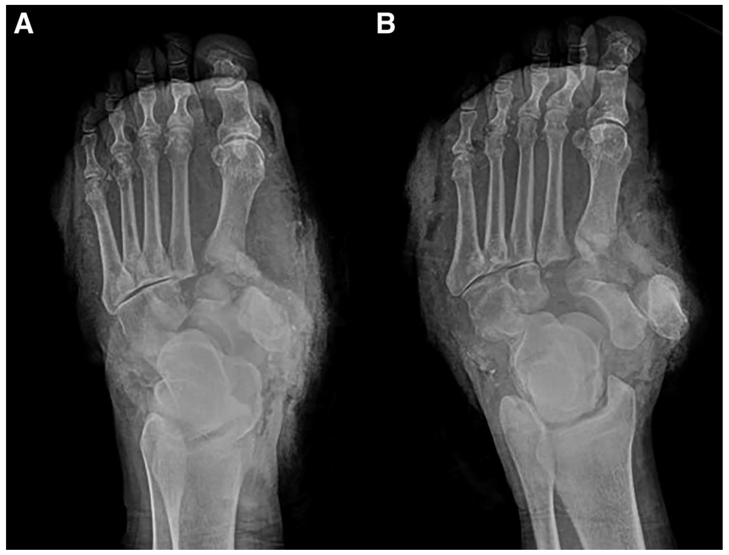
Figure 3.
Three-dimensional reconstructed computed tomographic images show extensive dislocation and inverted position of the medial cuneiform and mild displacement and plantar dislocation of the intermediate cuneiform. (A) Posterior, (B) lateral, and (C) anterior views.
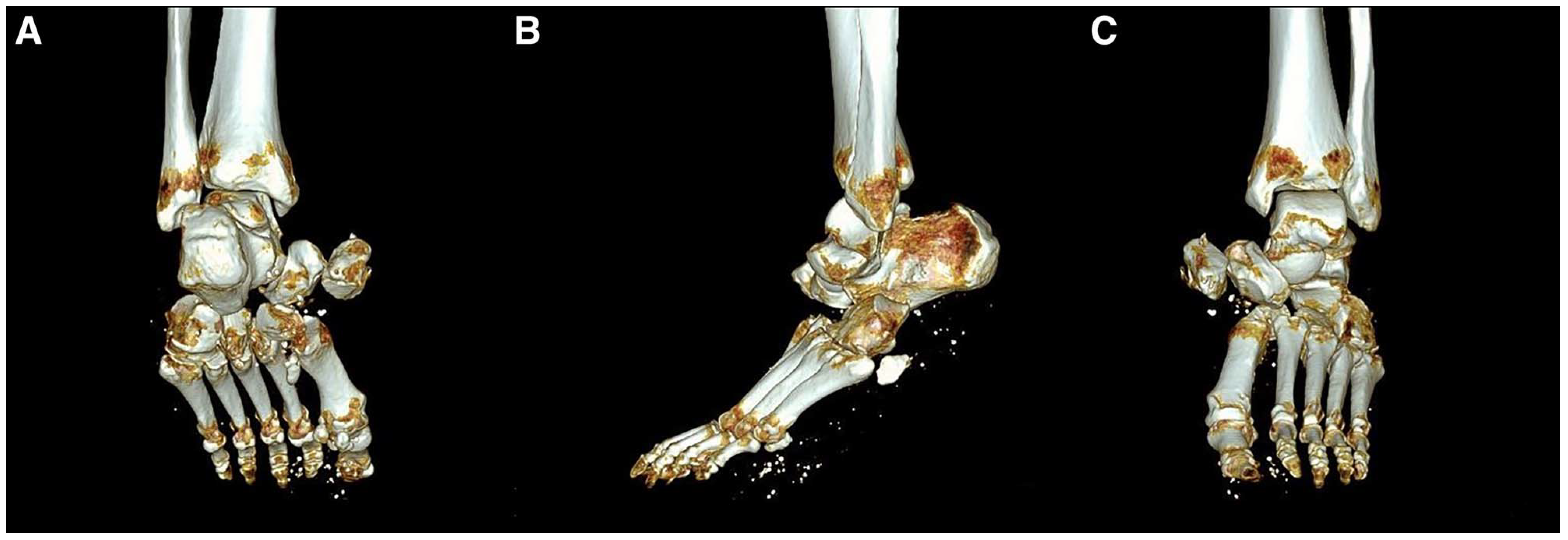
Figure 4.
Immediate postoperative plain radiographs show a relatively well-reduced state of the Lisfranc and Chopart joints and medial and intermediate cuneiforms. (A) Lateral, (B) anteroposterior, and (C) oblique views.
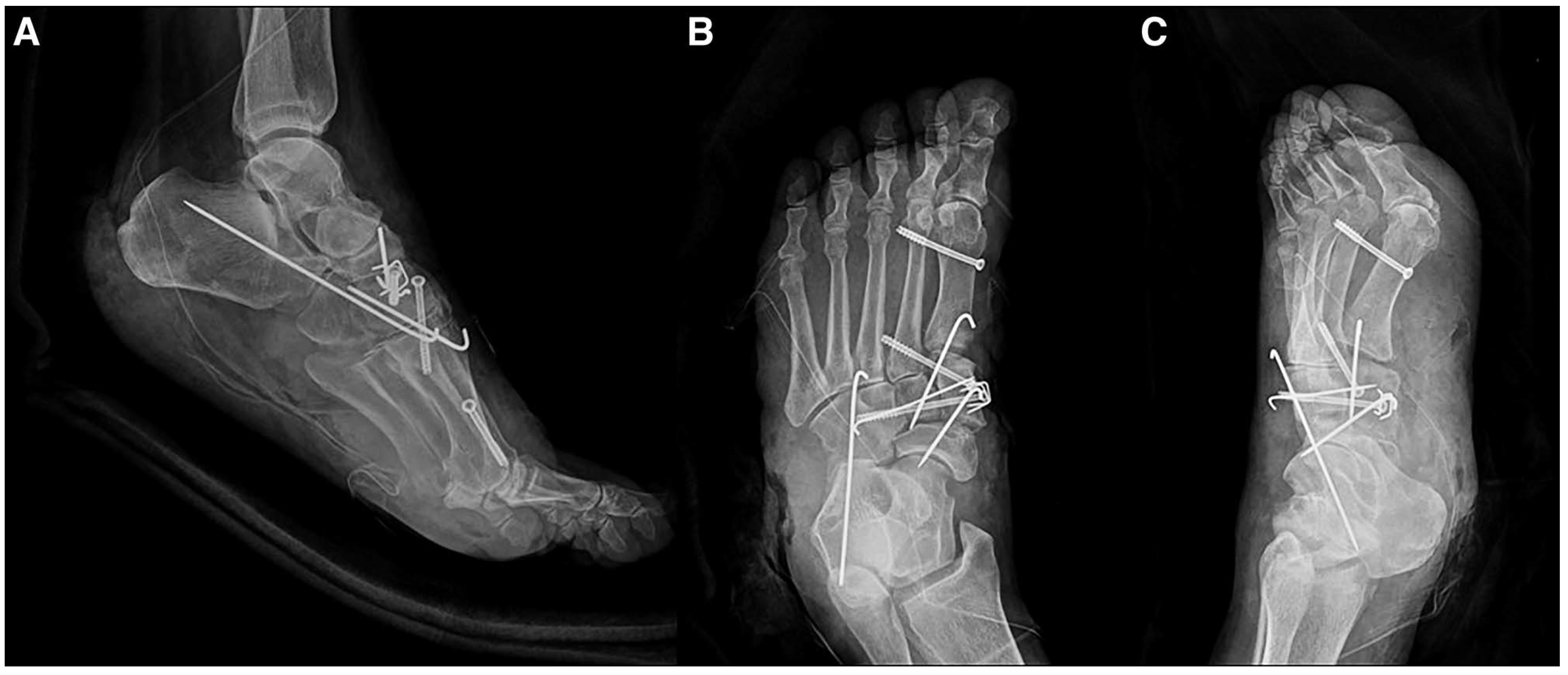
Figure 5.
Wound photographs obtained 7 days postoperatively (A and B) showing extensive skin necrosis on the initial soft-tissue degloving injury. Debridement of the necrotic tissue and a split-thickness skin graft (C and D) were performed.

Figure 6.
Postoperative plain radiographs show avascular necrosis and partial loss of the intermediate cuneiform at 6 months (A and B), progression at 12 months (C and D), and complete loss at 18 months (E and F).
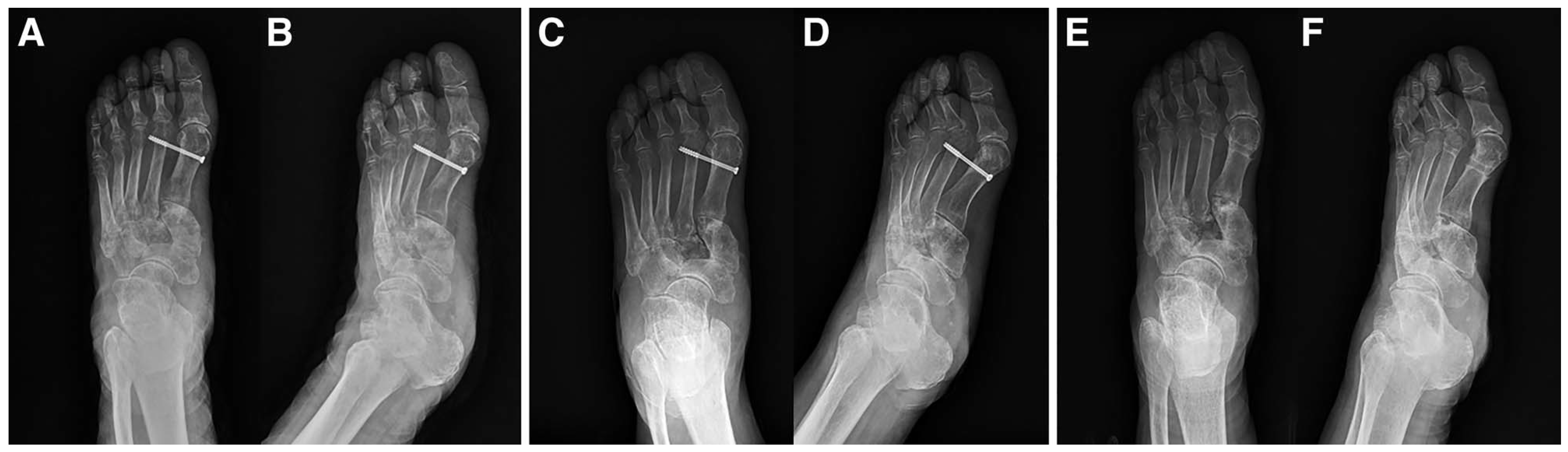
Figure 7.
Three-year postoperative plain radiographs show the maintenance of anatomical alignment of the Lisfranc and Chopart joints and the intercuneiform joint, with absence of the intermediate cuneiform. (A) Lateral, (B) anteroposterior, and (C) oblique views.
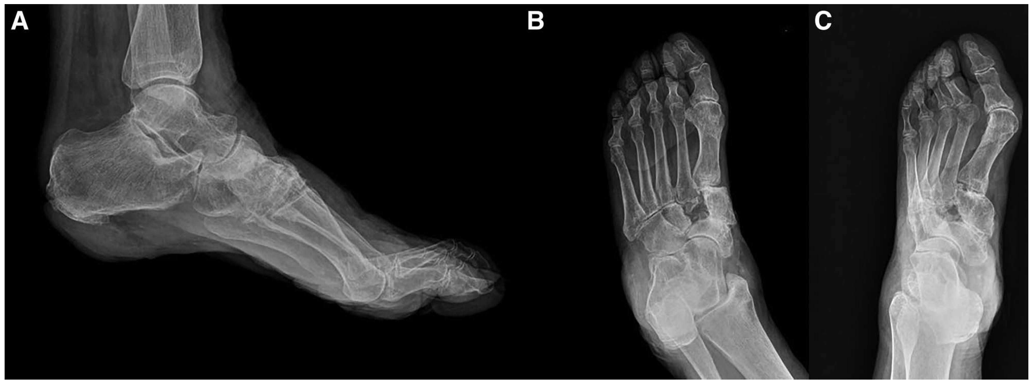
Figure 8.
Last follow-up photographs show a relatively healthy foot and completely healed soft tissue of the foot 12 months postoperatively (A and B) and 36 months postoperatively (C and D).
Discussion
The midfoot is a complex anatomical association of many bones and articulations, restrained by an even more complex network of ligaments, capsules, and fascia, which must work in concert to provide normal and painless locomotion [3]. Open injuries to the midfoot are dependent on the magnitude of the force applied and the application vector. A force that causes direct soft-tissue trauma can be accompanied by bone injury [5]. The treatment principles for this injury may include soft-tissue stabilization (primary goal), maintenance of the appropriate lateral and medial column length, maintenance of the appropriate relationship between the forefoot and the hindfoot to ensure a plantigrade foot, the use of stable internal fixation to maintain anatomical reductions or primary arthrodesis, and late weightbearing (which may decrease the incidence of complications) [1,6,7].
The patient in this case presented with severe injuries of the midfoot, including a left foot degloving injury, an open fracture of the navicular bone, dislocation of the medial and intermediate cuneiform, Chopart joint injury, and Lisfranc ligament injury. We performed an open reduction and internal fixation and tried to maintain normal foot anatomy as much as possible.
In fact, later follow-up examinations showed good maintenance of the talonavicular, calcaneocuboid, and intercuneiform joints and the split-thickness skin graft used to cover the medial, dorsal, and plantar sole areas after the patient underwent debridement of the necrotic skin. Despite our efforts to have the patient return to the clinic, approximately 6 months after surgery, the patient developed a pain accompanied by significant intermediate cuneiform collapse consistent with AVN. This case is the second report of posttraumatic AVN of the intermediate cuneiform. The first case was reported by McGlinchey [4]. However, in McGlinchey's report, the intermediate cuneiform was dorsally dislocated and 3-week postoperative plain radiographs showed local increases in bone density in the intermediate cuneiform, indicative of AVN. The AVN of the intermediate cuneiform returned to normal after another 10 months [4]. According to the literature, dislocation of the intermediate cuneiform is very uncommon. Dorsal dislocations are more common than plantar dislocations in cuneiform dislocations and usually are caused by axial loading and forced plantarflexion of the forefoot. Nashi and Banerjee 8] reported plantar displacement of the intermediate cuneiform as a result of the impact from a flagstone on the dorsum of the foot [9,10]. Similar to the results of Nishi et al 11], another report showed that when a flat piece of steel was used to apply force to the dorsal aspect of the midfoot to compress and decrease the transverse and longitudinal arches of the foot, a plantar dislocation of the intermediate cuneiform was produced.
Our case presents a similar mechanism of injury, with a direct force applied by an automobile. This mechanism of injury clearly explains both the plantar dislocation of the intermediate cuneiform and the plantar and medial dislocation of the medial cuneiform. Interestingly, even though the medial cuneiform was extensively dislocated medially, AVN did not develop, whereas in the intermediate cuneiform dislocated plantarward with a small displacement, AVN occurred. These differences can be explained by different sources of blood supply between the intermediate and medial cuneiforms. The intermediate cuneiform is supplied through its dorsal, medial, and lateral surfaces, mainly from the dorsal arterial network, whereas the medial cuneiform is well vascularized from multiple sources, including the deep and superficial medial plantar artery, direct contributions from the dorsalis pedis, and the plantar medial artery [12]. In the case presented here, the dorsalis pedis artery ruptured at the talus level and, while being repaired by arteriorrhaphy of the lateral branch of the dorsalis pedis artery with a venous graft, thromboses occurring at the branch that supplies the intermediate cuneiform could have occurred, causing the AVN. Moreover, the intermediate cuneiform is the smallest of the three cuneiforms, and it is wedge-shaped, with an apex on the plantar aspect and the broad surface dorsally. Therefore, it is possible that when the same force was applied to the dorsal aspect of the midfoot, the resultant intermediate cuneiform may have been more severely affected than the other cuneiforms.
Conclusions
In conclusion, dislocation of the medial and intermediate cuneiforms is very uncommon. This pattern of injury can develop many complications, including global shortening, collapse of the midfoot, persistent instability, deformity, or arthritis. This case is the first report of the total loss of the intermediate cuneiform by posttraumatic AVN. Therefore, AVN of the intermediate cuneiform should be considered as one of the complications of midfoot fractures and dislocations, and the potential development of this complication needs to be explained to the patient before surgery.
Financial Disclosure: This study was supported by a grant from the Biomedical Research Institute of Chonbuk National University Hospital (CNUH-BRI-2012-02-005).
Conflict of Interest: None reported.
References
- GrivasTBVasiliadisEDKoufopoulosG: Midfoot fractures. Clin Podiatr Med Surg23: 323, 2006.
- MawhinneyIMcCoyG:The crushed foot. J R Coll Surg Edinb40: 138, 1995.
- BenirschkeSKMeinbergEAndersonSA: Fractures and dislocations of the midfoot: Lisfranc and Chopart injuries. J Bone Joint Surg Am94: 1325, 2012.
- McGlincheyJ:Dislocation of the intermediate cuneiform bone. Injury12: 501, 1981.
- AdelaarRS:Complications of forefoot and midfoot fractures. Clin Orthop Relat Res391: 26, 2001.
- PylawkaTAndersenLB:Midfoot trauma. Curr Orthop Pract19: 228, 2008.
- SchildhauerTANorkSESangeorzanBJ:Temporary bridge plating of the medial column in severe midfoot injuries. J Orthop Trauma17: 513, 2003.
- NashiMBanerjeeB:Isolated plantar dislocation of the middle cuneiform—a case report. Injury28: 704, 1997.
- BertoldiLMolinariMSoldiniA: Isolated fracture-dislocation of the second cuneiform bone. Acta Orthop Scand62: 604, 1991.
- HubbellJGoldhagenPO'ConnorD: Isolated plantar fracture-dislocation of the middle cuneiform. Am J Orthop (Belle Mead NJ)27: 234, 1998.
- NishiHTakaoMUchioY: Isolated plantar dislocation of the intermediate cuneiform bone: a case report. J Bone Joint Surg Am86: 1772, 2004.
- KrausJCMcKeonKEJohnsonJE: Intraosseous and extraosseous blood supply to the medial cuneiform: implications for dorsal opening wedge plantarflexion osteotomy. Foot Ankle Int35: 394, 2014.
© 2020 American Podiatric Medical Association