


Abstract
Enchondroma is the most common benign cartilage bone tumor of the toes. In contrast, the foot is a rare region for chondrosarcoma, and the involvement of phalanges is extremely rare. In this article, we report an unusual case of intermediate chondrosarcoma involving the proximal phalanx of the great toe of a 52-year-old woman who was previously treated with curettage and bone grafting because of misinterpretation of enchondroma at a local hospital. She presented complaining of pain and swelling that she had experienced for a period of 1 year after the first operation. Radiography revealed a lytic lesion with a subtle punctuate calcification and endosteal scalloping in the proximal phalanx of the great toe. Gadolinium-enhanced magnetic resonance imaging confirmed soft-tissue involvement and cortical destruction. Staging evaluation with computed tomographic scan of the chest, abdomen, and pelvis was performed to ensure that there was no metastatic disease. Subsequently, a bone biopsy was performed, and the diagnosis was grade 2 chondrosarcoma. The patient was informed about the recurrence of the lesion and the clinical context on the basis of tumor biology of chondrosarcoma and was offered the option of either amputation or wide resection. She preferred the latter. The patient was treated with wide resection and underwent reconstruction with cement and Kirschner wire. She remains free of disease after 1 year of follow-up.
Primary chondrosarcoma (CHS) is the third most common primary malignancy of bone after plasmacytoma and osteosarcoma.[1] The most common skeletal sites are pelvic bones, followed by the proximal humerus, distal femur, and ribs. The small bones of the hands and feet are rarely affected (<1% of all cases of CHS).[2] When a tumor occurs in the feet, involvement of toes is extremely rare.[3] Although a positive prognosis is expected, there is little information about the biological behavior of phalangeal CHS. Phalangeal CHS acts as a locally aggressive lesion and, unlike CHS elsewhere, metastasis is rare. Chondrosarcomas are a group of tumors with a wide range of features and behavioral patterns ranging from slow-growing, nonmetastatic lesions to highly aggressive metastatic sarcomas.[4] Chondrosarcomas are innate cartilaginous malignant tumors in which the tumor matrix formation is completely chondroid. Chondrosarcomas are most common in men aged 30 to 60 years, with a slight female dominance for phalangeal chondrosarcomas. Chondrosarcomas typically slowly develop neoplasms. The most common symptoms are mild pain and tenderness. Local swelling with soft-tissue masses and pathologic fractures are other physical findings.
Chondrosarcoma remote metastasis passes through a hematogenous pathway and typically progresses straight to the lungs. The overall prognosis is related to lesion size, anatomical location, type, and histologic grade. The 5-year survival rate for patients with grade 1 lesions is 90%; the rate decreases to 29% for those with grade 3 tumors. Grade 1 lesions do not metastasize. The radiographic appearance of conventional CHSs reveals large radiolucent areas with thick walls. These radiolucent sites have trabeculation with multilocular medullary bone destruction in central regions. The most important radiographic findings that indicate whether a tumor is low grade or high grade are the pattern of calcification, the nature of the tumor margin, and the size of the soft-tissue mass. Magnetic resonance imaging (MRI) provides a complete view of intraosseous and soft-tissue sizes of chondrosarcomas. Cartilage regions of the tumor have moderate signal intensity on T1-weighted images relative to adjacent skeletal muscle and high signal intensity on T2-weighted images. Calcification appears as an area without signal.[5-8] Computed tomography and standard radiography are the best methods for imaging calcification. When strengthened with gadolinium, MRI can determine the location of the biopsy by identifying areas of necrosis within the lesion. These physical and radiographic findings distinguish malignant and nonmalignant tumors of the foot. The differential diagnosis of a standing chondrosarcoma in the foot includes benign lytic bone tumors such as enchondroma, malignant bone tumors metastasized from other regions or osteosarcoma, and infection.[9] In this article, we present a case of an intermediate grade primary CHS in the proximal phalanx of the great toe.
Case Report
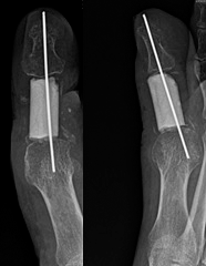
A 52-year-old woman was admitted to a state hospital with a swollen toe 2 years previously. She was diagnosed with enchondroma after clinical and radiologic evaluation, and a Tru-Cut (Merit Medical, South Jordan, Utah) biopsy was performed. Histopathologic examination also revealed enchondroma that had been treated by intramedullary curettage and bone grafting. The patient was referred to our hospital for local recurrence of the tumor 7 months after this procedure. Physical examination revealed a swollen right great toe. Radiography and MRI confirmed the endosteal scalloping, cortical destruction, and soft-tissue extension on the proximal phalanx of the great toe (Figs. 1 and 2). Computed tomography revealed that the chest and pelvis were free of disease. Blood tests were normal. A Tru-Cut biopsy revealed intermediate CHS. We discussed the clinical context with the patient on the basis of tumor biology of CHS, no evidence of metastasis, natural barrier effect of the tendinous sheath of tendons,[10] and the importance of the great toe in gait. It was suggested that she should undergo either wide resection or amputation. She preferred the former. Excision of the proximal phalanx of the great toe was performed. Flexor and extansor tendon sheaths are considered natural barriers to tumor extension. Marginal margin at the tendinous sheath and wide resection in other locations were performed. The resected proximal phalanx was reconstructed by bone cement and Kirschner wire (Fig. 3). The specimen was sent for histopathologic examination.
Figure 1.
Preoperative appearance of the patient's foot, previously treated by intralesional curettage and bone grafting, showing destructive cortex with endosteal scalloping.
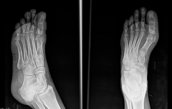
Figure 2.
Magnetic resonance imaging–confirmed soft-tissue invasion of the tumor and destruction of the cortex of the proximal phalanx.
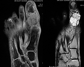
Figure 3.
The resected proximal phalanx was reconstructed by bone cement and Kirschner wire.
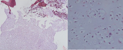
Histologic sections showed pleomorphism, lobular lesions composed of a combination of myxoid matrix and chondroid (Fig. 4). The cellularity was significantly higher than in normal cartilage. Chondroid cells were atypical. The final report confirmed intermediate grade CHS. The patient was referred to the oncology department. The patient underwent diagnostic testing with positron emission tomography, which did not reveal any metastasis. She is still free of disease after 1 year of follow-up.
Figure 4.
Pleomorfism, chondroid lobular lesions, and high cellularity.
Discussion
As a rule, CHSs are slow growing and tend to recur late. The metastatic potential and survival of CHS patients are correlated to the histologic grade. Prompt classification of the grade of CHS is important because the prognosis and treatment are different for different stages. Evans et al[7] used their own grading system, as follows: low-grade, low-level malignancy; intermediate-grade, moderately malignant; and high-grade, highly malignant. Reported 10-year survival rates were 83% for low-grade, 64% for intermediate-grade, and 29% for high-grade CHS. The incidence of metastatic disease is rare for low-grade, 10% for intermediate-grade, and 71% for high-grade CHS.[6-9]
Bovee et al[6] reported that phalangeal CHS recurs in 36% of cases but does not metastasize. Phalangeal CHS has a much better prognosis than CHS in other regions. Mohammadianpanah et al[9] described a case of phalangeal CHS at the proximal phalanx of the third toe that proceeded to an amputation at the metacarpophalangeal joint. Lo et al[11] reported on a case of a low-grade CHS at the base of the fifth metatarsal, lateral aspect of the cuboid, and third cuneiform. The patient proceeded to have a below-knee amputation. In the presented case, to the best of our knowledge, this is the only one in the literature in which an intermediate CHS was localized in the proximal phalanx of the great toe and treated by wide resection.
Conventional CHSs are divided into four histologic grades according to their appearance under a microscope. The classification is based primarily on the nuclear size of tumor cells, nuclear staining, and cellulite. Grade I (low-grade) tumors resemble normal cartilage, but may surround areas of lamellar bone, or show atypical cells including binucleate forms. Grade II (intermediate-grade) are more cellular, with a greater degree of nuclear atypia, hyperchromasia, and nuclear size. Grade III (high-grade) tumors have significant areas of marked pleomorphism, large cells with more hyperchromatic nuclei than grade II tumors, occasional giant cells, and abundant necrosis. Mitoses are frequently detected. Mesenchymal and dedifferentiated CHSs are considered to be grade IV tumors. Dedifferentiated CHSs, along with mesenchymal CHSs, are highly malignant, particularly aggressive (ie, rapidly growing and disturbing surrounding tissues), and carry a poor prognosis. Histologic grading, anatomical location, tumor size, and primary treatment success are the most effective criteria for determining the prognosis.[12] Chondrosarcoma is mostly resistant to chemotherapy and radiotherapy; thus, surgery is generally the preferred treatment.[13] In general, grade 1 lesions should be discussed with intralesional curettage; high-speed motor mechanical cleaning; and adjuvant surgical methods such as cryosurgery, phenol, high temperature, and bone cement combined therapy.[14,15]
In the presented case, we discussed the clinical context with the patient on the basis of the tumor biology of CHS, no evidence of metastasis, natural barrier effect on the tendinous sheath of tendons,[10] and the importance of the great toe in gait, and the patient was treated with mostly wide resection where available and at one point marginal margin. After 1-year follow-up, the patient had no evidence of local recurrence or metastasis.
The reported 10-year survival rates are 83%, 64%, and 29% for grades I, II, and III, respectively.[6] The treatment of choice for early CHS is radical resection. Chemotherapy is very ineffective and radiation therapy is usually used only for palliation in patients with tumors that are unsuitable for surgical resection.
Conclusions
The diagnosis of chondrogenic tumor remains a significant challenge for pathologists. Clinical and radiologic follow-up is extremely important when malignancy is suspected. Finally, the patient should be informed about the clinical context. We offer limb salvage for intermediate CHS of the phalanx of the foot with wide resection in selected cases on the basis of clinical context.
Financial Disclosure
None reported.
Conflicts of Interest
None reported.
References
- Unni KK: Dahlin's Bone Tumors: General Aspects and Data on 11,087 Cases, 5th Ed, Lippincott-Raven, Philadelphia, 1996.
- Bovee JV, van der Huel RO, Tamminiau AH, et al: Chondrosarcoma of the phalanx: a locally aggressive lesion with minimal metastatic potential. A report of 35 cases and a review of the literature. Cancer86: 1724, 1999.
- Murphey MD, Walker EA, Wilson AJ, et al: From the archives of the AFIP imaging of primary chondrosarcoma: radiologic-pathologic correlation. Radiographics23: 1245, 2003.
- Mendeszoon MJ, Mendeszoon ER, Snyder J: Chondrosarcoma of the foot: a rare occurrence in the distal phalanx of the fourth digit. Clin Surg2: 1382, 2017.
- Harkless L: Soft-tissue and bone tumors. Clin Podiatr Med Surg10: 587, 1993.
- Bovee JV, Cleton-Jansen AM, Taminiau AH, et al: Emerging pathways in the development of chondrosarcoma of bone and implications for targeted treatment. Lancet Oncol6: 599, 2005.
- Evans HL, Ayala AG, Romsdahl MM: Prognostic factors in chondrosarcoma of bone: a clinicopathologic analysis with emphasis on histological grading. Cancer40: 818, 1977.
- Kinoshita G, Maruoka T, Matsumoto M, et al: Bone and soft tissue tumors in the foot. J Bone Joint Surg Am84: 216, 2002.
- Mohammadianpanah M, Torabinejad S, Bagheri MH, et al. Primary chondrosarcoma of phalanx. Foot14: 159, 2004.
- Kawaguchi N, Ahmed AR, Matsumoto S, et al: The concept of curative margin in surgery for bone and soft tissue sarcoma. Clin Orthop Relat Res419: 165, 2004.
- Lo EP, Pollak R, Harvey CK: Chondrosarcoma of the foot. JAPMA90: 203, 2000.
- Fiorenza F, Abudu A, Grimer RJ, et al: Risk factors for survival and local control in chondrosarcoma of bone. J Bone Joint Surg Br84: 93, 2002.
- Somers J, Faber LP: Chondroma and chondrosarcoma. Semin Thorac Cardiovasc Surg11: 270, 1999.
- Hanna SA, Whittingham-Jones P, Sewell MD, et al: Outcome of intralesional curettage for low-grade chondrosarcoma of long bones. Eur J Surg Oncol35: 1343, 2009.
- Mohler DG, Chiu R, McCall DA, et al: Curettage and cryosurgery for low-grade cartilage tumors is associated with low recurrence and high function. Clin Orthop Relat Res468: 2765, 2010.
© 2019 American Podiatric Medical Association