


Abstract
Background: The spring ligament fibrocartilage complex (SLFC) is an important static foot stabilizer comprising the superomedial ligament (SML) and the inferior ligament, with anatomical variations (third ligament). The aim of this study was to describe the patterns of the lesions found during SLFC surgery, to allow direct comparison between the results with various surgical techniques. Methods: Fourteen consecutive patients with SLFC lesions were analyzed during surgical treatment. The mean patient age was 37.3 years, and the mean time from injury was 6.9 months. Intraoperative assessments and anatomical descriptions of the lesions were collected. Results: Three types of lesion were found. In 13 of 14 cases, only the superomedial ligament was involved: five superomedial ligament distentions and eight superomedial ligament ruptures. In one case, total SLFC (superomedial and inferior ligaments) rupture was observed. Conclusions: The first classification of SLFC lesions is presented, which is simple, consistent, and based on anatomical description.
The spring ligament fibrocartilage complex (SLFC) is one of the major static foot stabilizers.[1] Together with the cruciate ligament and the posterior tibial tendon, it forms both a connection between the hindfoot and midfoot and a mechanism that maintains the normal, longitudinal foot arch.[2,3] It is pertinent to note that the mechanical strength of this ligament, according to the literature, is much higher than that of the anterior cruciate ligament, achieving 477 N, whereas in the case of the anterior cruciate ligament, it is approximately 400 N.[4]
The spring ligament complex is recognized as a static supporter of the medial arch of the foot, serving as a “hammock” to the talar head. Spring ligament fibrocartilage complex chronic laxity or acute rupture may cause its insufficiency, and lead to talar bone plantarflexion and planovalgus deformity of the foot. The most common cause of chronic SLFC laxity is chronic dysfunction of the tibialis posterior tendon, whereas acute rupture is a result of midfoot injury. Both types of SLFC insufficiency when left untreated lead to flatfoot deformity, with abduction and pronation of the forefoot causing pain and shoe problems.
The anatomy of the ankle medial ligaments has long raised considerable doubts. There are different descriptions of SLFC morphology in the literature.[1,5,6] The majority of authors divide the SLFC into the upper, medial, and lower parts. There are, however, reports describing the third ligament, called the medioplantar oblique[1,5,6] (Fig. 1). In general, the SLFC is a complex structure comprising the superomedial ligament (SML) and the inferior and medioplantar oblique ligaments.[6]
Figure 1.
Specimen with the spring ligament fibrocartilage complex composed of the superomedial ligament (SML), the inferior ligament (IL), and a third ligament. A, Cadaveric specimen. B, Graphic description. (Reprinted with permission from Chirurgia Narządów Ruchu i Ortopedia Polska.)
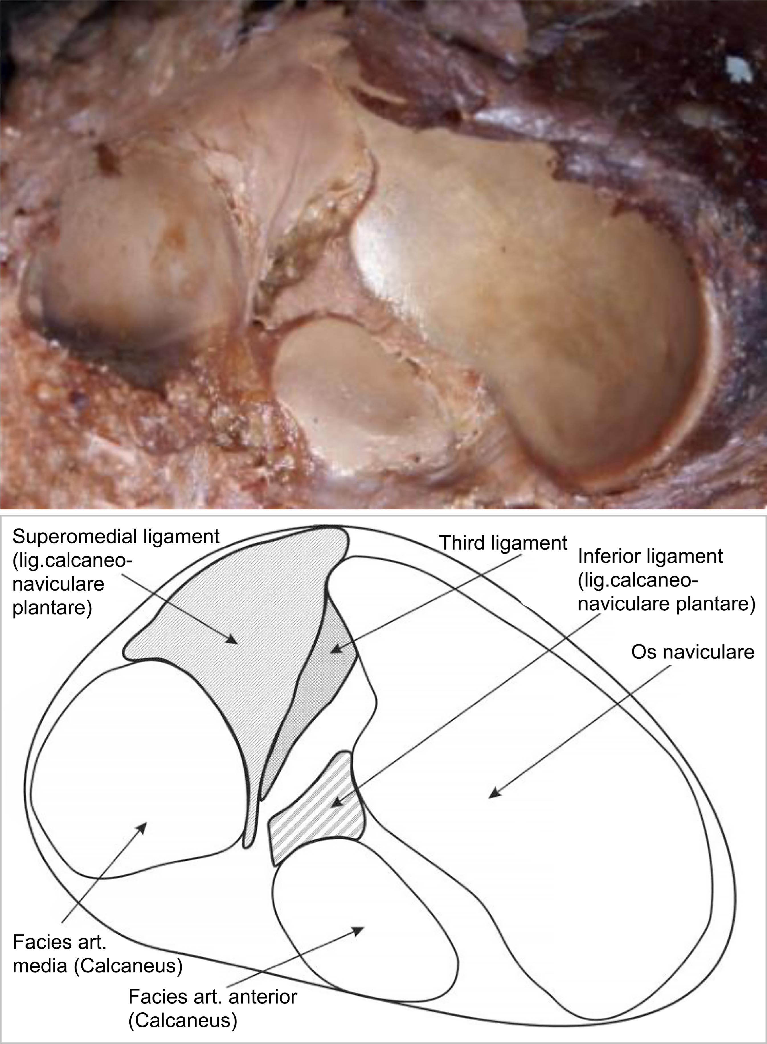
In the previous study performed at our university, Domzalski et al[6] dissected 10 cadaver feet and, in four cases, two components of the spring ligament complex: the superomedial calcaneonavicular ligament and the inferior calcaneonavicular ligament were identified. In the remaining six cases, the complex was composed of three ligaments, and we were able to identify the structure of the third ligament (ie, medioplantar oblique ligament). In this subgroup, in two cases, the spring ligament had its own fibrocartilage surface connected by tiny fibrous bands with a fibrocartilage articular surface.
Even though there has been controversy in the literature on the normal pattern of spring ligament anatomy, it is becoming clear that the ligament complex consists of three different parts that have been well described in anatomical cadaveric studies[7,8] in addition to radiologic studies[1,9,10] and clinical articles.[11] The medioplantar oblique ligament is clearly distinguished using magnetic resonance imaging and even ultrasonography.[9,11]
The significance of the spring ligament fibrocartilage complex is clearly visible in cases of acquired adult flatfoot deformity (AFFD). When structures such as the SLFC and tibialis posterior tendon, which support the longitudinal foot arch, are weak, the patient is predisposed to talar bone head dislocations and to foot arch lowering. According to the literature, the most common cause of AFFD is insufficiency of the dynamic stabilizer (tibialis posterior tendon), which subsequently leads to overtension of the other structures such as the SLFC.[1,12] There are also reports describing isolated SLFC lesions with AFFD deformation.[11,13]
Only one study reported successful results after conservative treatment of SLFC lesions.[14] The internal bracing method, anatomical reconstruction, and arthrodesis of the talonavicular joint were suggested as surgical treatment options.[13,15,16] Some authors did not believe in isolated SLFC repair, postulating the extension of the surgery into other stabilizers.[4] In all presented cases, intraoperative damage of the SLCF was noted, but without detailed anatomical description of this lesion.[4,11,13,15,16]
We decided to assess whether it was possible to correlate SLCF anatomy to damage types in patients with AFFD caused by an isolated lesion of the plantar calcaneonavicular ligament, aiming to categorize SLCF lesions to help find the best treatment options and to create classification for comparison with future studies. Our main goal was to unify and clarify the descriptions of SLFC lesions reported by the surgeon at the time of surgery.
Patients and Methods
Our study group included 18 consecutive patients admitted to our orthopedic department for acquired flatfoot during the course of treatment of the SLFC. The group consisted of six women and 12 men. The median age of the patients was 39 years (range, 21–52 years). Eleven left feet and seven right feet were involved. All recalled the traumatic event, associating it with the occurrence of AFFD. At the time of admission because of AFFD, the patients were not able to identify the exact type of foot injury. The median time from injury to admission was 6.9 months (range, 0.5–24 months). None of the admitted patients had received treatment for diabetes or any other metabolic reason. Charcot disease was not diagnosed in any of the patients. One of the male patients was a cigarette smoker. One of the women was on contraceptive hormonal therapy.
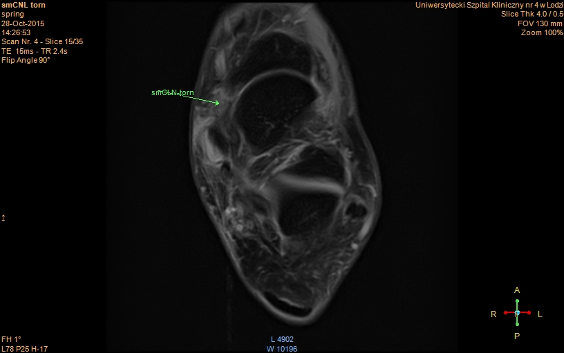
A clinical evaluation revealed a considerable decrease of the longitudinal arch of the foot with AFFD, concomitant with many toe signs, and valgization of the hindfoot. The midportion of the longitudinal arch was very painful on palpation, and decreased tension of the tibialis posterior muscle tendon was observed. After ultrasonography (Fig. 2) and magnetic resonance imaging (MRI) examination (Fig. 3), the diagnosis of a SLFC lesion was confirmed.
Figure 2.
Ultrasound image of the torn spring ligament.
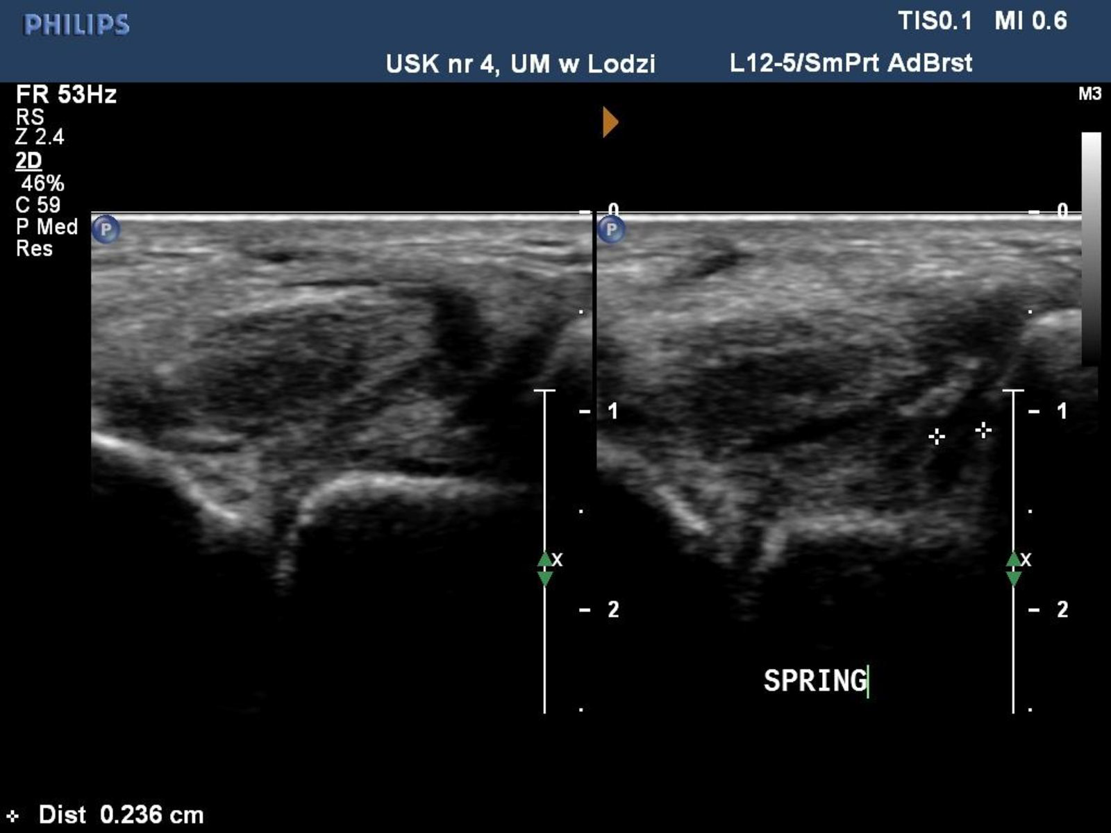
Figure 3.
Magnetic resonance image of the torn spring ligament (T2 axial plane).
All patients were submitted to surgical treatment. The operations were all performed by the same team, which had participated in the anatomical study of SLFC morphology.[6] The medial approach was used for all patients, slightly above the posterior tibial tendon. Then, after careful preparation of the tissue layers, the medial complex of the ankle ligaments was gradually exposed, followed by SLFC identification with reference of the posterior tibial tendon configuration. Then, when the SLFC was fully exposed, it was thoroughly inspected, and the character of the SLFC lesion was described, each time using the same terms, consistent with the above-mentioned description, published in 2007.[6] After exposition, visual inspection of the culprit lesion on the spring ligament was performed by the senior author (M.D.). In all cases, there was full agreement on the final verdict of classification type.
Results
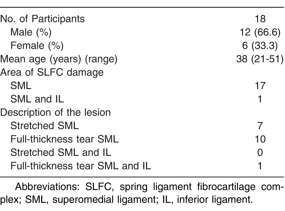
In all cases, an SLFC lesion was identified during surgery. In 17 of 18 operated cases, only the SML was involved, still leading to insufficiency of the entire complex.
Superomedial ligament lesions were not homogenous: in seven cases, the ligament was stretched out, and in 10 cases, the full rupture of this part of the SLFC was noted. In cases where the SML was stretched or distended, only its internal tendinous layer was disrupted but without involvement of the two external, chondrus layers (Table 1).
Table 1.
Characteristics of Treated Patients and Intraoperative Description of Spring Ligament Fibrocartilage Complex Lesions
Complete tear of the plantar calcaneonavicular ligament comprising both the SML and the inferior ligament was found in only one of the patients (Fig. 4). No isolated lesion of the inferior ligament or the “third ligament” was noted. The new classification system was suggested based on the SLFC morphology and the types of lesions observed in the study (Table 2).
Figure 4.
Intraoperative view of complete rupture of the spring ligament fibrocartilage complex (type III).
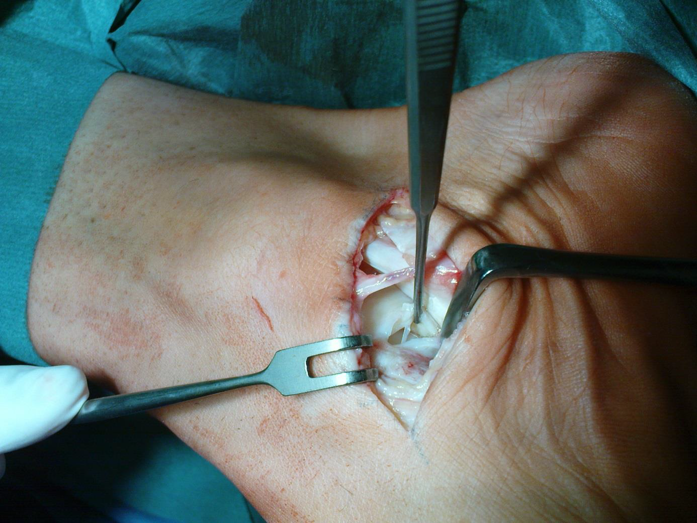
Table 2.
Classification of Spring Ligament Fibrocartilage Complex Lesions

Discussion
The role of the SLFC in the development of acquired flatfoot deformity is well established in the literature. This complex has been extensively studied regarding its mechanical strength and properties, and also anatomically.[1-6] Some studies have presented the results of various treatments of SLFC pathology without detailed description of the morphology of the lesion.[4,11,15,16]
In the literature, there is no consensus regarding the classification of SCFL lesions. In orthopedic technical reports and radiologic descriptions of plantar calcaneonavicular ligament lesions, no consistency regarding the systematization and the terms used by orthopedic surgeons to describe SLFC lesions was observed.[2,3,17,18] A classification system would facilitate communication and allow more precise presentation of both radiologic and orthopedic results.
Deland,[4] in a review of the literature dealing with AFFD treatment, described many therapeutic methods of SLFC lesions, and presented the results of these treatments as the great unknown. While analyzing various approaches and techniques, he did not, however, compare the applied methods or obtained results to the character of the complex lesion.[4] Tryfonidis et al[11] presented the analysis of treatment of nine cases with AFFD, also without a transparent system or classification of observed lesions.
Few authors used anatomical descriptions in their reports.[14,16] Masaragian et al[14] specified in their case report that the presented lesion comprised only the SML part. Palmanovich et al[16] stated that the SLFC injury was usually localized along the SML and was vertical. Unfortunately, a type II lesion of the SLFC (according to the proposed classification) is not found in the above-mentioned descriptions.
Toye et al[17] analyzed the SLFC injuries on MRI and reported that the majority of the observed lesions consisted of SML disruption, with a wavy appearance of the ligament during MRI examination. This finding corresponds well with our description of type II lesions because, during surgery, the SML ligament was loose and thin, with preserved continuity. A thinning of the ligament in patients with AFFD was also described by Williams et al[18] during MRI evaluation, indicating also that the SCFL lesions comprised only the SML section. Thinning phenomena may be explained by disruption of the middle layer of the SML, with preservation of the external cartilaginous layers.[6] In these MRI studies, no single case of total SCFL disruption (type III) was presented.[17,18]
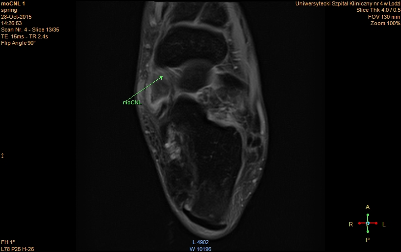
The absence of a standardized method of lesion description of SCFL is truly absent from the literature. In our study, each lesion was categorized by the same surgeon (M.D.), and the classification system was very useful for assessing pathology. Although the medioplantar oblique ligament is well described in anatomical studies[7,8] and was visible on preoperative magnetic resonance images (Fig. 5), we found distinguishing this part of the spring ligament using the standard surgical approach in live surgery very difficult in all cases. We believe this classification system is a potentially valuable tool in the assessment, management, and decision process regarding treatment options, and may help in stratifying decision making in such cases.
Figure 5.
Magnetic resonance image of the medioplantar oblique ligament of the spring ligament fibrocartilage complex (T2 axial plane).
The main weakness of our article is the limited number of patients included in the study. A similar study needs to be repeated and validated by other study groups describing a larger number of patients.
Conclusions
A new classification system of spring ligament fibrocartilage complex lesions was described. In our opinion, this system is simple, consistent with clinical and MRI findings, and based on anatomical studies.
Financial Disclosure
None reported.
Conflicts of Interest
None reported.
References
- Desai KR, Beltran LS, Bencardino JT, et al: The spring ligament recess of the talocalcaneonavicular joint: depiction on MR images with cadaveric and histologic correlation. AJR Am J Roentgenol196: 1145, 2011.
- Patil V, Ebraheim NA, Frogameni A, et al: Morphometric dimensions of the calcaneonavicular (spring) ligament. Foot Ankle Int28: 927, 2007.
- Rule J, Yao L, Seeger LL: Spring ligament of the ankle: normal MR anatomy. AJR Am J Roentgenol161: 1241, 1993.
- Deland JT. Spring ligament complex and flatfoot deformity: curse or blessing?Foot Ankle Int33: 239, 2012.
- Cromeens BP, Kirchhoff CA, Patterson RM, et al: An attachment-based description of the medial collateral and spring ligament complexes. Foot Ankle Int36: 710, 2015.
- Domzalski M, Kwapisz A, Król A, et al: The role of plantar calcaneonavicular ligament complex in the development of the adult flat foot: anatomical study [in Polish]. Chir Narzadow Ruchu Ortop Pol72: 265, 2007.
- Taniguchi A, Tanaka Y, Takakura Y, et al: Anatomy of the spring ligament. J Bone Joint Surg Am85: 2174, 2003.
- Tohno Y, Tohno S, Taniguchi A, et al: Characteristics of the three ligaments of human spring ligament complex from a viewpoint of elements. Biol Trace Elem Res146: 293, 2012.
- Mansour R, Jibri Z, Kamath S, et al: Persistent ankle pain following a sprain: a review of imaging. Emerg Radiol18: 211, 2011.
- Mansour R, Teh J, Sharp RJ, et al: Ultrasound assessment of the spring ligament complex. Eur Radiol18: 2670, 2008.
- Tryfonidis M, Jackson W, Mansour R, et al: Acquired adult flat foot due to isolated plantar calcaneonavicular (spring) ligament insufficiency with a normal tibialis posterior tendon. Foot Ankle Surg14: 89, 2008.
- Borton DC, Saxby TS: Tear of the plantar calcaneonavicular (spring) ligament causing flatfoot: a case report. J Bone Joint Surg Br79: 641, 1997.
- Weerts B, Warmerdam PE, Faber FWM: Isolated spring ligament rupture causing acute flatfoot deformity: case report. Foot Ankle Int33: 148, 2012.
- Masaragian HJ, Ricchetti HO, Testa C: Acute isolated rupture of the spring ligament: a case report and review of the literature. Foot Ankle Int34: 150, 2013.
- Acevedo J, Vora A: Anatomical reconstruction of the spring ligament complex “internal brace” augmentation. Foot Ankle Spec6: 441, 2013.
- Palmanovich E, Shabat S, Brin YS, et al: Anatomic reconstruction technique for a plantar calcaneonavicular (spring) ligament tear. J Foot Ankle Surg54: 1124, 2015.
- Toye LR, Helms CA, Hoffman BD, et al: MRI of spring ligament tears. AJR Am J Roentgenol184: 1475, 2005.
- Williams G, Widnall J, Evans P, et al: MRI features most often associated with surgically proven tears of the spring ligament complex. Skeletal Radiol42: 969, 2013.
© 2019 American Podiatric Medical Association