


Abstract
Background: The increased use of external interventions to diagnose and treat podiatric medical pathologies has warranted a greater understanding of the lower extremity in the transverse plane. Films, prosections, and plastinated cross sections have long been used alongside traditional instruction to assist students in anatomical studies. These methods, however, often fail to provide the tactile component of anatomical dissection and may obscure or exclude the requisite structures for identification by medical students. Such teaching techniques prove costly, time sensitive, and dated compared with the relatively less expensive and customizable nature of three-dimensional (3-D) printing. Methods: Limb length was measured on a cadaveric specimen, and eight cross sections of equal width were excised. Manual sketches of these sections were then digitized and 3-D printed. Results: Three-dimensional printing provides a safe and reproducible means to construct customizable cross sections of the lower extremity. Moreover, this method proves to be relatively inexpensive. Conclusions: Advancing traditional didactic teaching with the use of cost-effective 3-D printing can facilitate the visuospatial comprehension of lower-extremity anatomy. The evolution of 3-D printing enhances the clinical skills essential for the interpretation of imaging studies as well as planning for surgical procedures such as external fixation application.
Instruction in anatomy continues to provide the foundation for medical education.[1,2] This instruction continues to evolve as a result of emerging medical technologies and trends in preparedness among newly graduated physicians.[3,4] Supplementation of traditional didactic teaching with laboratory dissection and clinically relevant correlations, anatomy courses continue to play an essential role in the education of future health-care practitioners.[5] Improvements in anatomical instruction with an emphasis on new techniques will better prepare podiatric medical students for their transition into the clinical sciences.[6] To incorporate this component into an already full schedule, medical programs have introduced problem-based learning and plastinated prosections into existing curricula.[7,8,9] Programs have also incorporated modalities such as ultrasound and magnetic resonance imaging with traditional anatomical dissection, further increasing clinical relevance.[4,7,10,11,12] Consequently, such strategies that stressed the teaching of cross-sectional anatomy prepared students for radiographic studies and improved interpretation of tomographic images.[11,13]
By integrating these innovative and clinically relevant practices into the lower-extremity anatomy curriculum, the Barry University School of Podiatric Medicine has advanced the education of future lower-extremity specialists. Currently, cross sections of the lower extremity are not available for purchase by educational institutions. Tissue preservation protocols have made it possible to manually section, protect, and display specimens of interest.[14,15,16] However, the time commitment, cost, and concession of a cadaveric teaching model to produce these cross sections proves ineffective. Furthermore, the finite number of cross sections made available after tissue embalming and plastination may not be sufficient to adequately supply a classroom of students, some of whom have never taken a formal anatomy course. Such complications associated with the procurement and distribution of these indispensable study aids lend themselves to the reproducibility, accuracy, and affordability of three-dimensional (3-D) printing.
With the recent accessibility of commercially available 3-D printers, the medical field has launched numerous uses, from printing human organs to prostheses.[17] Although still in its infancy, 3-D printing has also proved useful in surgical planning and patient education. Moreover, this additive manufacturing technique has evolved and proved to be a viable option in the production of prosected anatomical specimens.[18,19] Such medical applications came to fruition because the information used in the computer-aided drafting software to communicate with the printer is easily compatible with the data present in computed tomography, magnetic resonance imaging, and other imaging modalities.[20] Similar digital modeling and printing techniques are currently used for the correction of foot and ankle deformities, taking advantage of the free, open source software available for printing.[21,22] In addition, 3-D printing has shown great potential in the fabrication of customized external fixators,[23] a technique that requires extensive cross-sectional anatomy knowledge and safe corridor pin placement.[24]
This article details the production of 3-D–printed lower-extremity cross sections and the savings attributed to the prints. We describe the potential uses of 3-D–printed cross sections in the instruction and evaluation of lower-extremity anatomy. We then recognize the current resources available for students and outline the advantages of this custom technique. Finally, we discuss the potential of cross sections to help prepare podiatric physicians and surgeons.
Materials and Methods
Cross Section Preparation
An embalmed, 65-year-old male cadaver right leg was selected from the specimens available after a one-semester lower-extremity anatomy dissection course. The limb length of the cadaver was measured as the distance from the anterior superior iliac spine (ASIS) to the medial malleolus. Incision sites were indicated transversely with a permanent marker around the leg at distances 20, 30, 40, 50, 60, 70, 80, 90, and 100 cm inferior to the ASIS (Fig. 1A). String was then tied around the leg both above and below each demarcated cross section to circumferentially hold the tissue together during dissection. A No. 24 blade was then used to follow the incision sites, cutting through soft tissue down to bone. Next, the bone was parted along this same incision site using a bone saw. With each cross section separated, a plexiglass shield was placed on top of the most inferior partition, and a dry-erase marker was used to trace the underlying tissue proper. On completion of each overlaying trace, the transcribed plexiglass was placed over a white background and imaged (Fig. 1B).
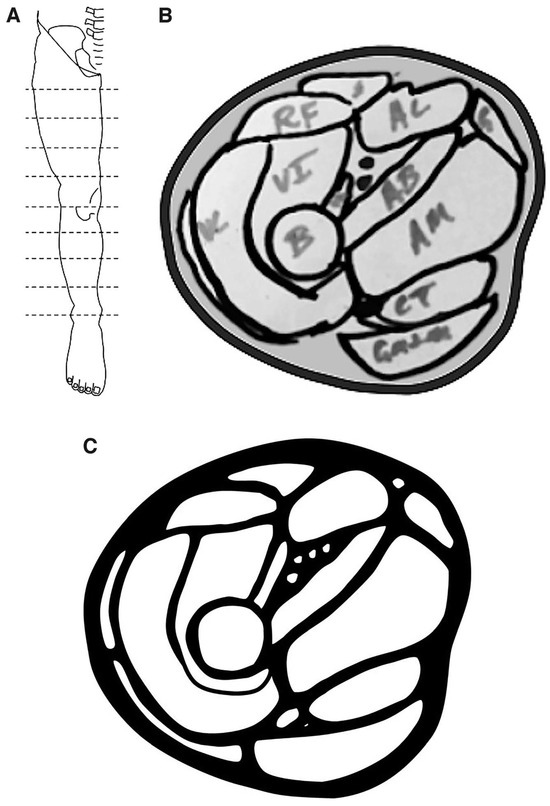
Figure 1.
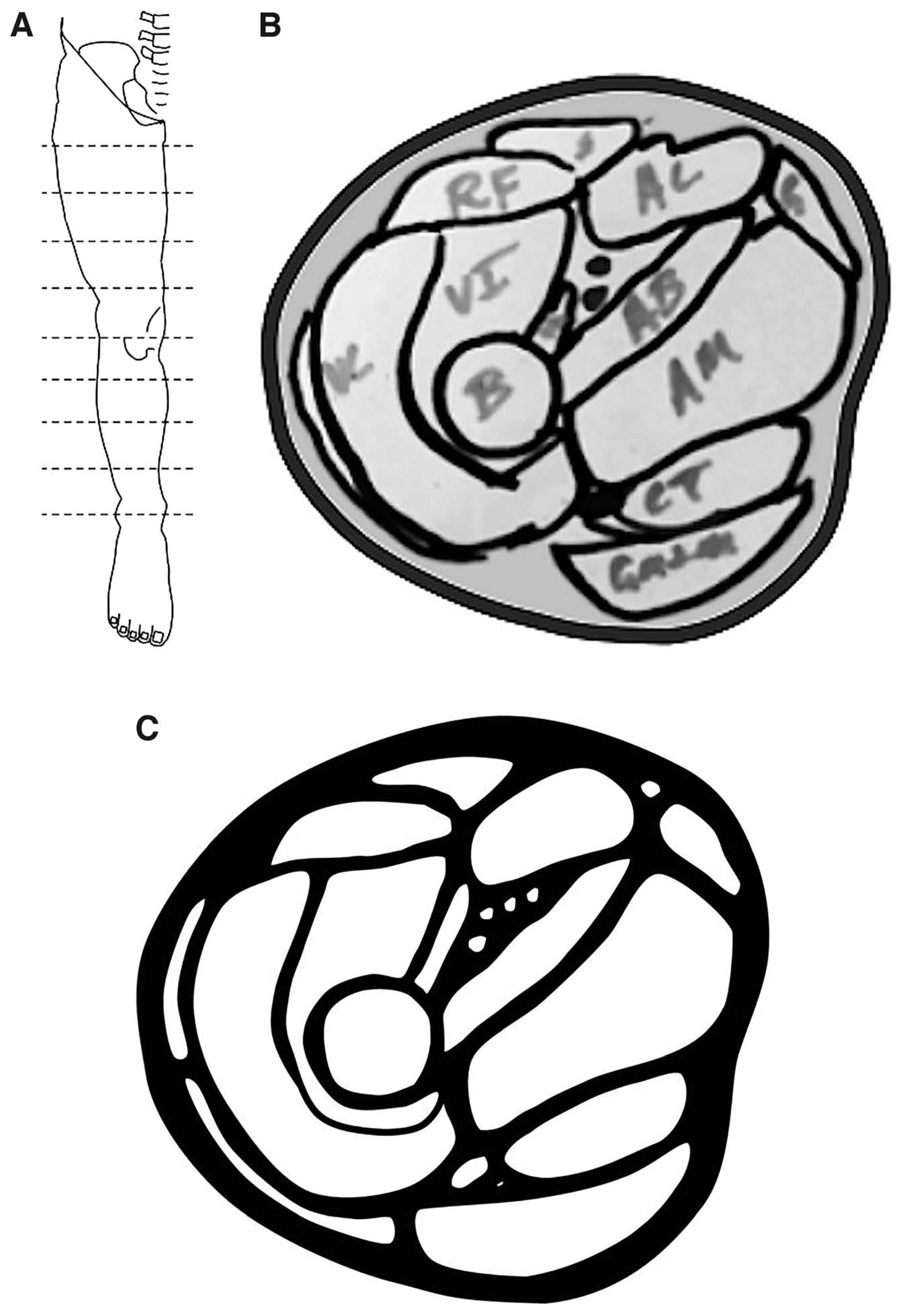
The positions of cross-sectional cuts (A) used for sketches (B) and digitized drafts (C).
Computer-Aided Drafting and Printing
Each image was then imported into Microsoft PowerPoint (Microsoft Corp, Redmond, Washington) and manually digitized using the shapes feature (Fig. 1C). The new digital image was then converted into a negative image and saved as a Joint Photographic Exports Group (JPEG) file. This JPEG file was then converted to a STereoLithography (STL) file using Selva3D, an online file converter. Finally, the STL file was imported into the free 3-D modeling software CURA, sized to the desired dimensions, and saved as a G Code file. This saved GCode file was sent to an Alunar 3-D desktop printer (Alunar; Shenzhen, China) equipped with black 1.75-mm-diameter polylactic acid filament (Q3D) (Quintessential Universal Building Device, Inc; Little Rock, Arkansas). Each file was printed under settings optimized for the size and detail of each cross section. This method provided black and tan cross-sectional stencils with delineated spaces representing unique features such as muscles, nerves, bones, and vasculature. Such optional features may also be printed and inserted into the voids at the instructor's discretion.
Results
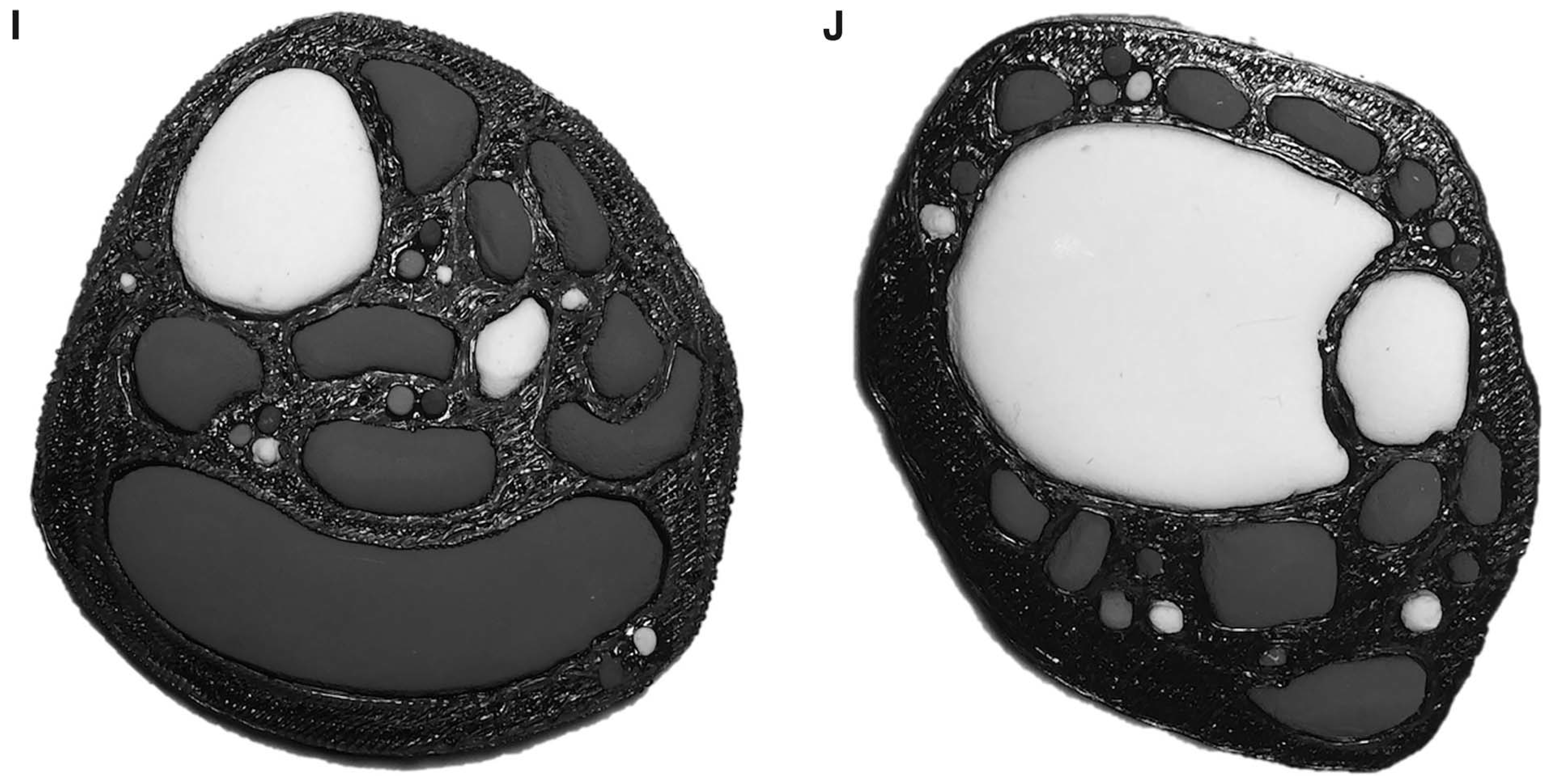
Information associated with the cost of materials and dimensions specific for each cross section at the corresponding distance from the ASIS needed for reproduction are reported in Table 1. Note that costs incurred from printing in other colors were not taken into account because this method focuses on printing a negative cross section, which can be left empty (Fig. 2A) or filled in (Fig. 2 B–J) at the user's discretion. The cost of the cadaver used to model each section was not included because the specimen was previously used for anatomical dissection. Furthermore, reprints of these sections would not require a new cadaver but simply the archived file.
Table 1 .
3-D–Printed Cross Section Production Parameters
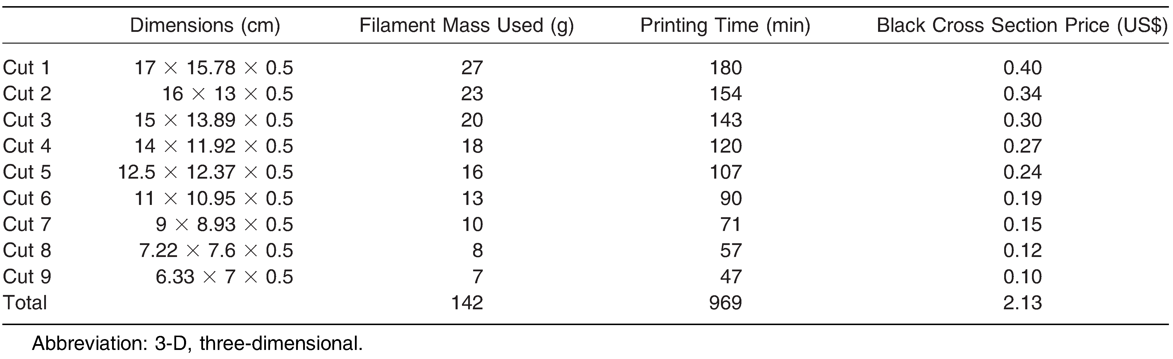
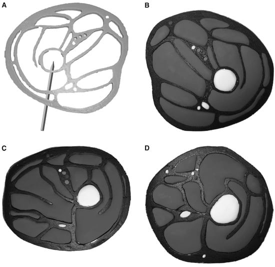
Figure 2.
Three-dimensional–printed cross sections. A, Example negative cross section with proper surgical fixation half pin placement. B-J, Cross sections filled with modeling clay representing bones, muscles, nerves, and vasculature. (continued on next page)
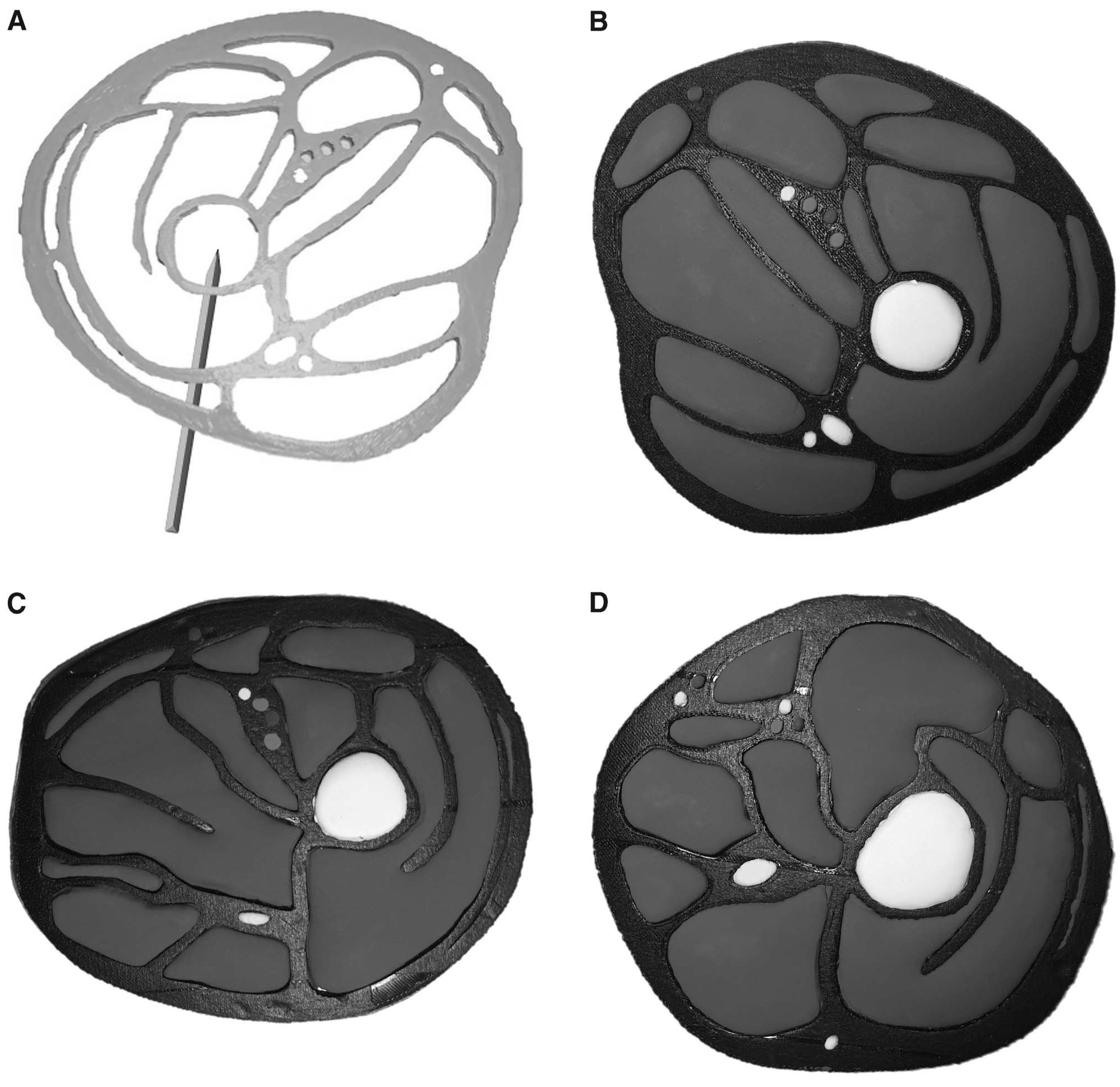
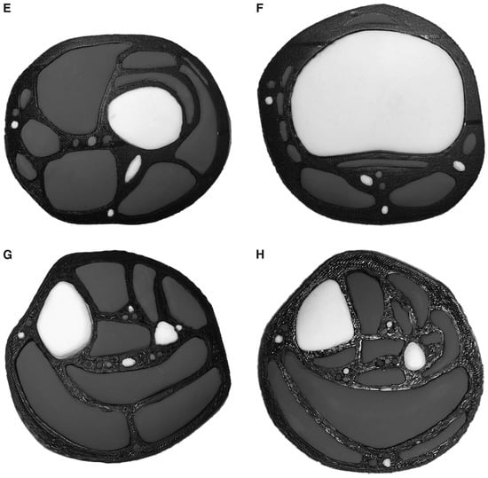
Figure 2.
Continued.
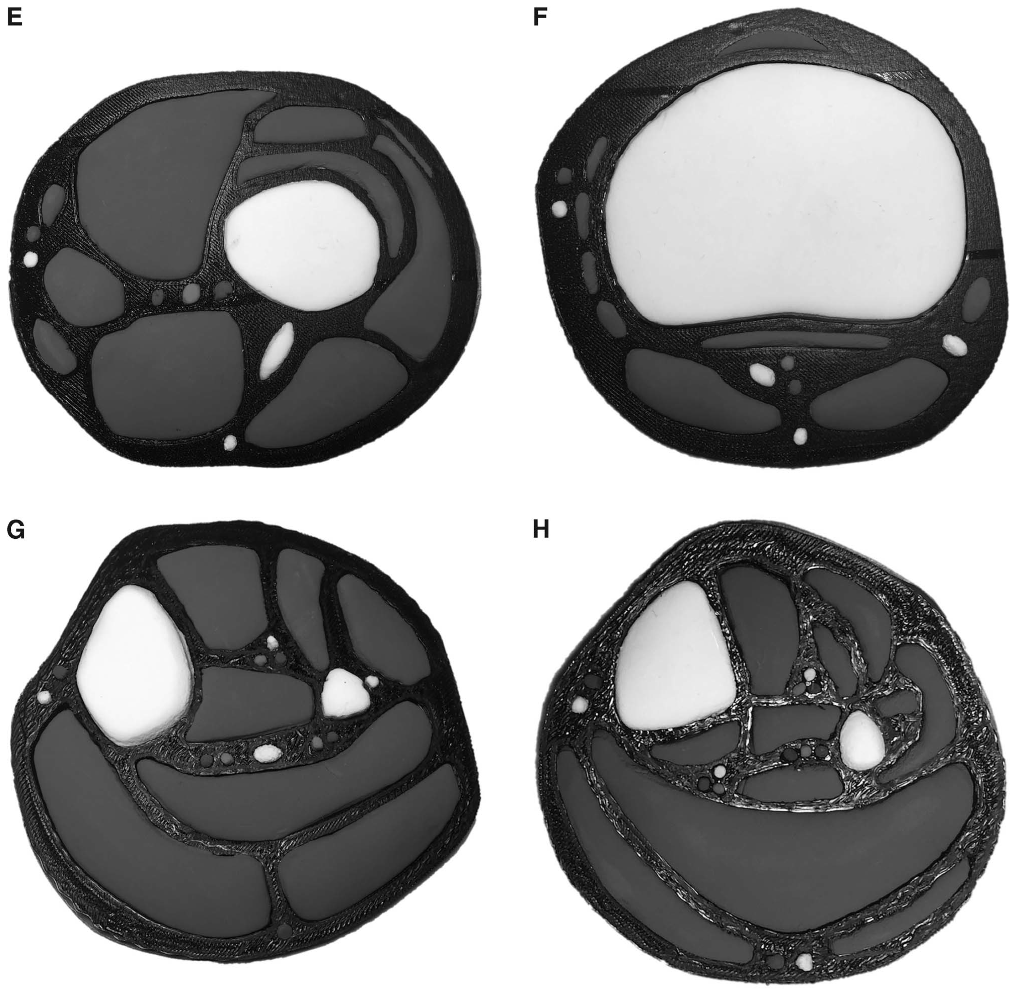
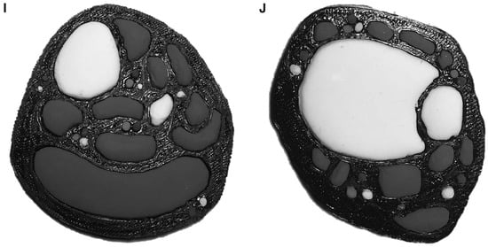
Figure 2.
Continued.
The initial startup cost for printing black cross sections reached just short of $304, with the printer incurring most of the price. Under optimized settings, cross sections finished printing at most 3 hours after starting or in as little as 47 minutes for the smallest section (Table 1). The area occupied by the printed sections ranged from 268.26 cm2 to 44.31 cm2, all with a 5-cm height. Sections were printed using as little as 7 g and at most 27 g of filament (Table 1). Accounting for the price of filament used in each print, the total cost for a set of nine cross sections reached $2.13 for the black filament.
Discussion
This project is the first to demonstrate the cost-effectiveness and use of 3-D printing in podiatric medical education. Priced less than $5, 3-D–printed cross sections provide an inexpensive investment to embrace a lower-extremity specialist's training. These sections represent a fraction of the cost associated with cadaver dissection and plastication.[25] Moreover, polylactic acid filament poses no chemical hazard to the student, unlike the toxic cadaveric cross section counterpart available to students after dissection. With a thickness of 0.5 cm and minimal weight, storage and transportation of these sections becomes feasible, making at-home study with these tools possible.
Because sections are digitally saved, 3-D printed cross sections can be reproduced at the discretion of the instructor, ending the confusion associated with natural variations and less than optimal dissections. On the other hand, because sections are customizable to fit learning objectives, desired anatomical variations may be added with ease. Three-dimensional cross sections require no user input once the printer starts and minimal time to complete printing, thereby allowing more time to be devoted to instruction rather than to specimen preparation. As a result of this affordability and ease of production, each student would be able to obtain a personal and affordable set of cross sections to complement his or her studies.
In an effort to prepare future podiatric physicians, cross sections can be modified to teach and assess students' understanding of anatomical as well as surgical principles. Combined with the printed negatives to teach the cross-sectional anatomy of the lower extremity (Fig. 2A), empty spaces can be filled in using plastic prints or modeling clay during evaluation (Fig. 2 B-J). Furthermore, to supplement anatomical understanding with clinical applications, surgical equipment such as Kirschner wires may be inserted through the prints to introduce and evaluate understanding of proper hardware placement, as illustrated in (Fig. 2A). Introducing such surgical planning methods in the transverse plane is difficult for students to appreciate using currently available imaging atlases, plastinated cross sections, and traditionally dissected cadaveric specimens. The 3-D–printed cross sections provide a unique solution to training future physicians.
By supplementing lower-extremity anatomy courses with 3-D–printed cross sections, students will begin to apply their structural understanding with functional implications in all three planes. Such an introduction will provide the framework that will be integral for the comprehension of future podiatric medical education coursework and, ultimately, practice. Extensive preoperative planning is becoming more prevalent in present-day patient care. Comprehensive understanding of the anatomy at the surgical site is imperative to limit possible adverse events. The use of this new, inexpensive, and practical teaching method with 3-D–printed cross sections provides a means to advance the podiatric medicine profession, starting with the foundation that is lower-extremity anatomy.
Conclusions
Advances in 3-D printing technology are revolutionizing medicine from patient education to surgical planning. The capability to view anatomy and practice procedures in all three dimensions more completely prepares the physician for practice. This technology may, in turn, be used to educate podiatric medical students during their introductory coursework, bridging the gap between the basic medical sciences and clinical experience. As an affordable and reproducible method of instruction, 3-D printing gives the instructor more opportunity to teach structure variations of interest to the podiatric medicine profession normally obscured in dissection or cut from textbooks. Moreover, students will have the ability to study from these sections at their leisure, outside of laboratory dissection, in part because of the nonhazardous production materials. Most importantly, 3-D cross sections enhance the learning experience by providing students with a model that allows them to visualize and think in the cardinal plane, inherent in both podiatric medical imaging and surgical planning.
Financial Disclosure
None reported.
Conflict of Interest
None reported.
References
- Korf HW, Wicht H, Snipes RL, et al: The dissection course: necessary and indispensable for teaching anatomy to medical students. Ann Anat190: 16, 2008.
- Drake RL, McBride JM, Lachman N, et al: Medical education in the anatomical sciences: the winds of change continue to blow. Anat Sci Educ2: 253, 2009.
- McMenamin PG, Quayle MR, McHenry CR, et al: The production of anatomical teaching resources using three-dimensional (3D) printing technology. Anat Sci Edu7: 479, 2014.
- Brown B, Adhikari S, Marx J, et al: Introduction of ultrasound into gross anatomy curriculum: perceptions of medical students. J Emerg Med43: 1098, 2012.
- Rizzolo LJ, Stewart WB: Should we continue teaching anatomy by dissection when ... Anat Rec B New Anat289: 215, 2006.
- D'Antoni AV, DiLandro AC, Chusid ED, et al: Psychometric properties and podiatric medical student perceptions of USMLE-style items in a general anatomy course. JAPMA102: 517, 2012.
- Teichgräber UK, Meyer JM, Poulsen Nautrup C, et al: Ultrasound anatomy: a practical teaching system in human gross anatomy. Med Educ30: 296, 1996.
- Johnson EO, Charchanti AV, Troupis TG: Modernization of an anatomy class: from conceptualization to implementation: a case for integrated multimodal-multidisciplinary teaching. Anat Sci Educ5: 354, 2012.
- Baker EW, Slott PA, Terracio L, et al: An innovative method for teaching anatomy in the predoctoral dental curriculum. J Dent Educ77: 1498, 2013.
- Magiros M, Kekic M, Doran GA: Learning relational anatomy by correlating thin plastinated sections and magnetic resonance images: preparation of specimens. Acta Anat (Basel)158: 37, 1997.
- Moscova M, Bryce DA, Sindhusake D, et al: Integration of medical imaging including ultrasound into a new clinical anatomy curriculum. Anat Sci Educ8: 205, 2015.
- Orsbon CP, Kaiser RS, Ross CF: Physician opinions about an anatomy core curriculum: a case for medical imaging and vertical integration. Anat Sci Educ7: 251, 2014.
- De Barros N, Rodrigues CJ, Junqueira AJQ, et al: The value of teaching sectional anatomy to improve CT scan interpretation. Clin Anat14: 36, 2001.
- Peppler RD, Kwasigroch TE, Hougland MW: Cross-sectional anatomy: preparation of teaching specimens. Anat Rec206: 341, 1983.
- von Hagens G: Impregnation of soft biological specimens with thermosetting resins and elastomers. Anat Rec194: 247, 1979.
- von Hagens G, Tiedemann K, Kriz W: The current potential of plastination. Anat Embryol (Berl)175: 411, 1987.
- Radenkovic D, Solouk A, Seifalian A: Personalized development of human organs using 3D printing technology. Med Hypotheses87: 30, 2016.
- AlAli AB, Griffin MF, Butler PE: Three-dimensional printing surgical applications. Eplasty15: e37, 2015.
- Vaccarezza M, Papa V: 3D printing: a valuable resource in human anatomy education. Anat Sci Int90: 64, 2015.
- Rengier F, Mehndiratta A, Von Tengg-Kobligk H, et al: 3D printing based on imaging data: review of medical applications. Int J Comput Assist Radiol Sur5: 335, 2010.
- Siddiqui NA, Lamm BM: Digital planning for foot and ankle deformity correction: Evans osteotomy. J Foot Ankle Surg53: 700, 2014.
- Jastifer JR, Gustafson PA: Three-dimensional printing and surgical simulation for preoperative planning of deformity correction in foot and ankle surgery. J Foot Ankle Surg56: 191, 2017.
- Qiao F, Li D, Jin Z, et al: Application of 3D printed customized external fixator in fracture reduction. Injury46: 1150, 2015.
- Nayagam S: Safe corridors in external fixation: the lower leg (tibia, fibula, hindfoot and forefoot). Strategies Trauma Limb Reconstr2: 105, 2007.
- Raja DS, Sultana B: Potential health hazards for students exposed to formaldehyde in the gross anatomy laboratory. J Environ Health74: 36, 2011.
© 2018 American Podiatric Medical Association