


Abstract
Background: To date, there is conflicting evidence that high-end “motion control” running shoes can correct and control rearfoot pronation. Many methods have been used to evaluate the efficacy of motion control footwear in reducing hindfoot pronation during gait, including stop-motion photography, three-dimensional camera kinematic analysis, and three-dimensional bone modeling using computed tomography. Until now, there have been no radiographic studies that examined the effect of motion control running shoes on the static posture of the foot. Murley et al devised a reliable system that correlated noninvasive clinical examinations to radiographic values that correspond to foot pronation. The aim of this prospective investigation was to determine whether motion control running shoes are able to produce a significant difference in pronation through a radiographic study, using the angular relationships as described by Murley et al, in two different shoe conditions as compared to the barefoot condition in female subjects. Methods: This prospective study screened 28 female subjects ranging in age from 22 to 27 years on the basis of arch height index. The 24 subjects with a standing arch height index less than 0.370 were invited to participate in the study. Unilateral weightbearing dorsoplantar and lateral foot radiographs were taken in barefoot, neutral shoe, and motion control shoe conditions. Calcaneal inclination angle, calcaneal–first metatarsal (CFMA) angle, talonavicular coverage angle (TNCA), and talus–second metatarsal angle were measured in each condition by two independent observers using the Opal-Ortho PACS software package and then averaged. Angles were compared to barefoot baseline values using paired t tests. Results: The motion control running shoe produced average decreases of 2.64% in CFMA, 12.62% in TNCA, 5.3% in talus–second metatarsal angle and an average increase of 1.3% in calcaneal inclination angle. Statistically significant (P > .05) improvements in CFMA were noted in both the motion control (P < .000) and neutral shoe conditions (P < .000) when compared to barefoot, whereas TNCA improved only in the motion control shoe condition as compared to barefoot (P = .003). Conclusions: This investigation found evidence that the particular models of motion control running shoes studied could correct foot pronation in the transverse and sagittal planes in stance. Motion control running shoes improved CFMA and TNCA from the barefoot condition and were more effective in correcting pronation compared with neutral running shoes in this radiographic study simulating static foot posture in stance.
Hindfoot pronation serves many important functions during the gait cycle; however, excessive, abnormal pronation, which continues late into the stance phase of gait, may increase the activity of the extrinsic musculature and ligaments of the foot, leading to strain or injury of such structures.[1,2] It is thought that such stresses affect the ability of the foot to undergo proper propulsion into the toe-off phase of gait and, over time, may contribute to the some of the manifestations of overuse injuries.[2] A study by Willems et al[3] demonstrated that subjects with increased pronation had a higher possibility of developing exercise-related lower leg pain during shod running. In their study, plantar pressure measurements and three-dimensional kinematic analysis was used to evaluate gait patterns for pronation, and exercise-related lower leg pain included pain secondary to conditions such as compartment syndrome, shin splints, stress fracture, and medial tibial stress syndrome.[3] Additionally, it is believed that continued subtalar joint pronation into midstance may be implicated in the development of such conditions as plantar fasciitis, hallux limitus or hallux valgus, neuroma, or sesamoiditis.[1]
Female runners are reported to be twice as likely to sustain certain running injuries such as patellofemoral pain syndrome, iliotibial band friction syndrome, tibial stress fractures, gluteus medius injuries, and sacroiliac injuries compared with their male counterparts.[4,5,6] This can be attributed largely to anatomical differences that directly affect the biomechanical patterns they display. Three-dimensional kinematic studies have demonstrated that female runners display greater hip frontal and transverse plane negative work, greater peak hip internal rotation and adduction,[4] twice the gluteus maximus activity, as well as significantly different lower extremity mechanics in the frontal and transverse planes compared with male runners.[7]
Even though reduction of foot pronation is a very common clinical approach to manage such sports injuries, to date, there is conflicting evidence that high-end “motion control” footwear can correct foot posture and control rearfoot pronation.[8] Currently, there is little evidence to back the manufacturers' claims, yet many physicians recommend these shoes to patients as a way to treat overpronation and prevent its associated pathologies.[8] Many methods have been used to estimate the position of the subtalar joint during midstance and evaluate the efficacy of motion control footwear in reducing hindfoot pronation during gait, including stop motion photography,[9] three-camera kinematic analysis,[10] and three-dimensional (3-D) bone modeling using computerized tomography.[11] Murley et al[12] established a reliable radiographic protocol for classifying normal and flat arched feet. Four radiologic measurements taken from dorsoplantar and lateral radiographs—the talus–second metatarsal angle (TSMA), talonavicular coverage angle (TNCA), calcaneal inclination angle (CIA), and calcaneal–first metatarsal angle (CFMA)—were found to have a direct relationship to clinical evaluations of pronation and arch height. The efficacy of motion control running shoes in reducing rearfoot pronation in static stance has never been studied radiographically. Radiographs are generally considered to be the gold standard for evaluating the alignment of the foot in static stance and thus are commonly used to confirm clinical foot posture.[12] The aim of this prospective investigation was to determine whether motion control running shoes are able to produce a significant difference in pronation through a radiographic study, using the angular relationships as described by Murley et al,[12] in two different shoe conditions as compared to the barefoot condition in female subjects. It was hypothesized that motion control shoes would be able to produce significant differences in the radiographic angles correlating with pronation compared with the barefoot condition.
Materials and Methods
Ethical approval for this investigation was obtained through the Institutional Review Board of Temple University (Protocol 23184) and the Temple University Medical Radiation Committee. We solicited the participation of active female subjects between the ages of 18 and 30 years. We chose to focus our investigation on women only because they are inherently more susceptible to certain kinetic and kinematic forces.[4,7] Inclusion criteria included arch height index (AHI) less than 0.370, body mass index (BMI) between 18.5 and 28 kg/m2, age between 18 and 30 years, and those willing and able to consent to radiographic evaluation. Subjects were excluded from the present study if they possessed forefoot or tarsal deformity that would impede radiographic measurements and if they were pregnant or planning to become pregnant within 5 months of the study. All subjects who met the inclusion criteria and expressed interest in participating in the study were invited by the research coordinator to be screened for inclusion. Candidates who signed the informed consent document were screened at the Foot & Ankle Institute on the basis of AHI and BMI. AHI was chosen as a screening measure for this study because it accounts for the same parameters used by Murley et al for screening in a consistent and reliable manner.[13]
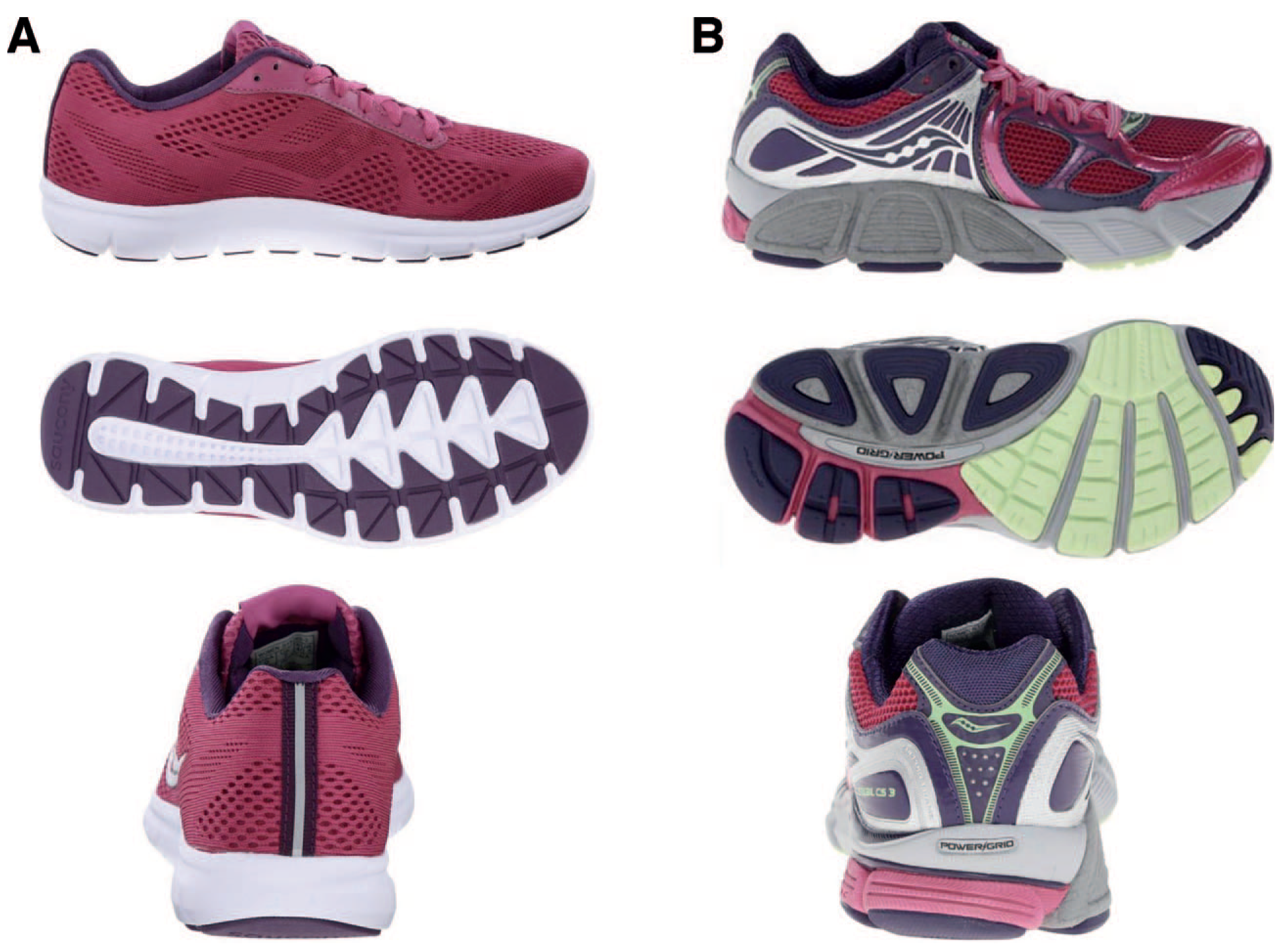
Twenty-eight female subjects aged 22 to 27 years were screened for participation, and the 24 subjects with a standing AHI less than 0.370 and a BMI between 18.5 and 28 kg/m2 were invited to participate in the study and scheduled for a separate visit to have a series of radiographs taken. After participants produced a negative result on a urine pregnancy test, unilateral weightbearing dorsoplantar and lateral digital foot radiographs of the foot with the lowest AHI were taken in barefoot, neutral shoe (Saucony Ideal; Saucony, Lexington, Massachusetts), and motion control shoe (Saucony Stabil) conditions. The Ideal was chosen because it represents a shoe that has more than adequate foam for cushioning but lacks any appointments for controlling foot motion or posture and thus is a part of Saucony's “neutral” category of running shoes. It is designed with mesh upper and memory foam liner with high to normal arch (Fig. 1A). Conversely, the Stabil has many features associated with the motion control category such as a straight last, stiff rearfoot, lateral heel flare, dual-density mid foot, and a large medial flare and thus is a part of Saucony's “stability” category of running shoes and is also made with added midfoot cushioning (Fig. 1B). Although heel-to-toe drop has been shown to have no effect on rearfoot pronation,[9] both shoe conditions had an 8-mm drop and were chosen for consistency. A single investigator measured the resting intramalleolar distance of each subject before the barefoot assessment, and the subjects were required to reproduce the same distance for each trial so that the subjects' stance position did not confound our results. Radiographs were marked with a subject identifying code and uploaded onto a secure server.
Figure 1.
Illustration of shoe features.
Using the Opal-Ortho PACS software package (Konica Minolta Medical Imaging, East Garner, North Carolina), CIA, CFMA, TNCA, and TSMA were measured in each condition by two independent observers. The respective measurements of each observer for each of the four angles were then averaged to determine four angular measurement for each subject. Angular measurements were then compared for each subject in the neutral shoe versus barefoot conditions, the motion control shoe versus barefoot conditions, and the neutral versus motion control shoe conditions, and a difference was obtained in angular measurements between each condition. It was considered an improvement in the angular relationship if either the CIA increased or the CFMA, TNCA, or TSMA decreased, as these values represent a change in angle from a foot posture consistent with a pronated foot type and trending toward values consistent with a rectus foot posture and thus were expressed as a positive value. If the intervention did not improve the angle, a negative value was expressed. Angles in the neutral shoe and motion control shoe were compared to barefoot baselines using paired, two-tailed t tests. The improvements in the neutral shoe condition were then compared to the improvements noted in the motion control shoe using the same measure.
Results
Of the 24 subjects tested, 66% saw an increase in calcaneal inclination in the neutral shoe versus 50% in the motion control shoe; 79.2% of subjects demonstrated a reduction in CFMA in the neutral shoe and 91.06% saw an improvement in this angle in the motion control shoe. For the transverse plane assessment, 41.66% of the subjects demonstrated a decrease in TNCA and 70.83% demonstrated improvement in this angle in the motion control shoe. The TMSA improved in only 33.33% of subjects in the neutral shoe but showed a decrease in 62.5% of the subjects in the motion control shoe condition. A comparison of the initial measurements yielded an average magnitude of disagreement between the observers of 0.6° for CIA, 0.84° for CFMA, 2.17° for TNCA, and 1.4° for TSMA.
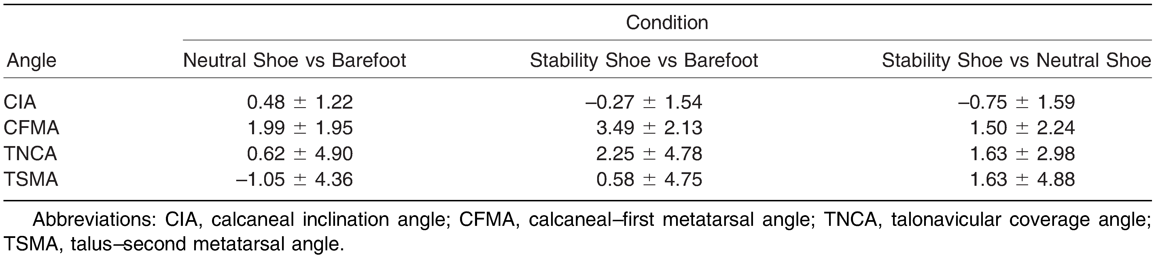
Overall, subjects in the neutral shoe condition demonstrated an average increase in CIA of 0.48 ± 1.22° (2.24%). There was an average decrease of 1.99 ± 1.95° (1.50%) in CFMA. The transverse plane measurements revealed average angular improvements of 0.62 ± 4.90° (5.51%) decrease in TNCA and –1.05 ± 4.36° (–15.62%) decrease in TSMA. In the motion control shoe condition, the subjects displayed an average decrease of –0.27 ± 1.54° (1.3%) in CIA and an average decrease of 3.49 ± 2.13° (2.64%) in CFMA. The motion control shoe produced average improvements in the transverse plane angles of 2.25 ± 4.78° (12.62%) decrease in TNCA and 0.58 ± 4.75° (5.32%) decrease in TSMA compared with the barefoot conditions.
Variability was seen across subjects in the ability of the shoes to affect the measured angles. Some subjects experienced improvement in the measured angular relationships, whereas other subjects did not see any improvements in the same shoe conditions. The degree to which the angles improved was also highly variable between the 24 subjects. The largest angular improvement in the data collected for the CIA from the barefoot to neutral shoe conditions was an increase of 2.75° as seen in one subject (Table 1). Comparably, the motion control shoe demonstrated the ability to increase CIA only as much as 1.50° as compared to the barefoot condition as seen in another subject (Table 1). The largest decrease in CIA observed was 1.85° in the neutral shoe, whereas a decrease of as 4.89° was seen in the motion control shoe versus the barefoot condition. The neutral shoe was able to improve CFMA as much as 5.80° compared with the barefoot condition in one subject. The greatest improvements seen in the motion control shoe from neutral were demonstrated by a decrease of 7.45° in CFMA. A 1.95° increase in CFMA was demonstrated in the neutral shoe versus the barefoot condition as compared to a 2.00° increase in the motion control shoe versus the barefoot condition. The transverse plane assessment revealed larger extremes, with maximum decreases in TNCA of 11.95° and 14.15° in the neutral and motion control shoe conditions, respectively, when compared to baseline. These same shoe conditions caused an increase in TNCA in other subjects as large as 10.80°) in the neutral shoe and 7.20° in the motion control shoe compared with barefoot baseline values. The neutral shoe condition produced an improvement in TSMA of 7.15°, whereas the motion control shoe produced an improvement of 8.90°. Conversely, an increase of 10.85° in TSMA was observed in the neutral shoe, compared with an increase of 10.95° in the motion control shoe.
Table 1.
Mean ± SD Improvements in Angles in Each Shoe Condition (Degrees)
Discussion
The purpose of this study was to determine whether motion control running shoes are able to produce a statistically significant difference in pronation through a radiographic study, using the angular relationships in two different shoe conditions compared with the barefoot condition in female subjects. It was hypothesized that motion control shoes would be able to produce significant differences in the radiographic angles correlating with pronation compared with the barefoot condition. The present study demonstrates statistically significant differences in angular relationships in both the sagittal and transverse planes.
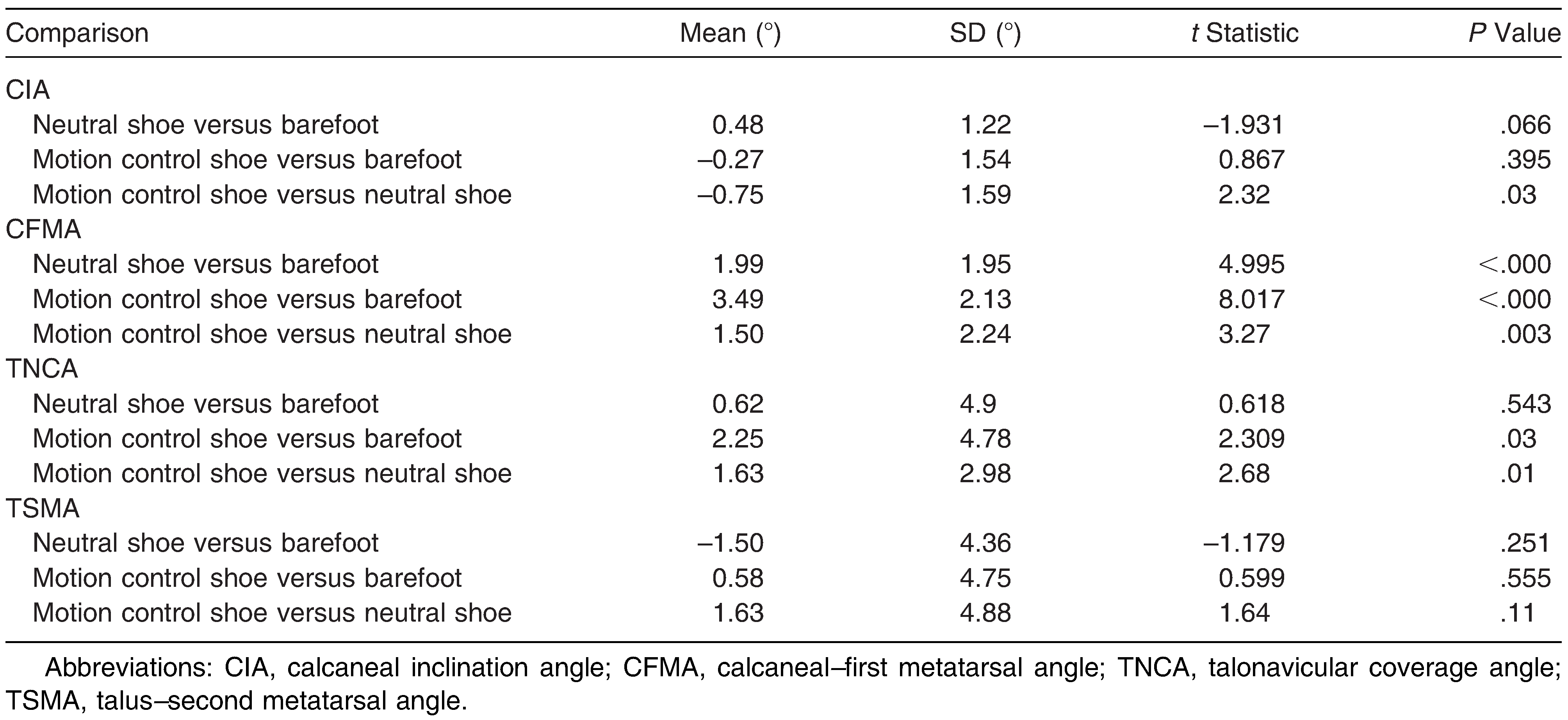
In the sagittal plane, statistically significant differences in the CFMA could be appreciated in both shoe conditions compared with the barefoot condition and also between the two conditions themselves (Table 2). In the transverse plane, statistically significant differences between measures in TNCA in the motion control shoe versus the barefoot and the two shod conditions were seen. This study is the first of its kind to demonstrate the ability of motion control shoes to decrease hindfoot pronation in stance radiographically. It is possible that such angular decreases could translate dynamically to earlier re-supination of the foot during the gait cycle, thus decreasing potential strain on the musculature and ligamentous structures in the foot, although the extent of such effects remains unclear at this point.
Table 2.
Statistical Comparison of Improvements in Angles in Each Shoe Condition
There were four subjects (aged 21–24 years) included in the study cohort whose AHIs were considered to be within a rectus range (0.345–0.370). All of these subjects showed improvements in CFMA, TNCA, and TSMA in the motion control shoe condition compared with barefoot measurements, and three of the four showed an improvement in CIA. In this condition, none of the angles improved enough to place the subject in the supinatory range. They also showed a lower percentage of variation between subjects in measured angular relationships compared with the pronatory foot type group. This demonstrates that motion control shoes may have some utility in people with a rectus or mild pronatory foot type.
It is important to consider that the shoes in question are designed to attenuate dynamic motion of the foot during running. The purpose of the current investigation was to determine whether the shoe features designed to control motion could affect static foot posture. It is logical to reason that angular relationships measured may experience different changes when the foot is fully weightbearing in dynamic motion.
Many other studies have been performed to determine the efficacy of motion control running shoes in reducing pronation; however, the modalities and markers used to measure the ability of these shoes to affect pronation vary widely, and there is conflicting evidence to date. In a study by Hoffman et al,[14] dynamic biplane radiographic imaging and 3-D bone modeling using computed tomography was used to determine navicular drop, an indirect measure of pronation, in multiple shoe conditions in subjects while running. It was found that the motion control shoes resulted in a slower rate of navicular drop compared with the barefoot and minimalist shoe conditions studied, but no statistically significant differences could be appreciated with regard to magnitude of navicular drop while wearing the motion control shoes.[14] Another study by Cheung and Ng[15] sought to determine the ability of motion control shoes to control rearfoot movement in a group of 25 female subjects with a pronatory foot type, with an additional focus on the state of fatigue of the lower extremity muscles using 3-D kinematic evaluation. They demonstrated that the degree of rearfoot movement was reduced when running in a motion control shoe compared with the neutral shoe irrespective of the level of muscular fatigue.[15] Other studies suggest that motion control shoes do not necessarily limit the range of joint motion in the rearfoot but work by causing deceleration of the midfoot during running.[13] Cheung et al[16] conducted a systematic review of the literature on motion control shoes including 14 studies encompassing a total of 288 patients. Study methods included two-dimensional and 3-D kinematic analysis, use of force plates, and insole sensors. A statistically significant difference was seen in the kinematic studies in the ability of the motion control shoes to reduce pronation. Vertical impact loading was also significantly lessened in subjects when wearing motion control shoes.[16] These studies focus primarily on dynamic investigations of the footwear, thus making it difficult to directly compare the present results of this static study to the results of previous studies.
As with any scientific investigation, critical readers are encouraged to review the study design and results and reach their own conclusions. Data were collected from a limited number of subjects (N = 24); therefore, these results might not be representative of a broader population sampling. It is important to note that although the statistical strength of some measurements was weak, there were subjects whose outcomes were drastically different from the average. High-magnitude outliers, either because their angular values differed greatly from the mean or because of having a majority of values that differed slightly from the mean, have a large impact on such a small sample size and detract from an otherwise strong correlation.
Because an average improvement of only 1.50° was observed in the sagittal plane from neutral shoe to motion control shoe, it is unclear just how much of a benefit one model offers over the other. The clinical significance of such a decrease is unknown. It is also worth noting that the structural features of these shoes are unique only to these specific models. Many manufacturers produce several shoes that are classified into neutral and motion control shoe categories each year, and each shoe has specific features that are designed to work together to deliver the desired function. It is impossible to test each shoe and gauge its efficacy, and it is illogical to assume that the results of this investigation would apply to all shoes in the described categories. Overall, the most meaningful endpoint to consider should not be whether a specific angular measurement has been achieved, but whether a certain shoe can effectively reduce pain and discomfort in a patient.
This study also measured angles in only the sagittal and the transverse planes. When assessing foot posture and pronation clinically and describing etiologies of foot conditions, clinicians often focus on the frontal plane effects of overpronation. Clinicians must therefore estimate the effects of sagittal and transverse plane relationships on frontal plane parameters. It is probable that changes in both sagittal and transverse planes will affect the frontal plane. This is an area where future research would be valuable to reveal the extent of interplay between the planes.
Conclusions
As evidenced by improvements in TNCA and CFMA in this investigation, motion control running shoes can reduce static pronation in the sagittal and transverse planes. Motion control running shoes improved CFMA and TNCA compared with the barefoot condition, and were more effective in correcting pronation compared with neutral running shoes in this study. However, the clinical significance of these changes remains unknown. Excessive rearfoot pronation puts increased strain on specific muscles and ligaments and increases the incidence of exercise-related lower leg pain during shod running. Although measuring dynamic foot pronation is inherently difficult, there are established objective radiographic relationships in the literature to quantify static pronation. Future studies correlating static radiographic markers of pronation with dynamic, clinical trials of subjects running in motion control shoes could better elucidate the efficacy of motion control running shoes in controlling pronation and decreasing lower extremity injury.
Our study shows that there is radiographic evidence that motion control running shoes can reduce pronation in the sagittal and transverse planes in stance. Motion control running shoes display the ability to correct the sagittal component of pronation twice as much as neutral running shoes. The difference in static measurements may indicate a difference in the shoes' propensity to attenuate motion.
Financial Disclosure
Funding was provided by the American Podiatric Medical Student Association and the Temple University School of Podiatric Medicine Student Government Association.
Conflict of Interest
None reported.
References
- Kirby K. Biomechanics of the normal and abnormal foot. JAPMA90: 30, 2000.
- McPoil G, Knecht HG. Biomechanics of the foot in walking: a function approach. J Orthop Sports Phys Ther7: 69, 1985.
- Willems TM, Witvrouw E, De Cock A, et al: Gait-related risk factors for exercise-related lower-leg pain during shod running. Med Sci Sports Exerc39: 330, 2007.
- Ferber R, Davis IM, Williams DS III: Gender differences in lower extremity mechanics during running. Clin Biomech (Bristol, Avon)18: 350, 2003.
- Boling M, Padua D, Marshall S, et al: Gender differences in the incidence and prevalence of patellofemoral pain syndrome. Scand J Med Sci Sports20: 725, 2010.
- Taunton J: A retrospective case-control analysis of 2002 running injuries. Br J Sports Med36: 95, 2002.
- Chumanov ES, Wall-Scheffler C, Heiderscheit BC: Gender differences in walking and running on level and inclined surfaces. Clin Biomech (Bristol, Avon)23: 1260, 2008.
- Richards CE, Magin PJ, Callister R: Is your prescription of distance running shoes evidence-based?Br J Sports Med43: 159, 2009.
- Clarke T, Frederik E, Hamill C: The effects of shoe design parameters on rearfoot control in running. Med Sci Sports Exerc15: 376, 1983.
- Cheung RT, Wong MY, Ng GY: Effects of motion control footwear on running: a systematic review. J Sports Sci29: 1311, 2011.
- Peltz C, Haladik J, Hoffman S, et al: Effects of footwear on three-dimensional tibiotalar and subtalar joint motion during running. J Biomech47: 2647, 2014.
- Murley GS, Menz HB, Landorf KB: A protocol for classifying normal- and flat-arched foot posture for research studies using clinical and radiographic measurements. J Foot Ankle Res2: 22, 2009.
- Butler RJ, Hillstrom H, Song J, et al: Arch height index measurement system: establishment of reliability and normative values. JAPMA98: 102, 2008.
- Hoffman S, Peltz C, Haladik J, et al: Dynamic in-vivo assessment of navicular drop while running in barefoot, minimalist, and motion control footwear conditions. Gait Posture41: 825, 2015.
- Cheung RTH, Ng GYF. Efficacy of motion control shoes for reducing excessive rearfoot motion in fatigued runners. Phys Ther Sport8: 75, 2007.
- Cheung RT, Wong MY, Ng GY: Effects of motion control footwear on running: a systematic review. J Sports Sci29: 1311, 2011.
© 2018 American Podiatric Medical Association