


Abstract
Background: We sought to determine the prevalence of lower-extremity arterial calcification in a cohort of patients with diabetes and associated foot pathology receiving inpatient treatment at an urban US tertiary health-care system. Methods: The primary outcome measure was defined as either radiographic evidence of vessel calcification or noninvasive vascular testing that resulted in any reporting of vessel noncompressibility or an ankle-brachial index greater than 1.1. Radiographic evidence of vessel calcification was defined as radiodense calcification in the proximal first intermetatarsal space (deep plantar perforating artery), anterior ankle (anterior tibial artery), or posterior ankle (posterior tibial artery) on dorsoplantar and lateral foot projections. Results: Of the 367 individuals included in the study, 359 underwent radiography, with radiographic evidence of calcification in 192 (53.5%). Noninvasive vascular testing was performed on 265 participants, with any reporting of noncompressibility or an ankle-brachial index greater than 1.1 observed in 153 (57.7%). Ninety-four participants (25.6%) demonstrated evidence of arterial calcification on the radiographs and noninvasive testing, meaning that 251 participants (68.4%) demonstrated evidence of arterial calcification on at least one test, including 63.6% of participants classified as black/African American race, 65.4% as white race, and 78.3% as Hispanic/Latino ethnicity. Conclusions: The results of this investigation increase the body of knowledge with respect to the evaluation and treatment of diabetic foot disease and may lead to future investigations on the topic of lower-extremity arterial calcification.
A significant percentage of the patient population with diabetes mellitus is at risk for the development of foot pathology, and the treatment of these patients forms a substantial portion of US health-care expenditure.[1,2,3,4,5] One of the consequences of the diabetes mellitus disease process that contributes to this foot pathology is peripheral arterial disease, specifically in the form of lower-extremity occlusive disease and the development of medial arterial calcific sclerosis.[6,7,8,9] The latter involves vessel calcification with the potential to affect both patient evaluation and clinical outcomes. First, the subsequent incompressibility of the arteries has been shown to lead to inaccurate testing results when considering segmental Doppler examinations, photoplethysmography, and the ankle-brachial index (ABI).[10,11,12] These tests are critical to the diagnosis of peripheral arterial disease and contribute to medical decision making with respect to assessment of healing potential.[13,14,15] Second, the presence of arterial calcification has been associated with poor clinical outcomes, including an increased risk of ulceration development, major amputation, and mortality.[16,17,18,19,20,21,22,23,24,25,26,27]
Despite this, prevalence rates of arterial calcification in the diabetic foot and ankle have not been well documented. Aragon-Sanchez and Lazaro-Martinez[28] observed radiographic arterial calcification in 59.4% of 165 inpatients admitted for diabetic foot pathology at a hospital in Spain. They found that radiographic calcification was associated with duration of diabetes, retinopathy, albuminuria, and peripheral arterial disease. Young et al,[20] in a patient population in England, found radiographic evidence of calcification in 78.8% of 54 ankles in neuropathic diabetic patients with an ulceration, 61.9% of 40 ankles in neuropathic diabetic patients without an ulceration, and 25.0% of 43 ankles in nonneuropathic diabetic patients. They also observed a positive correlation between radiographic calcification and increasing ABI. In another study enrolling 122 patients in England, Sharma et al[29] found a prevalence of radiographic calcification of 59.0% in diabetic patients, with no differences between those with Charcot's neuroarthropathy, osteomyelitis, and uncomplicated ulceration.
The objective of this investigation was to determine the prevalence of lower-extremity arterial calcification based on radiographs and noninvasive vascular testing in a cohort of patients with diabetes and foot pathology receiving inpatient treatment at an urban US tertiary health-care system.
Methods
Following institutional review board approval, an observational retrospective medical record review was performed on all inpatients seen in consultation by the Foot and Ankle Surgery Service at Temple University Hospital (Philadelphia, Pennsylvania) over a 12-month data collection period (July 1, 2014, to June 30, 2015). The inclusion criteria of the study cohort consisted of patients with the diagnosis of diabetes mellitus and seen in consultation for a diabetic foot issue (acute or chronic wound/ulceration, soft-tissue infection, bone/joint infection, gangrene, history of minor/major amputation, or Charcot's neuroarthropathy). The medical record was reviewed for patient age, sex, race (American Indian/Alaskan Native, black/African American, white, Native Hawaiian/Pacific Islander, Asian, or other), ethnicity (Hispanic/Latino or not Hispanic/Latino), most recent hemoglobin A1c (HbA1c) level, history of smoking, presence of end-stage renal disease (ESRD), performance of foot plain film radiographs, and performance of noninvasive vascular testing at the time of admission.
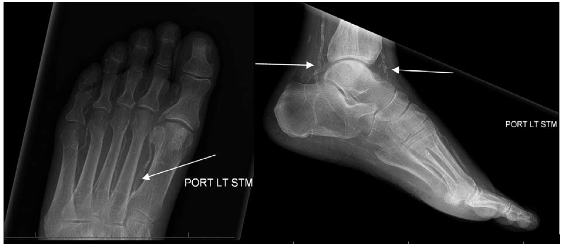
The primary outcome measure of this investigation was the prevalence of medial arterial calcific sclerosis, and this was defined as either radiographic evidence of vessel calcification or noninvasive vascular testing, which resulted in any reporting of vessel noncompressibility or an ABI from either the anterior tibial artery or posterior tibial artery less than 1.1. Radiographic evidence of vessel calcification was defined as radiodense calcification in the proximal first intermetatarsal space (deep plantar perforating artery), anterior ankle (anterior tibial artery), or posterior ankle (posterior tibial artery) on dorsoplantar and lateral projections (Fig. 1).
Figure 1.
Radiographic evidence of vessel calcification (white arrows) was defined as radiodense calcification in the proximal first intermetatarsal space (deep plantar perforating artery), anterior ankle (anterior tibial artery), or posterior ankle (posterior tibial artery) on dorsoplantar and lateral projections.
Data were stored in a password-protected personal computer for subsequent statistical analysis. All of the statistical analyses were performed using SAS Software, Version 9.2 (SAS Institute Inc, Cary, North Carolina). Descriptive statistics were calculated and consisted of the mean, SD, range, frequency count, and odds ratio (OR) analysis.
Results
Three hundred sixty-seven participants met the investigation inclusion criteria. The mean ± SD age was 58.0 ± 12.2 years (range, 20–93 years), and 218 (59.4%) were male. In terms of race, 196 (53.4%) were classified as black/African American, 78 (21.3%) as white, 1 (0.3%) as Asian, and 92 (25.1%) as other. In terms of ethnicity, 89 (24.3%) were Hispanic/Latino. A total of 117 participants (31.9%) had a smoking history, and 73 (19.9%) had ESRD. Data on HbA1c were available from 307 participants, with a mean ± SD of 8.9% ± 2.6% (range, 4.6%–17.5%).
Plain film radiographs of the foot were performed on 359 participants (97.8%). There was radiographic evidence of calcification in 192 (53.5%) of these 359 participants (52.3% of all 367 participants). This included positive findings in 178 dorsoplantar radiographs (49.6%) in the proximal first intermetatarsal space, 162 lateral radiographs (45.1%) at the anterior ankle, and 160 lateral radiographs (44.6%) at the posterior ankle.
Noninvasive vascular testing was performed on 265 participants (72.2%). Any reporting of noncompressibility or an ABI greater than 1.1 was observed in 153 (57.7%) of the 265 tests performed (41.7% of all 367 participants).
Ninety-four participants (25.6%) demonstrated evidence of arterial calcification on the radiographs and noninvasive testing, meaning that 251 participants (68.4%) demonstrated evidence of arterial calcification on at least one test. This included 63.6% of participants classified as black/African American race, 65.4% classified as white race, and 78.3% classified as Hispanic/Latino ethnicity.
We observed that participants with ESRD were more likely to have evidence of vessel calcification than were those without ESRD (86.3% versus 63.9%; OR, 3.55; 95% confidence interval [CI], 1.68–7.71; P < .001). Participants with a history of smoking were less likely to have evidence of vessel calcification than those without a smoking history (57.3% versus 73.6%; OR, 0.481; 95% CI, 0.295-0.783; P < .003). Participants with an HbA1c level of at least 8.0% were not more or less likely to have evidence of vessel calcification than were those with an HbA1c level less than 8.0% (67.1% versus 69.6%; OR, 0.889; 95% CI, 0.559–1.415; P = .682).
Discussion
The results of this investigation provide podiatric physicians with information about the high prevalence of arterial calcification in diabetic patients with foot pathology. We observed that most patients (68.4%) demonstrated evidence of arterial calcification on at least one test, with positive radiographic findings present in 53.5% of participants. Interestingly, evidence of calcification was observed on both tests in only 25.6% of participants, indicating that both radiographs and noninvasive vascular testing may provide independently important diagnostic information on this topic. These findings are consistent with previously published reports of prevalence rates[20,28,29] but provide unique information for a new patient population.
We also observed that patients with ESRD were more likely to have evidence of arterial calcification (OR, 3.55; 95% CI, 1.68–7.71), whereas patients with a positive smoking history were less likely to demonstrate evidence of arterial calcification (OR, 0.481; 95% CI, 0.295–0.783). We found no association with HbA1c levels greater or less than 8.0% (OR, 0.889; 95% CI, 0.559–1.415).
We believe that this information should have an effect on the interpretation of noninvasive vascular testing and decision making with respect to healing potential in patients with diabetes. These findings indicate that most individuals with diabetes with foot pathology are at risk for falsely elevated ABIs. Podiatric physicians should be aware of this when interpreting the results of this test regarding the diagnosis of peripheral arterial disease and for assessment of healing potential of tissue loss and partial foot amputation. These findings also provide evidence that most diabetic patients with foot pathology are at risk for poor clinical outcomes. Lew et al[16] observed a U-shaped relationship between the ABI and lower-extremity comorbidity, with elevated ABIs associated with poor outcomes. Others have similarly observed that arterial calcification and elevated ABIs are associated with amputation and mortality.[17-27]
As with any scientific investigation, critical readers are encouraged to review and assess the study design and specific results to reach their own independent conclusions; the preceding represents our conclusions based on the data. We also embrace the fact that all investigations have limitations, and this one had several to consider. First, data were collected from a limited number of patients in an urban environment, and, therefore, these results may not be representative of a broader population sampling. More to this point, more than 50% of the cohort was classified as black/African American race and nearly 25% were of Hispanic/Latino ethnicity. Although this could be considered a limitation, it could also be considered a study strength as previous publications on this topic have not specifically reported on these patient populations.[20,28,29] Second, we looked for radiographic evidence of calcification at only three specific locations about the foot and ankle. These were locations where we have observed in clinical practice that calcification is most common, but it is possible that evaluation of other anatomical areas might produce different results. We are unaware of any established diagnostic criteria for defining the presence of lower-extremity arterial calcification. Third, we defined arterial calcification on noninvasive vascular testing based on an ABI reading greater than 1.1. Others might define a different threshold that might produce different results. Finally, we made no attempt to associate arterial calcification with clinical outcome and instead only attempted to provide descriptive statistics. A prospective investigation of the effect of arterial calcification on specific podiatric medical outcomes might represent an interesting avenue for future investigation, as would investigations further clarifying specific patient risk factors for the development of lower-extremity arterial calcification.
Conclusions
The results of this investigation provide original data on the high prevalence of arterial calcification in a unique patient cohort of an urban US tertiary care hospital. These results increase the body of knowledge with respect to the evaluation and treatment of diabetic foot disease and may lead to future investigations on the topic of lower-extremity arterial calcification.
Financial Disclosure
None reported.
Conflict of Interest
None reported.
References
- Hingorani A, LaMuraglia GM, Henke P, et al: The management of the diabetic foot: a clinical practice guideline by the Society for Vascular Surgery in collaboration with the American Podiatric Medical Association and the Society for Vascular Medicine. J Vasc Surg63: 3S, 2016.
- Boulton AJ: The diabetic foot: grand overview, epidemiology and pathogenesis. Diabetes Metab Res Rev24: S3, 2008.
- Markakis K, Bowling FL, Boulton AJ: The diabetic foot in 2015: an overview. Diabetes Metab Res Rev32: 1669, 2016.
- Carls GS, Gibson TB, Driver VR, et al: The economic value of specialized lower-extremity medical care by podiatric physicians in the treatment of diabetic foot ulcers. JAPMA101: 93, 2011.
- Harrington C, Zagari MJ, Corea J, et al: A cost analysis of diabetic lower-extremity ulcers. Diabetes Care23: 1333, 2000.
- Faglia E: Characteristics of peripheral arterial disease and its relevance to the diabetic population. Int J Low Extrem Wounds10: 152, 2011.
- Jude EB, Eleftheriadou I, Tentolouris N: Peripheral arterial disease in diabetes: a review. Diabet Med27: 4, 2010.
- Allison MA, Criqui MH, Wright CM: Patterns and risk factors for systemic calcified atherosclerosis. Arterioscler Thromb Vasc Biol24: 331, 2004.
- Wu M, Rementer C, Giachelli CM: Vascular calcification: an update on mechanisms and challenges in treatment. Calcif Tissue Int93: 365, 2013.
- Aerden D, Massaad D, von Kemp K, et al: The ankle-brachial index and the diabetic foot: a troublesome marriage. Ann Vasc Surg25: 770, 2011.
- Aboyans V, Ho E, Denenberg O, et al: The association between elevated ankle systolic pressures and peripheral occlusive arterial disease in diabetic and nondiabetic subjects. J Vasc Surg48: 1197, 2008.
- Zhang H, Li XY, Si YJ, et al: Manifestation of lower extremity arthrosclerosis in diabetic patients with high ankle-brachial index. Chin Med J (Engl)123: 890, 2010.
- Apelqvist J, Castenfors J, Larsson J, et al: Prognostic value of systolic ankle and toe blood pressure levels in outcome of diabetic foot ulcer. Diabetes Care12: 373, 1989.
- Brownrigg JR, Hinchliffe RJ, Apelqvist J, et al: Performance of prognostic markers in the prediction of wound healing or amputation among patients with foot ulcers in diabetes: a systematic review. Diabetes Metab Res Rev32: 128, 2016.
- Gibbons GW, Wheelock FCJr, Hoar CSJr, et al: Predicting success of forefoot amputations in diabetics by noninvasive testing. Arch Surg114: 1034, 1979.
- Lew E, Nicolosi N, Botek G: Lower extremity amputation risk factors associated with elevated ankle brachial indices and radiographic arterial calcification. J Foot Ankle Surg54: 473, 2015.
- Everhart JE, Pettitt DJ, Knowler WC, et al: Medial arterial calcification and its association with mortality and complications of diabetes. Diabetologia31: 16, 1988.
- Guzman RJ, Bian A, Shintani A, et al: Association of foot ulcer with tibial artery calcification in independent of peripheral occlusive disease in type 2 diabetes. Diabetes Res Clin Pract99: 281, 2013.
- Edmonds ME, Morrison N, Laws JW, et al: Medial arterial calcification and diabetic neuropathy. Br Med J (Clin Res Ed)284: 928, 1982.
- Young MJ, Adams JE, Anderson GF, et al: Medial arterial calcification in the feet of diabetic patients and matches non-diabetic control subjects. Diabetologia36: 615, 1993.
- Silvestro A, Diehm N, Savolainen H, et al: Falsely high ankle-brachial index predicts major amputation in critical limb ischemia. Vasc Med11: 69, 2006.
- David Smith C, Gavin Bilmen J, Iqbal S, et al: Medial artery calcification as an indicator of diabetic vascular disease. Foot Ankle Int29: 185, 2008.
- Chantelau E, Lee KM, Jungblut R: Association of below-knee atherosclerosis to medial arterial calcification in diabetes mellitus. Diabetes Res Clin Pract29: 169, 1995.
- Mayfield JA, Caps MT, Boyko EJ, et al: Relationship of the medial arterial calcinosis to autonomic neuropathy and adverse outcomes in a diabetic veteran population. J Diabetes Complications16: 165, 2002.
- Arain FA, Ye Z, Bailey KR, et al: Survival in patients with poorly compressible leg arteries. J Am Coll Cardiol59: 400, 2012.
- Guzman RJ, Brinkley DM, Schumacher PM, et al: Tibial artery calcification as a marker of amputation risk in patients with peripheral arterial disease. J Am Coll Cardiol51: 1967, 2008.
- Adragao T, Pires A, Branco P, et al: Ankle-brachial index, vascular calcifications and mortality in dialysis patients. Nephrol Dial Transplant27: 318, 2012.
- Aragon-Sanchez J, Lazaro-Martinez JL: Factors associated with calcification in the pedal arteries in patients with diabetes and neuropathy admitted for foot disease and its clinical significance. Int J Low Extrem Wounds12: 252, 2013.
- Sharma A, Scammell BE, Fairbairn KJ, et al: Prevalence of calcification in the pedal arteries in diabetes complicated by foot disease. Diabetes Care33: e66, 2010.
© 2018 American Podiatric Medical Association