


Abstract
A case of a repaired failed toe arthrodesis is presented. A novel and previously unreported approach using a percutaneously placed Kirschner wire coupled with a pulsed electromagnetic field achieved healing of a painful pseudoarthrosis at 54 days. With a percutaneous technique, open debridement of the failed arthrodesis site can be avoided.
Arthrodesis of the proximal interphalangeal joint is a common procedure performed by podiatric and orthopedic surgeons to correct hammer toe and other deformities of the toes.[1] Classically, arthrodesis was performed by fixation of the interphalangeal joint with Kirschner wires[2] or cannulated screws.[3] Regardless of the type of fixation technique, failure to achieve arthrodesis still has been reported.[3,4] Newer procedures for arthrodesis site stabilization have been designed to overcome these difficulties. Such novel methods include resorbable rods, allograft bone pins, screw fixation, bone staples, and one- or two-piece metallic fixation systems, such as the Smart Toe (MMI-USA Inc, Memphis, Tennessee) and the StayFuse (Wright Medical, Memphis, Tennessee).[5,6] Although the literature is sparse on the rate of nonunions in hammer toe surgery and the percentage of nonunions that become symptomatic, a study in 2013 by Scholl et al[7] addressed the frequency of these occurrences. This study compared an intramedullary implant with a buried Kirschner wire, with an osseous union rate of 68.9% with the intramedullary device and 82.1% with the buried Kirschner wires.[7] The symptomatic nonunions that required additional surgical revision comprised 8.6% of the surgeries using the intramedullary device and 10.7% of the surgeries using the buried Kirschner wires for fixation.[7]
Bone growth stimulators have been reported and used since the 1800s specifically to accelerate fracture healing.[8-10] In today's current practice, bone stimulators are divided into two groups: electromagnetic and ultrasound.[11] Pulsed electromagnetic field (PEMF) therapy was popularized in the 1970s, with more than 250 published clinical research studies and 200 basic science studies supporting the efficacy of PEMF therapy in fracture healing.[12]
It has been demonstrated that a PEMF accelerates the process of fracture healing. The precise mechanism of osteogenesis enhanced by PEMF therapy is yet to be determined; however, several studies reported the stimulatory effect of PEMF therapy not only on cell proliferation but also on osteoblast differentiation and matrix mineralization.[13-15] On the molecular level, exposure of osteoblasts to PEMFs generates an electric field gradient across the cell membrane, promoting osteoblastic activity.[16,17] On the other hand, PEMF therapy was also reported to slow down bone resorption mediated by inhibition of osteoclastogenesis and increased osteoclast apoptosis.[18,19] Several reports demonstrated that PEMF therapy upregulates the negative regulator of osteoclast, osteoprotegerin, and downregulates the osteoclast stimulator, with the net effect of increased bone mass.[19-21]
Failure of digital arthrodesis leading to a painful pseudoarthrosis mandates a surgical modification. The usual process typically involves reopening the interphalangeal joint to debride the interposing fibrous tissue and prepare the osseous surfaces for implantation of the new hardware to fixate the arthrodesis site. Opening the joint again increases the possibility of additional undesirable scar tissue, leading to nonunion, shortening of the toe, infection, and prolonged healing periods.
In this case report, we present a case of failed arthrodesis in the second toe after cannulated screw fixation of the proximal interphalangeal joint. To avoid reopening the failed arthrodesis site, we performed a temporary percutaneous external fixation after removal of the cannulated screw. We simultaneously used PEMF therapy to promote osteogenesis of the arthrodesis site, and solid arthrodesis was observed by radiography after 8 weeks.
Case Report
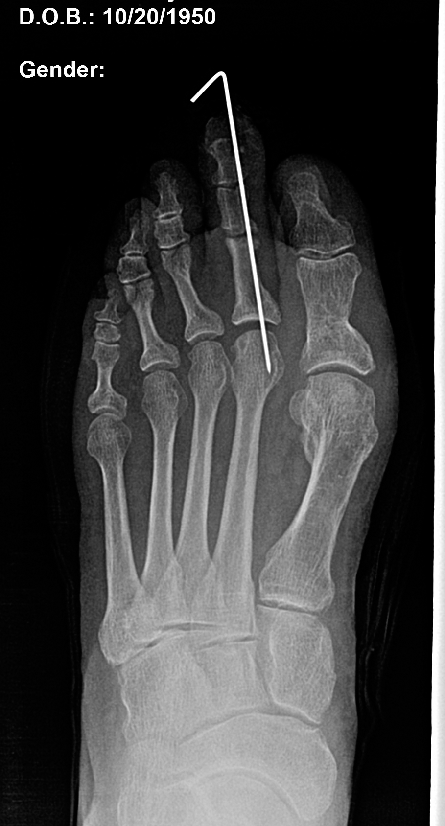
A 65-year-old woman had previously undergone an arthrodesis with a 2.0-mm cannulated screw to fixate the proximal interphalangeal joint of her painful second toe in the left foot (Fig. 1). During 5 months postoperatively she continued to have substantial throbbing pain, erythema, and edema at the proximal interphalangeal joint. Digital radiographs showed distraction of the proximal interphalangeal joint with lateral drift of the medial and distal phalanges leading to deviation of the screw in the proximal phalanx (Fig. 2). The patient returned to the operating room, and the cannulated screw was removed. Under C-arm guidance, the second toe was adjusted to the correct position and fixated percutaneously with a single 0.45 Kirschner wire (Fig. 3). Placement was obtained in the solid medial bone structure of the toe, as the central portion of the bones were devoid of solid substance after removal of the cannulated screw. At day 10 postoperatively, a noninvasive type of PEMF bone stimulator was dispensed with the electromagnetic field concentrated over the second toe. On day 15 the Kirschner wire loosened and had to be removed. Additional splintage to the second toe was maintained with flexible 1-inch tape wrapped concentrically, then buddy splinted to the hallux.
Figure 1.
Preoperative radiograph shows a fixed cannulated screw in the proximal interphalangeal joint.
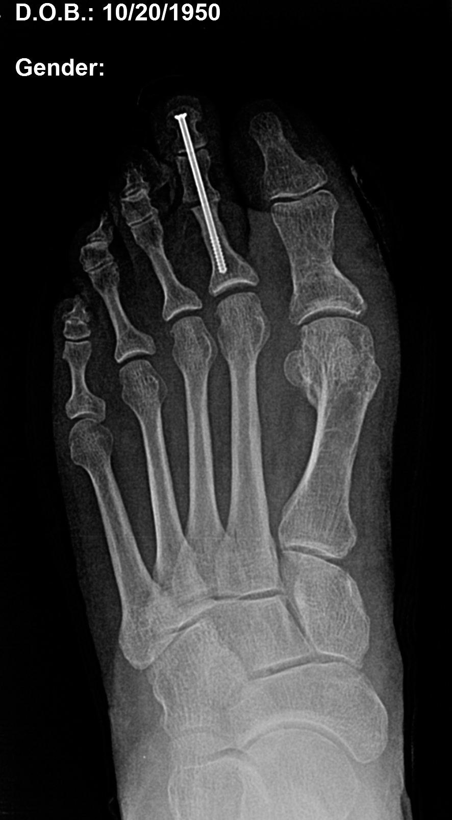
Figure 2.
Preoperative radiograph displays failed arthrodesis of the proximal interphalangeal joint with displacement of the cannulated screw.
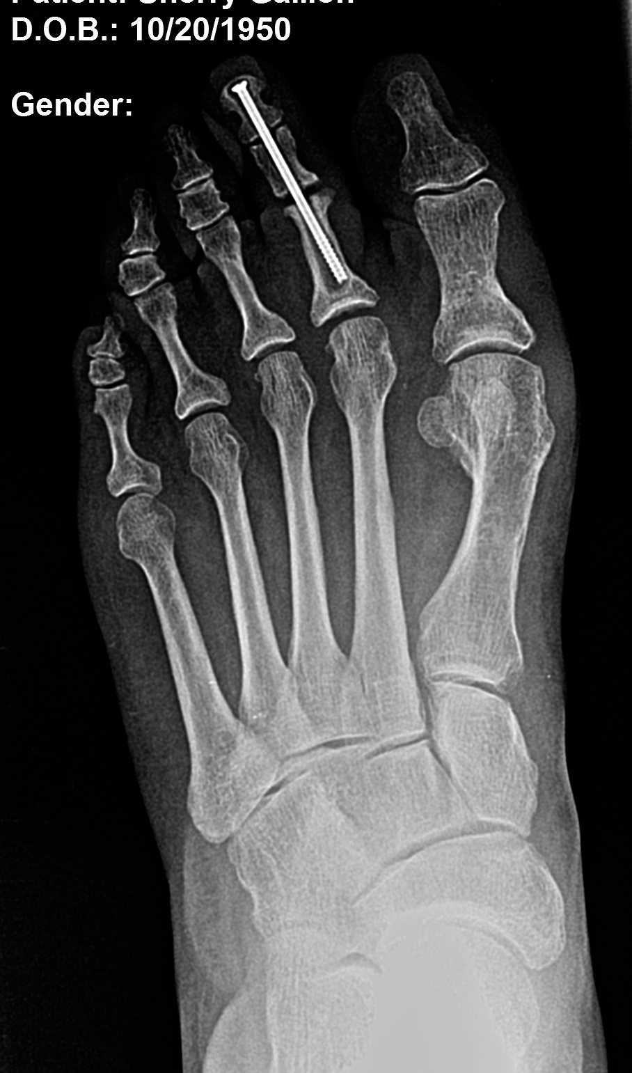
Figure 3.
Radiograph on postoperative day 6 displays removal of the screw and insertion of the Kirschner wire in the proximal interphalangeal joint.
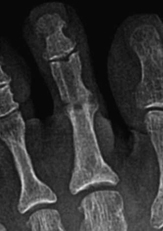
Digital radiographs taken on day 27 postoperatively showed substantial osteogenesis at the arthrodesis site (Fig. 4). Radiographs on day 54 postoperatively showed solid bony bridging at the arthrodesis site (Figs. 5 and 6). Clinically, her second toe was in excellent alignment, with complete resolution of her pain and edema.
Figure 4.
Radiograph on postoperative day 27 shows removal of the Kirschner wire and increased osteogenesis at the proximal interphalangeal joint.
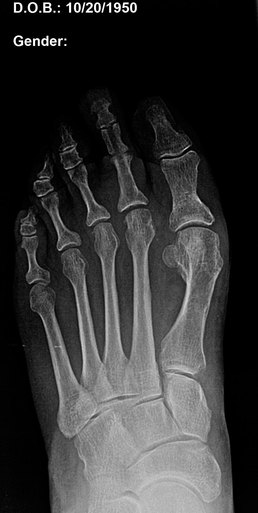
Figure 5.
Radiograph on postoperative day 54 displays successful arthrodesis and joint union of the proximal interphalangeal joint.
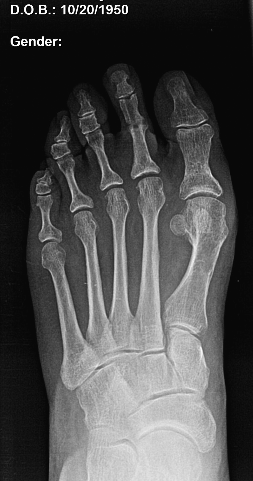
Figure 6.
Magnified radiograph on postoperative day 54 displays successful arthrodesis and joint union of the proximal interphalangeal joint. (x2)
Discussion
We presented a case of failed arthrodesis in the proximal interphalangeal joint of the second toe after cannulated screw fixation. The revisional surgery was performed percutaneously in both the removal of the failed cannulated screw and the placement of the 0.45 Kirschner wire with the toe in the corrected position. Performing this repair percutaneously eliminated the need to reopen the proximal interphalangeal joint, with the potential complications of shortening of the toe, infection at the proximal interphalangeal joint, or recurrent hardware failure of nonunion. By using a percutaneously placed Kirschner wire and noninvasive PEMF bone stimulator, solid evidence of arthrodesis was verified by radiography on day 54 postoperatively, and the patient's symptoms of swelling, pain, and deformity were eliminated.
Funding
None reported.
Conflicts of Interest
None reported.
References
- Coughlin M.“Lesser Toe Deformities,” inSurgery of the Foot and Ankle, 8th Ed, edited byMCoughlin, RMann, CSaltzman, p 363, Mosby, Philadelphia, 2007.
- Granowitz S.Vainio K.Proximal interphalangeal joint arthrodesis in rheumatoid arthritis: a follow-up study of 122 operations. Acta Orthop Scand37: 301, 1966.
- Casterini R.Farsetti P.Tarantino U.et al: Arthrodesis of the toe joints with an intramedullary cannulated screw for correction of hammertoe deformity. Foot Ankle Int25: 256, 2004.
- Bennett GL.Kay DB.Sabatta J.First metatarsophalangeal joint arthrodesis: an evaluation of hardware failure. Foot Ankle Int26: 593, 2005.
- Guelfi M.Pantalone A.Daniel JC.et al: Arthrodesis of proximal inter-phalangeal joint for hammertoe: intramedullary device options. J Orthop Traumatol16: 269, 2015.
- Witt BL.Hyer CF.Treatment of hammertoe deformity using a one-piece intramedullary device: a case series. J Foot Ankle Surg51: 450, 2012.
- Scholl A.McCarty J.Scholl D.et al: Smart Toe® implant versus buried Kirschner wire for proximal interphalangeal joint arthrodesis: a comparative study. J Foot Ankle Surg52: 580, 2013.
- Paterson DC.Carter RF.Maxwell GM.et al: Electrical bone-growth stimulation in an experimental model of delayed union. Lancet1: 1278, 1977.
- Paterson DC.Hillier TM.Carter RF.et al: Experimental delayed union of the dog tibia and its use in assessing the effect of an electrical bone growth stimulator. Clin Orthop Relat Res128: 340, 1977.
- Paterson DC.Lewis GN.Cass CA.Treatment of delayed union and nonunion with an implanted direct current stimulator. Clin Orthop Relat Res148: 117, 1980.
- Li JK.Lin JC.Liu HC.et al: Comparison of ultrasound and electromagnetic field effects on osteoblast growth. Ultrasound Med Biol32: 769, 2006.
- Ryaby JT.Clinical effects of electromagnetic and electric fields on fracture healing. Clin Orthop Relat Res355(suppl): S205, 1998.
- Tsai MT.Chang WH.Chang K.et al: Pulsed electromagnetic fields affect osteoblast proliferation and differentiation in bone tissue engineering. Bioelectromagnetics28: 519, 2007.
- Yang W.Huo XL.Song T.Effects of extremely low frequency pulsed electromagnetic field on different-derived osteoblast-like cells [in Chinese]. Zhonghua Lao Dong Wei Sheng Zhi Ye Bing Za Zhi25: 710, 2007.
- Diniz P.Shomura K.Soejima K.et al: Effects of pulsed electromagnetic field (PEMF) stimulation on bone tissue like formation are dependent on the maturation stages of the osteoblasts. Bioelectromagnetics23: 398, 2002.
- Panagopoulos DJ.Karabarbounis A.Margaritis LH.Mechanism for action of electromagnetic fields on cells. Biochem Biophys Res Commun298: 95, 2002.
- Yan JL.Zhou J.Ma HP.et al: Pulsed electromagnetic fields promote osteoblast mineralization and maturation needing the existence of primary cilia. Mol Cell Endocrinol404: 132, 2015.
- Chang K.Chang WH.Huang S.et al: Pulsed electromagnetic fields stimulation affects osteoclast formation by modulation of osteoprotegerin, RANK ligand and macrophage colony-stimulating factor. J Orthop Res23: 1308, 2005.
- Chang K.Chang WH.Tsai MT.et al: Pulsed electromagnetic fields accelerate apoptotic rate in osteoclasts. Connect Tissue Res47: 222, 2006.
- Chen J.He HC.Xia QJ.et al: Effects of pulsed electromagnetic fields on the mRNA expression of RANK and CAII in ovariectomized rat osteoclast-like cell. Connect Tissue Res51: 1, 2010.
- Schwartz Z.Fisher M.Lohmann CH.et al: Osteoprotegerin (OPG) production by cells in the osteoblast lineage is regulated by pulsed electromagnetic fields in cultures grown on calcium phosphate substrates. Ann Biomed Eng37: 437, 2009.
© 2018 American Podiatric Medical Association