


Abstract
Background: Understanding the concept of kinematic coupling is essential when selecting the appropriate therapeutic strategy and grasping mechanisms for the occurrence of injuries. A previous study reported that kinematic coupling between the rearfoot and shank during running and walking were different. However, because foot mobility involves not only the rearfoot but also the midfoot or forefoot, kinematic coupling is likely to occur among the rearfoot, midfoot, and forefoot segments. We investigated changes in kinematic coupling among the rearfoot, midfoot, and forefoot segments during running and walking. Methods: Ten healthy young men were instructed to run (2.5 ms–1) and walk (1.3 ms–1) on a treadmill at speeds set by the examiner. The three-dimensional joint angles of the rearfoot, midfoot, and forefoot were calculated based on the Leardini foot model Kinematic coupling was evaluated with the absolute value of the cross-correlation coefficients and coupling angles obtained by using a vector coding technique. Results: The cross-correlation coefficient between rearfoot eversion/inversion and midfoot dorsiflexion/plantarflexion was significantly higher during running (r = 0.79) than during walking (r = 0.58), suggesting that running requires stronger kinematic coupling between rearfoot eversion/inversion and midfoot plantarflexion/dorsiflexion than walking. Furthermore, the coupling angle between midfoot eversion/inversion and forefoot eversion/inversion was significantly less during running (30.0°) than during walking (40.7°) (P < .05). Hence, the magnitude of midfoot frontal plane excursion during running was greater than that during walking. Conclusions: Excessive rearfoot eversion during running is likely to lead to excessive midfoot dorsiflexion, and such abnormal kinematic coupling between the rearfoot and midfoot may be associated with mechanisms for the occurrence of injuries.
The foot complex is intricate, and traditional gait analyses consider the foot as a single rigid body with no intrinsic motion.[1,2] Recently, intrafoot kinematics during walking have been successfully quantified by using noninvasive three-dimensional photogrammetry.[3,4,5]
Kinematic analysis of the foot with multiple segments may be necessary to gain insight into the occurrence of injuries. Specifically, the midfoot consists of the navicular, the cuboid, and three cuneiform bones[6] and exhibits many clinical problems, such as flatfoot and pes cavus. In addition, these foot deformities are likely to cause running injuries.[7,8] However, many previous studies[4,9,10] have evaluated intrafoot kinematics by using a foot model with two segments, such as the rearfoot and forefoot without investigating midfoot motion. Leardini et al[3] proposed a foot model that can evaluate not only rearfoot and forefoot motion but also midfoot motion. They showed that the midfoot has three-dimensional movement during walking. The motion of the medial longitudinal arch also plays a role in shock attenuation during dynamic activities, and the midfoot is associated with that motion.[11] Thus, evaluation of midfoot motion is clinically essential for evaluation of dynamic motion, such as walking and running.
Furthermore, information about the kinematic coupling among intersegments provides additional insight into our understanding of the mechanisms for the occurrence of injuries. Kinematic coupling indicates that the motion of one joint affects the motion of the adjacent joint, and the disruption of kinematic coupling between segments has been associated with the occurrence of injuries.[12] Nigg et al[13] evaluated kinematic coupling between the shank and the rearfoot during running. According to this study, rearfoot eversion is accompanied by shank internal rotation during the first half of the stance phase, with these motions reversed during the latter half of the stance phase.
On the other hand, differences between running and walking influence kinematic coupling between the shank and the rearfoot. Pohl et al[10] investigated kinematic coupling between the shank and the rearfoot during running and walking across different running speeds. Although different running speeds did not change kinematic coupling between the shank and the rearfoot, this kinematic coupling became weak during walking relative to running. Therefore, kinematic coupling among the rearfoot, midfoot, and forefoot segments is likely to change between running and walking. However, although the motion of the midfoot is important during gait,[3] a previous study[10] did not evaluate the midfoot. Quantifying kinematic coupling including the midfoot segment may provide basic information to grasp the mechanisms for the occurrence of injuries.
This study aimed to investigate changes in kinematic coupling among the rearfoot, midfoot, and forefoot segments during running and walking.
Materials and Methods
Participants
Ten healthy men (mean ± SD: height, 171.8 ± 5.1 cm; body mass, 65.5 ± 8.3 kg; age, 22.7 ± 3.2 years) participated in this study. The inclusion criteria were no lower-limb injury or pain until now and no obvious anatomical foot malalignment while static standing. Furthermore, the present study targeted obvious rearfoot strikers. All of the participants provided informed consent before participation. The Niigata University of Health and Welfare (Niigata, Japan) internal review board approved this study.
Experimental Protocol
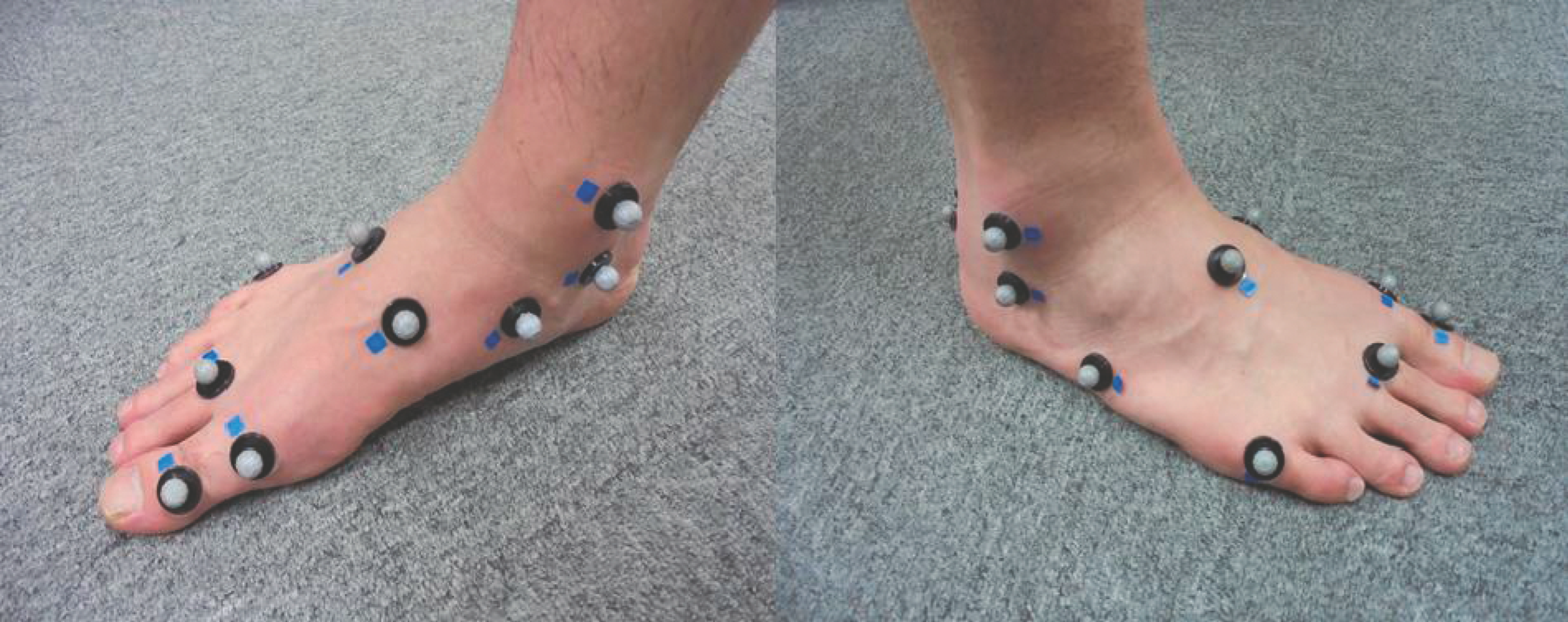
The reflective markers (9 mm in diameter) were fixed to the right shank and foot at the tibial tuberosity, fibula head, medial malleolus, lateral malleolus, Achilles tendon attachment, sustentaculum tali, peroneal tubercle, navicular bone, cuboid, first metatarsal base, first metatarsal head, second metatarsal base, second metatarsal head, fifth metatarsal head, and head of the proximal phalanx of the hallux (Fig. 1). The marker attachment was based on the 3D Foot model.[3] The reflective markers were measured using a three-dimensional motion analysis system (Vicon Motion Systems Ltd, Oxford, England) that included 13 infrared cameras with a sampling rate of 100 Hz.
Figure 1.
Anteromedial (left) and anterolateral (right) views of the marker set.
Before data acquisition, anthropometric data were measured from participants while static standing in the anatomical position. The men performed walking and running barefoot on a treadmill (Autorunner AR-100; Minato Medical Science Co Ltd, Osaka, Japan). The treadmill was set to normal walking speed (1.3 ms–1) or normal slow running speed (2.5 ms–1) according to a previous study.[10] The participants began with at least 1 min of walking or running to accustom themselves to the treadmill speeds before collection of data. After the examiner confirmed that the participants were accustomed to the motion by oral cueing, each task was performed. The speeds of walking and running are permuted randomly, and a short period of rest was given between trials. Data collection was performed for two trials of walking and running, and ten strides were measured for each trial. The analysis was performed during the stance phase from right heel contact to ipsilateral toe-off.
Data Analysis
Captured raw marker trajectory data in the static standing stance and tasks were filtered using a second-order zero-lag Butterworth low-pass filter with a 6-Hz cutoff frequency. The three-dimensional joint angles of the rearfoot, midfoot, and forefoot were calculated from filtered marker trajectories using the 3D Foot model.[3] The proposed 3D Foot model has equivalent repeatability with the Oxford model.[14] Moreover, repeatability using this model has been confirmed in foot deformities[15] and adult patients.[16] The shank coordinate system was defined as described by Cappozzo et al.[17]
The three-dimensional joint angles were calculated using cardan angles, with the distal segment expressed relative to the adjacent proximal segment. Hence, the joint angles were calculated as plantarflexion/dorsiflexion (PF/DF), eversion/inversion (EV/IN), and abduction/adduction (AB/AD) of the rearfoot with respect to the shank, of the midfoot with respect to the rearfoot, and of the forefoot with respect to the midfoot. After calculation of the joint angles in each task, the data were time normalized to the stance phase (100 data points) in each trial. These data were ensemble averaged for each participant.
The present study used cross-correlation coefficients[18] to evaluate kinematic coupling between any two joint angles in the stance phase. The assessment of kinematic coupling was as follows: rearfoot in the frontal plane and midfoot in the sagittal plane, rearfoot in the frontal plane and midfoot in the frontal plane, rearfoot in the frontal plane and midfoot in the transverse plane, midfoot in the frontal plane and forefoot in the sagittal plane, midfoot in the frontal plane and forefoot in the frontal plane, and midfoot in the frontal plane and forefoot in the transverse plane. Because the frontal plane motions of the segments may be associated with the occurrence of running injuries,[19] the present study focused on distal segment rotations with respect to the frontal plane motion of the proximal segment and were in accordance with the study by Pohl et al.[10] An absolute value of cross-correlation coefficient between 0.7 and 1.0 demonstrates strong coupling, between 0.3 and 0.69 demonstrates moderate coupling, and between 0 and 0.29 suggests weak or no coupling.[10] Herein, the kinematic coupling between two segments is expressed as follows: eg, the kinematic coupling between rearfoot EV/IN and midfoot DF/PF means rearfoot EV coupled with midfoot DF and rearfoot IN coupled with midfoot PF.
For the cross-correlation part of this study, the distal angular displacement was shifted with respect to the proximal angular displacement up to ±20% of the stance phase of the gait cycle, and the highest cross-correlation coefficient was extracted. This peak coefficient value, phase-shift at the time point, and time to peak joint angles were calculated for each participant. The time to peak joint angles was calculated as the index of phase-shift. The phase-shift is represented as a percentage of the stance phase. A positive phase-shift represents kinematic coupling from the distal to proximal segment. A negative phase-shift represents kinematic coupling from the proximal to distal segment.
However, a limitation of cross-correlation technique is that it provides only an indication of the temporal similarity between two curves. Thus, to determine whether the coupling values obtained were from joint rotations of similar angular excursion magnitudes or whether one rotation underwent a greater excursion than the other, the coupling angles (θ) were determined by the vector coding technique.[10]
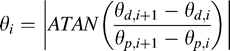 where i represents 1, 2, ... n; θp and θd, the proximal and distal joint angles, respectively; and n, data points. The coupling angles were averaged over the entire stance phase as described by Pohl et al.[10] A coupling angle of 45° indicates that each joint rotation underwent equal amounts of angular displacement. An angle greater than 45° indicates greater movement at the distal relative to the proximal joint. In addition, the peak joint angle excursion value was calculated for each participant to discuss this coupling angle and was defined as the difference between the maximum value and the value at touchdown.[10] Finally, all of the variables were ensemble averaged for all of the participants. The data analysis was conducted using Scilab Release 5.5.0 software (Scilab Enterprises, Inria, Versailles, France).
Statistical Analysis
A Shapiro-Wilk test was used to evaluate the normal distribution of joint angle excursion, time to peak joint angles, and coupling angles in each task. The Wilcoxon signed rank test was used for the following variables: time to peak rearfoot EV, forefoot DF, forefoot EV, forefoot AD, and midfoot EV excursion. The paired t test was used for variables including other data. Statistical significance was set at 5%. The statistical analysis was performed using R Version 3.0.0 (The R Foundation).
Results
Angular Displacements of Foot Segments and Kinematic Variables
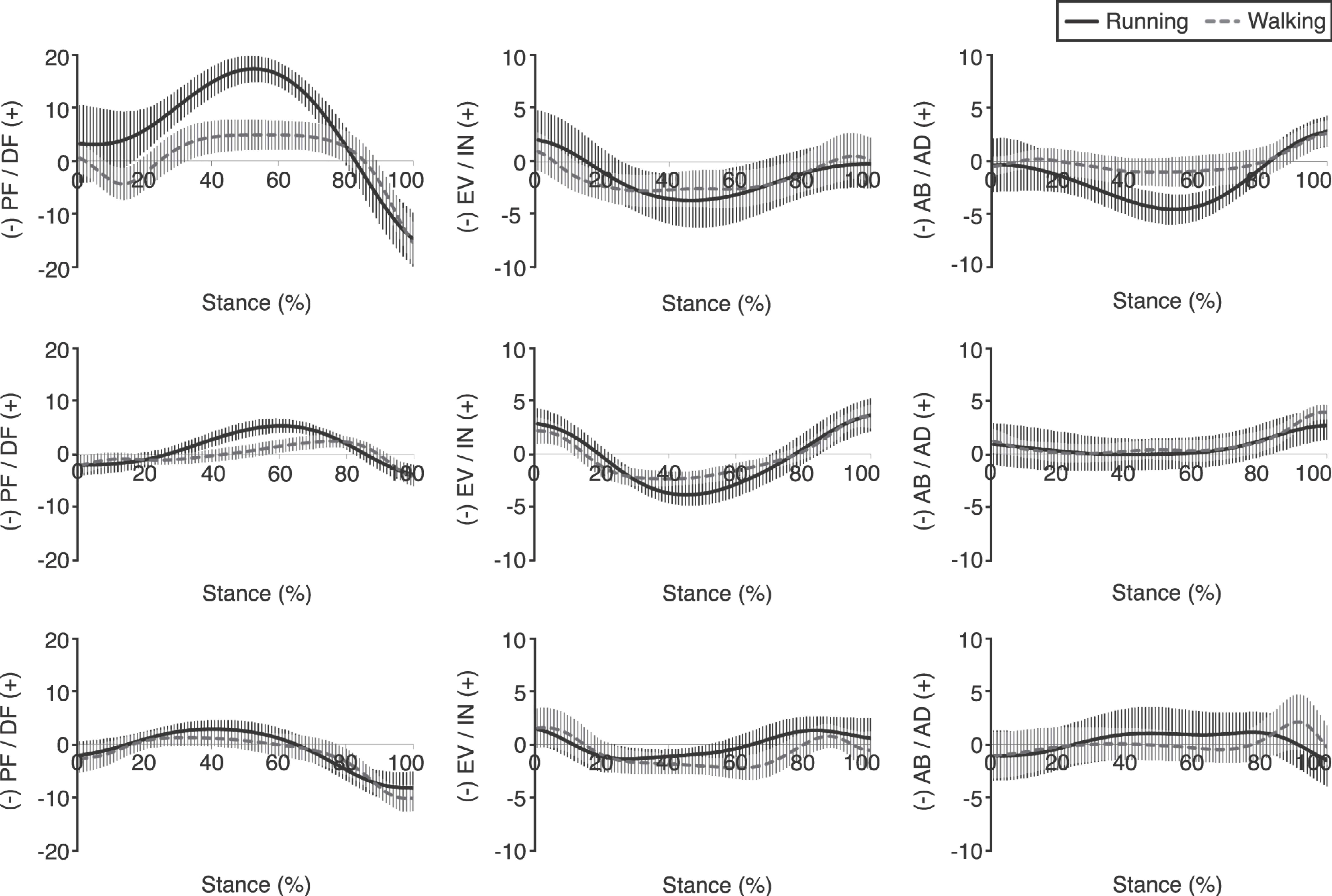
The angular displacements of the rearfoot, midfoot, and forefoot are shown in Figure 2. In the rearfoot, DF, EV, and AB excursion during running (14.1°, −5.8°, and −4.2°, respectively) were significantly higher than during walking (4.8°, −4.1°, and −0.9°, respectively; P < .05). Time to peak rearfoot DF, EV, and AB did not change between running (52.7%, 52.1%, and 55.1%, respectively) and walking (53.8%, 37.1%, and 46.5%, respectively). Although midfoot DF excursion values during running (1.1°) and walking (1.5°) were not statistically significantly different, time to peak midfoot DF during running (60.5%) occurred significantly earlier than during walking (75.7%; P < .05). Midfoot EV excursion and time to peak midfoot EV during running (−6.8° and 45.3%) were significantly higher and slower, respectively, than during walking (−4.7° and 40.7%; P < .05). Although time to peak forefoot EV was not significantly different during running (47.0%) and walking (61.6%), forefoot EV excursion during running (−3.3°) was significantly lower than during walking (−4.2°; P < .05).
Figure 2.
The joint angles of the rearfoot, midfoot, and forefoot during running and walking. The first, second, and third columns show plantarflexion/dorsiflexion (PF/DF), eversion/inversion (EV/IN), and abduction/adduction (AB/AD), respectively. The vertical bars show the SD.
Kinematic Coupling Between Adjacent Foot Segments
The cross-correlation coefficients for the rearfoot, midfoot, and forefoot are shown in Table 1. Kinematic coupling between rearfoot EV/IN and midfoot DF/PF was strong during running (r = 0.79) and moderate during walking (r = 0.58). Although this kinematic coupling occurred from rearfoot to midfoot during running and walking, the time lag of the kinematic coupling was shorter during running than during walking (−6.5% and −10.4% of stance, respectively).
Table 1.
Cross-Correlation Coefficient and Phase-Shift Between Rearfoot Eversion/Inversion (EV/IN) and Midfoot Motion in Each Plane and Between Midfoot EV/IN and Forefoot Motion in Each Plane
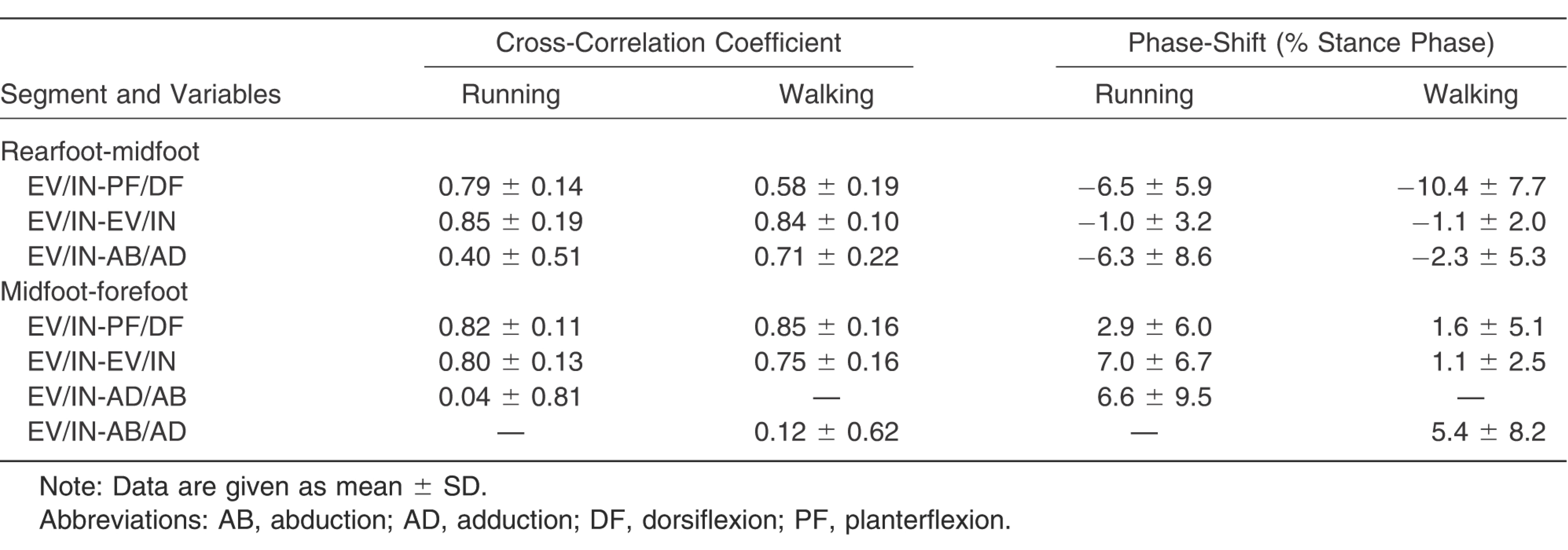
During running and walking, strong coupling was observed between rearfoot EV/IN and midfoot EV/IN (r = 0.85 and 0.84, respectively), between midfoot EV/IN and forefoot PF/DF (r = 0.82 and 0.85, respectively), and between midfoot EV/IN and forefoot EV/IN (r = 0.80 and 0.75, respectively). Although strength of kinematic coupling between rearfoot EV/IN and midfoot AB/AD differed during running and walking, the SD of the cross-correlation coefficient was high (0.51 and 0.22, respectively). Similarly, the SD of the cross-correlation coefficient for kinematic coupling between midfoot EV/IN and forefoot AD/AB during running and between midfoot EV/IN and forefoot AB/AD during walking was high (0.81 and 0.62, respectively).
The Coupling Angle Between Adjacent Foot Segments
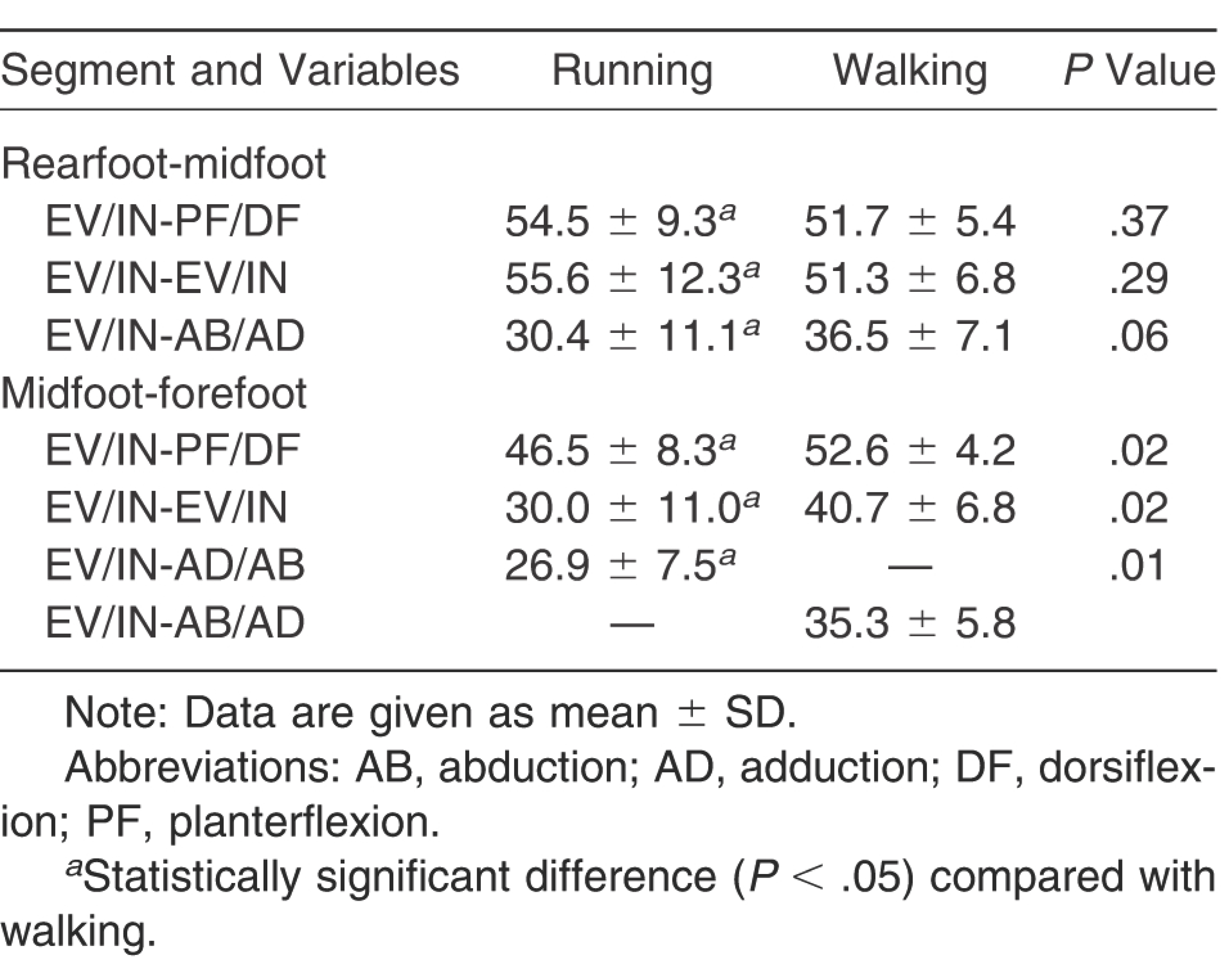
The coupling angles of the rearfoot, midfoot, and forefoot are presented in Table 2. The coupling angle between midfoot EV/IN and forefoot EV/IN and between midfoot EV/IN and forefoot AD/AB during running (30.0° and 26.9°, respectively) suggested that the magnitude of midfoot frontal plane excursion exceeded that of the forefoot frontal and transverse plane excursions, and coupling angles during running were significantly different than those during walking (40.7° and 35.3°, respectively; P < .05). Although the coupling angle between midfoot EV/IN and forefoot DF/PF during running (46.5°) indicated similar excursion magnitudes in two motions, the coupling angle during walking (52.6°) demonstrated a forefoot DF/PF excursion magnitude larger than that during midfoot EV/IN rotation, which was significantly different during running and walking (P < .05).
Table 2.
Coupling Angles Between Rearfoot Eversion/Inversion (EV/IN) and Midfoot Motion in Each Plane and Between Midfoot EV/IN and Forefoot Motion in Each Plane
Discussion
The present study is the first to quantify kinematic coupling including the midfoot segment. It was shown that kinematic coupling between rearfoot EV/IN and midfoot DF/PF was different during running and walking. During running, after the rearfoot touchdown inverted position, eversion occurs until 52% of the stance phase, with subsequent inversion to the terminal stance phase. This rearfoot motion corresponds with that described in a previous study.[20] Also, after the midfoot touches down during plantarflexion, dorsiflexion occurs until 60% of the stance phase, followed by plantarflexion to the terminal stance phase during running. In view of the time lag, the waveform patterns of rearfoot EV/IN and midfoot PF/DF during running were similar.
During walking, although the rearfoot everts until 37.1% of the stance phase, the everted position is maintained at a constant period, and then again inverts until approximately 70% of the stance phase. Furthermore, after the midfoot touchdown plantarflexion position, dorsiflexion occurs until 75% of the stance phase, with subsequent plantarflexion to the terminal stance phase during running. This kinematic pattern of the rearfoot and midfoot during walking closely matched previously published results.[3] Even if the time lag by using cross-correlation coefficients is considered, the waveform pattern of rearfoot EV/IN and midfoot PF/DF did not correspond with that during the stance phase. Therefore, kinematic coupling during walking was moderate. A previous study[10] reported that kinematic coupling is influenced by the stiffness of the soft tissues around the joint. In addition, midfoot PF/DF related to deformation of the medial longitudinal arch is thought to contribute to shock attenuation and propulsion.[21] Because running demands more shock attenuation and stronger propulsion than walking, it is hypothesized that kinematic coupling between rearfoot EV/IN and midfoot DF/PF becomes stronger during running in the stance phase.
In the present study, kinematic coupling between rearfoot EV/IN and midfoot EV/IN and between midfoot EV/IN and forefoot EV/IN was strong during running and walking. In contrast, previous studies[9,10] have reported weak or absent kinematic coupling between rearfoot EV/IN and forefoot EV/IN during running and walking. However, these studies did not analyze the midfoot or calculate the angles of the forefoot with respect to the rearfoot. The midfoot moves during walking in the stance phase,[3] and motion was confirmed to occur during walking at the navicular and cuboid bone, including the midfoot segment using three-dimensional motion analysis with an intracortical pin.[22,23] The present findings provide deeper insight into kinematic coupling among the rearfoot, midfoot, and forefoot segments.
The coupling angle between midfoot EV/IN and forefoot EV/IN was statistically significantly lower during running than during walking. The frontal plane motion of the midfoot exceeded that of the forefoot more evidently during running than during walking, indicating that midfoot EV excursion was significantly higher during running than during walking and that forefoot EV excursion was significantly lower during running than during walking. Hence, although kinematic coupling between midfoot EV/IN and forefoot EV/IN was strong under both conditions, midfoot EV/IN during running was greater than forefoot EV/IN during walking.
The SDs of the cross-correlation coefficient, the coupling angle, and the phase-shift showed high values with the following movements: rearfoot EV/IN and midfoot AB/AD during running and walking, midfoot EV/IN and midfoot AD/AB during running, and midfoot EV/IN and midfoot AB/AD during running. These results may indicate that kinematic coupling and phase-shift were subject dependent. Interpretation of this result requires investigation of foot types and kinetic analysis.
Hintermann et al[24] stated that calcaneal eversion resulted in significant internal tibial rotation; however, internal tibial rotation did not induce calcaneal eversion in the in vitro study. The results of the present study suggest that rearfoot EV results in not only internal shank rotation[10] but also midfoot DF during running in the stance phase. Tiberio[12] showed that excessive subtalar joint pronation induces conditions such as patella femoral pain syndrome from running. Thus, excessive eversion of the rearfoot during running is likely to lead to excessive dorsiflexion of the midfoot, and the abnormal kinematic coupling between the rearfoot and midfoot may be associated with mechanisms for the occurrence of injuries for posterior tibialis tendon dysfunction and medial tibial stress syndrome, for which abnormal foot motion is a risk factor. The results of the present study would be useful for prescription of foot orthoses and treatment in the clinical setting.
There are some limitations to the present study. First, kinematic coupling and the coupling angle were analyzed through the stance phase in accordance with previous studies.[9,10,25] However, the role of the foot during running is functionally different at the first half and the latter half of stance.[26] Thus, if evaluated for each phase separately, kinematic coupling and the coupling angle may differ from those in the present results. Second, the present study targeted rearfoot strikers while barefoot. However, Pohl and Buckley[9] reported that the differences in strike patterns (forefoot and toe strike) influenced the joint angle of foot segments, and Eslami et al[27] reported that barefoot running and shod (sandal) running conditions showed different kinematic coupling intersegments. Finally, because the present study did not quantify foot posture by using an objective foot assessment, such as arch index or arch height, the initial foot assessment may be subjective and depend on the examiner. These limitations need to be investigated in the future.
Acknowledgment
Members of the Institute for Human Movement and Medical Sciences for their assistance.
Financial Disclosure
None reported.
Conflict of Interest
None reported.
References
- Cappozzo A.Leo T.Pedotti A.A general computing method for the analysis of human locomotion. J Biomech8: 307, 1975.
- Kadaba MP.Ramakrishnan HK.Wootten ME.et al: Repeatability of kinematic, kinetic, and electromyographic data in normal adult gait. J Orthop Res7: 849, 1989.
- Leardini A.Benedetti MG.Berti L.et al: Rear-foot, mid-foot and fore-foot motion during the stance phase of gait. Gait Posture25: 453, 2007.
- Carson MC.Harrington ME.Thompson N.et al: Kinematic analysis of a multi-segment foot model for research and clinical applications: a repeatability analysis. J Biomech34: 1299, 2001.
- MacWilliams BA.Cowley M.Nicholson DE.Foot kinematics and kinetics during adolescent gait. Gait Posture17: 214, 2003.
- Grivas TB.Vasiliadis ED.Koufopoulos G.et al: Midfoot fractures. Clin Podiatr Med Surg23: 323, 2006.
- Moen MH.Bongers T.Bakker EW.et al: Risk factors and prognostic indicators for medial tibial stress syndrome. Scand J Med Sci Sports22: 34, 2012.
- Huang YC.Wang LY.Wang HC.et al: The relationship between the flexible flatfoot and plantar fasciitis: ultrasonographic evaluation. Chang Gung Med J27: 443, 2004.
- Pohl MB.Buckley JG.Changes in foot and shank coupling due to alterations in foot strike pattern during running. Clin Biomech (Bristol, Avon)23: 334, 2008.
- Pohl MB.Messenger N.Buckley JG.Forefoot, rearfoot and shank coupling: effect of variations in speed and mode of gait. Gait Posture25: 295, 2007.
- Powell DW.Long B.Milner CE.et al: Frontal plane multi-segment foot kinematics in high- and low-arched females during dynamic loading tasks. Hum Mov Sci30: 105, 2011.
- Tiberio D.The effect of excessive subtalar joint pronation on patellofemoral mechanics: a theoretical model. J Orthop Sports Phys Ther9: 160, 1987.
- Nigg BM.Cole GK.Nachbauer W.Effects of arch height of the foot on angular motion of the lower extremities in running. J Biomech26: 909, 1993.
- Mahaffey R.Morrison SC.Drechsler WI.et al: Evaluation of multi-segmental kinematic modelling in the paediatric foot using three concurrent foot models. J Foot Ankle Res6: 43, 2013.
- Deschamps K.Staes F.Bruyninckx H.et al: Repeatability of a 3D multi-segment foot model protocol in presence of foot deformities. Gait Posture36: 635, 2012.
- Caravaggi P.Benedetti MG.Berti L.et al: Repeatability of a multi-segment foot protocol in adult subjects. Gait Posture33: 133, 2011.
- Cappozzo A.Catani F.Croce UD.et al: Position and orientation in space of bones during movement: anatomical frame definition and determination. Clin Biomech (Bristol, Avon)10: 171, 1995.
- Li L.Caldwell GE.Coefficient of cross correlation and the time domain correspondence. J Electromyogr Kinesiol9: 385, 1999.
- Boling MC.Padua DA.Marshall SW.et al: A prospective investigation of biomechanical risk factors for patellofemoral pain syndrome: the Joint Undertaking to Monitor and Prevent ACL Injury (JUMP-ACL) cohort. Am J Sports Med37: 2108, 2009.
- McClay I.Manal K.A comparison of three-dimensional lower extremity kinematics during running between excessive pronators and normals. Clin Biomech (Bristol, Avon)13: 195, 1998.
- Barnes A.Wheat J.Milner C.Association between foot type and tibial stress injuries: a systematic review. Br J Sports Med42: 93, 2008.
- Lundgren P.Nester C.Liu A.et al: Invasive in vivo measurement of rear-, mid- and forefoot motion during walking. Gait Posture28: 93, 2008.
- Nester CJ.Liu AM.Ward E.et al. In vitro study of foot kinematics using a dynamic walking cadaver model. J Biomech40: 1927, 2007.
- Hintermann B.Nigg BM.Sommer C.et al: Transfer of movement between calcaneus and tibia in vitro. Clin Biomech (Bristol, Avon)9: 349, 1994.
- Pohl MB.Messenger N.Buckley JG.Changes in foot and lower limb coupling due to systematic variations in step width. Clin Biomech (Bristol, Avon)21: 175, 2006.
- Dugan SA.Bhat KP.Biomechanics and analysis of running gait. Phys Med Rehabil Clin N Am16: 603, 2005.
- Eslami M.Begon M.Farahpour N.et al: Forefoot-rearfoot coupling patterns and tibial internal rotation during stance phase of barefoot versus shod running. Clin Biomech (Bristol, Avon)22: 74, 2007.
© 2018 American Podiatric Medical Association