


Abstract
Background: This study investigated the resistance of bacteria isolated from diabetic foot ulcers (DFUs) to antibiotics frequently used in the management of the diabetic foot infections, at a range of pH values (pH 6.5, 7.5, and 8.5) known to exist in DFU wound fluid. This study aimed to determine whether changes (or atypical stasis) in wound fluid pH modulate the antibiotic resistance of DFU isolates, with potential implications in relation to the suppression/eradication of bacterial infections in DFUs. Methods: Thirty bacterial isolates were recovered from DFU wound fluid, including Staphylococcus spp, Staphylococcus aureus, Escherichia coli, Streptococcus spp, Pseudomonas spp, and Pseudomonas aeruginosa. The resistances of these isolates to a panel of antibiotics currently used in the treatment of infected or potentially infected DFUs, ie, ciprofloxacin, amoxicillin-clavulanate, doxycycline, and piperacillin-tazobactam, at the previously mentioned pH values were determined by a modification of the Kirby-Bauer assay. Results: The resistance of DFU isolates to clinically relevant antibiotics was significantly affected by the pH levels in DFU wound fluid. Conclusions: These findings highlight the importance of a more comprehensive understanding of the conditions in DFUs to inform clinical decision making in the selection and application of antibiotics in treating these difficult-to-heal wounds. The scale of the differences in the efficacies of antibiotics at the different pH values examined is likely to be sufficient to suggest reconsideration of the antibiotics of choice in the treatment of DFU infection.
Antibiotics are widely used to kill harmful bacteria at the local and systemic levels. Such extensive use of antibiotics, and their selective effect on bacterial resistance to antibiotics, means that many important pathogens are becoming increasingly resistant to antibiotics, which is a major public health concern.[1,2,3] This situation has relevant implications for the effective treatment of DFUs because unresolved infection in these wounds can often lead to major amputation.[4] Infection in DFUs is different from infection in acute wounds. Acute wounds display a range of signs and symptoms, including the presence of friable granulation tissue with a tendency to bleed, inflammation, redness, heat, unexpected pain, or increased exudate. However, the identification and diagnosis of infection in DFUs is more difficult because, due to neuropathy, these patients lack pain/sensation and have a diminished response to infection and a reduced inflammatory response. Thus, inflammation may be observed only when infection spreads to bone or more widely throughout the body.[5,6,7] The elimination of infection is well known to be a complex process and an integral factor in the healing of DFUs. Thus, when a DFU is classified as infected, immediate topical or systemic antibiotic therapy is recommended,[8] despite the absence of adequate evidence of the efficacy of antibiotic therapy in the treatment and management of DFUs.
pH is believed to affect the efficacy of antibiotics, although the exact mechanism(s) of such inhibition is not yet fully understood.[9] A variety of studies have related pH and antibiotic efficacy. For example, several studies have suggested that real-time monitoring of wound pH has the potential to detect wound infection.[10,11] Percival et al[12] reported that lowering the pH from 8.5 to 5.5 enhanced the bactericidal activity of silver dressings. However, less is known about the efficacy of antibiotics under the atypical conditions that exist in DFUs. Currently, suspected infections in DFUs are treated with oral or intravenous antibiotics, but no consideration is given to the pH of the DFU wound environment in which they are intended to act, probably because wound pH measurement is not part of routine wound assessment.
Standard laboratory procedures for establishing antibiotic efficacy are performed using media at or near pH 7,[13] despite the fact that pH values in chronic ulcers (including DFUs) are known to be very different from those in acute wounds.[2,12,14] Such differences, and the much longer-term persistence of these conditions in chronic wounds, increase uncertainties in relation to the efficacy of antibiotics in these often infected and frequently difficult-to-resolve wounds. Such effects may contribute to the frequently reported difficulties in eradicating infections from DFUs.[7,15]
Clinically Relevant Antibiotics and DFU Bacteria
Potentially infecting bacteria commonly found on the surface of DFUs include gram-positive (Staphylococcus spp and Streptococcus spp) and gram-negative (Escherichia coli and Pseudomonas spp) bacteria. Pseudomonas aeruginosa is of particular increasing concern owing to its ability to become multidrug resistant.[16] Efforts to eliminate infections involving such pathogens include oral or intravenous antibiotic therapies using a range of agents, as discussed by Peters et al.[17] Commonly prescribed antibiotics used to treat suspected infected DFUs include amoxicillin-clavulanate, doxycycline, and ciprofloxacin.[18] When Pseudomonas spp, including P aeruginosa, infection is suspected, oral ciprofloxacin or intravenous piperacillin-tazobactam is prescribed.
This study explored the susceptibility of clinically isolated DFU bacteria to antibiotics frequently used in podiatric medical practice at a range of acid and alkaline pH values (pH 6.5, 7.5, and 8.5). In doing so, it aimed to provide a greater understanding of the effects (if any) of such pH conditions on the efficacy of antibiotics routinely used in the management of DFU infection.
Methods
A representative sample of 30 clinically isolated DFU bacteria was used in this study. The microbiological isolates examined in this study were recovered during a collaborative research project between Ulster University (Newtownabbey, County Antrim, Ireland) and the High Risk Foot Clinic at Belfast City Hospital (Belfast, Ireland). These clinical DFU isolates, including Staphylococcus aureus, other Staphylococcus spp, Escherichia coli, Streptococcus spp, P aeruginosa, and other Pseudomonas spp, were recovered, characterized, and studied in the Ulster University microbiology laboratories using standard UK clinical microbiological protocols.
Isolates of DFU were cultured in tryptone soya broth (CM0129; Oxoid Ltd, Basingstoke, England) and incubated overnight at 37°C. Lawns of the resultant cultures (1×10⁷ cfu/mL) were spread on the surfaces of simple gel models consisting of Mueller-Hinton agar (MHA) plates adjusted to mean ± SD pH values of 6.5 ± 0.2, 7.5 ± 0.2, or 8.5 ± 0.2. The pH values chosen for this study are in line with pH values known to exist in the fluid of chronic wounds, including DFUs.[11,19,20] pH adjustment was accomplished as follows: briefly, the necessary range of buffered MHA was achieved by preparing and sterilizing MHA and filter sterilizing double-strength phosphate-diphosphate and citric acid pair buffer solutions. These solutions were aseptically mixed to form a range of single-strength MHA plates. The pH of the MHA was confirmed using a pH/millivolt/temperature meter (model 6250; Jenco Instruments Inc, San Diego, California) and a micro-combination pH electrode (model PHR-146; Lazar Research Laboratories Inc, Los Angeles, California).
Antibiotic discs (Oxoid Ltd) (Table 1) were placed on the surface of inoculated MHA plates (in triplicate) using sterile forceps and were incubated for 24 hours. Isolates of S aureus (n = 8), Staphylococcus spp (n = 6), and E coli (n = 4) were exposed to ciprofloxacin, amoxicillin-clavulanate (AM30), and doxycycline (DO30). Streptococcus spp (n = 7) isolates were exposed to AM30 and DO30, and P aeruginosa (n = 2) and Pseudomonas spp (n = 3) isolates were exposed to ciprofloxacin (CIP5) and piperacillin-tazobactam (PIP36).
Table 1.
Antibiotics and Target Bacterial Species/Groups
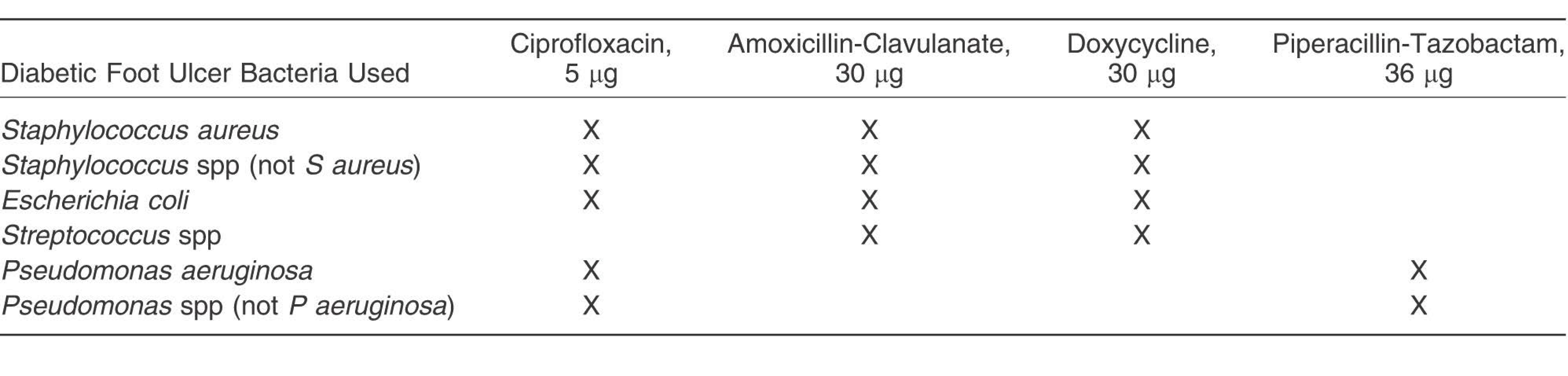
The antibiotics and the concentrations of their use against target bacterial species or groups are presented in Table 1. These agents and their concentrations were in line with Clinical and Laboratory Standards Institute protocols and represent concentrations used in clinical practice.[13] The Kirby-Bauer assay[21] was used to determine the antibiotic resistance profiles of the bacteria at pH 6.5, 7.5, and 8.5. In this assay, the diameter of the clear area surrounding the antibiotic disc after incubation, known as the zone of inhibition (ZOI), was measured twice using a transparent plastic ruler calibrated in millimeters. The mean ZOI was recorded. The edge of the ZOI is where the concentration of antibiotic that has diffused into the agar during incubation is sufficient to inhibit bacterial growth, thus the larger the ZOI the less resistant the bacteria is to the antibiotic in question. Each ZOI around the antibiotic disc was measured after 24-hour incubation at 37°C.[13]
One-way analysis of variance (P ≤ .05) was used to establish whether there were any statistically significant differences in ZOIs among the three pH values (6.5, 7.5, and 8.5) for each set of bacteria and challenge antibiotics. The Student t test was also used to determine whether there were any statistically significant differences in ZOIs between the three groups of pH values, ie, pH 6.5 and 7.5, pH 6.5 and 8.5, or pH 7.5 and 8.5. All of the data were analyzed using Microsoft Excel software, version 2010 (Microsoft Corp, Redmond, Washington), in which P ≤ .05 was reported as being statistically significant.
Results
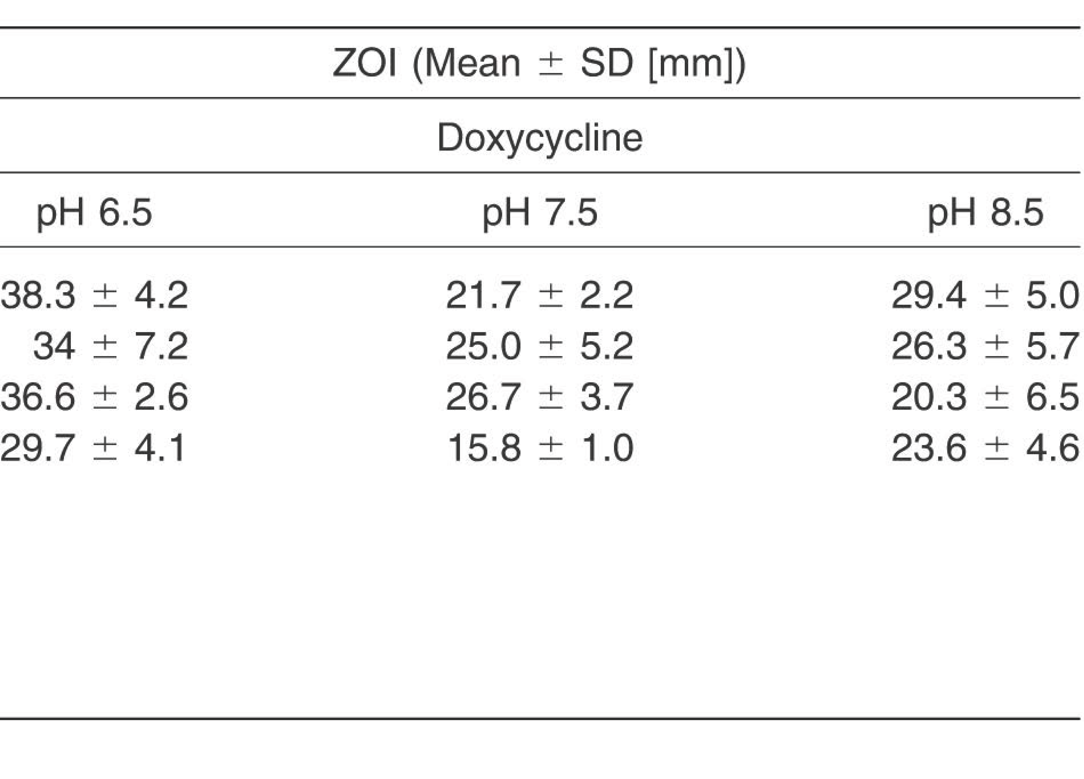
The resultant ZOIs are presented in Table 2.
Table 2.
Mean ZOIs of DFU Bacteria When Exposed to Antibiotics at DFU-Observed pH Values
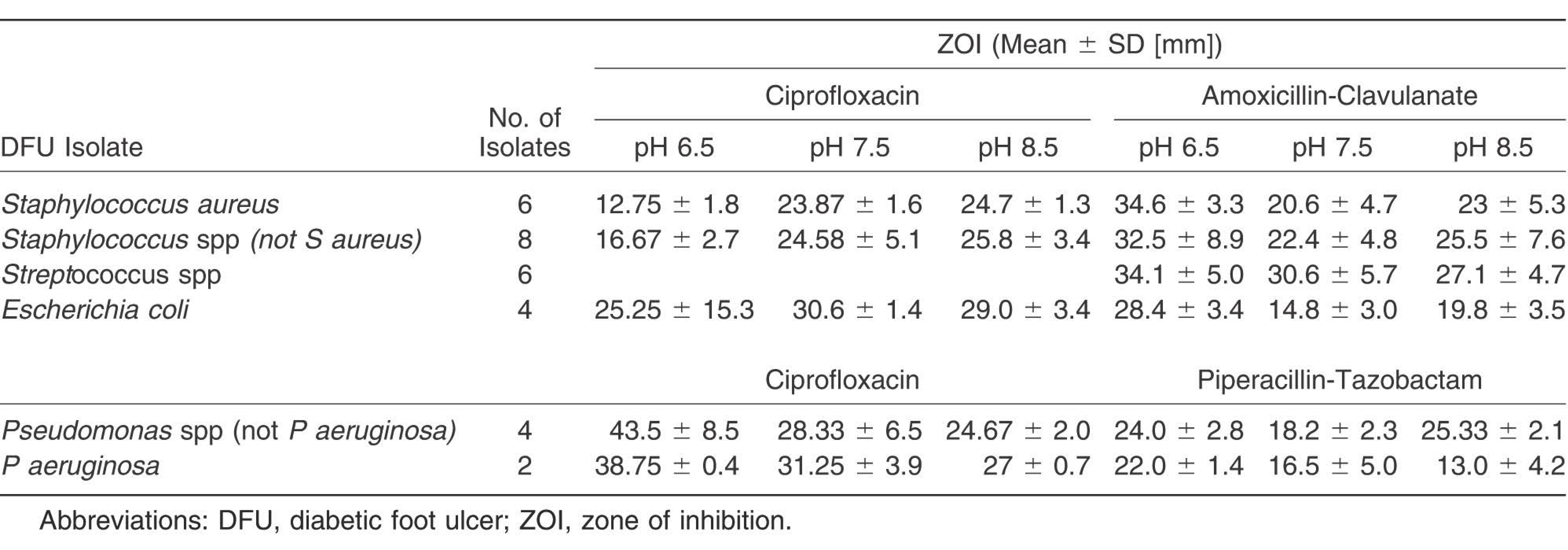
Table 2.
extended
Resistance of Gram-Positive Bacteria
No statistically significant differences were observed among mean ZOIs of Staphylococcus spp, including S aureus, after exposure to CIP5 at pH 7.5 and 8.5 (24.69 and 23.87 mm, respectively). However, the mean ZOIs at these pH values were significantly larger than the CIP5-associated ZOIs at pH 6.5 (12.75 mm; P < .05). All of the Staphylococcus spp exposed to AM30 and DO30 displayed larger ZOIs at pH 6.5 than at pH 7.5 and 8.5 (P < .05) (Table 2). Streptococcus spp displayed similar patterns as Staphylococcus spp, but the only significant differences (P = .004) between staphylococcal and streptococcal ZOIs were observed on DO30 plates.
Resistance of Gram-Negative Bacteria
Some nonsignificant differences (P = .95) were observed among E coli isolates exposed to CIP5 across the examined pH range. This observation may be related to the atypically large standard deviation (15.3 mm) among E coli isolates exposed to CIP5 at pH 6.5. In contrast, the ZOIs observed when E coli was exposed to AM30 or DO30 at pH 6.5 were larger (P = .014 and .007, respectively) than at pH 7.5 and 8.5. Larger ZOIs were observed among Pseudomonas spp (not including P aeruginosa) isolates in pH 6.5 CIP5 plates than in pH 7.5 or 8.5 plates. Significant differences were observed between Pseudomonas. spp (not including P aeruginosa) isolates exposed to PIP36 at pH 6.5 and 7.5 (P = .023) and between these isolates exposed to PIP36 at pH 7.5 and 8.5 (P = .007). Because only two DFU P aeruginosa isolates were examined, it was not possible to perform any analysis of the data; the results, however, displayed an overall trend of larger ZOIs at lower pH values.
Discussion
The study noted that the resistance of DFU isolates to clinically relevant antibiotics was affected by the pH levels in DFU wound fluid. A variety of isolates in this study displayed increased resistance at a lower pH (6.5); for example, all Staphylococcus and E coli isolates recovered were more resistant to CIP5 at pH 6.5 than at pH 7.5 and 8.5. Such greater resistance to CIP5 under acidic conditions concurs with findings from Emrich et al,[22] who observed bacteria to have greater resistance to CIP5 at pH 5.8 than at pH 7.2. It is important to note that the panels of bacteria examined in the study by Emrich et al[22] were isolated from sources other than DFUs.
In contrast, several gram-negative and gram-positive isolates displayed less resistance to AM30, DO30, and PIP36 at pH 6.5. This is in agreement with findings from Falagas et al,[23] who demonstrated that clinical isolates of Bacteroides fragilis are less resistant under acidic conditions. It is also in agreement with a more recent study[24] that investigated the effects of pH on antibiotic efficacy against a range of uropathogens (E coli, Klebsiella pneumonia, Proteus mirabilis, Enterococcus faecalis, and Staphylococcus spp). These authors[24] noted that some of the uropathogens were less resistant under acidic conditions. They did, however, also note some antibiotic/bacterial interactions that were largely unaffected by pH. For example, pH had little effect on uropathogen resistance to amoxicillin-clavulanate, which differs from the findings of the present study. Comparison of these studies suggests that there may be differences between the pH responses of some uropathogens and the pH responses of DFU-sourced isolates.
Some of the results obtained in this study suggest greater diversity in the group of E coli isolates from DFUs. For example, the observed range of resistances (as demonstrated by the much larger standard deviation value calculated) when the isolates were subjected to CIP5 at pH 6.5 was much larger than in any other bacterial/antibiotic combination. Such diversity may signal the presence of a wide range of E coli biotypes contaminating/colonizing the sampled DFUs. Irrespective of the sources of such organisms, their presence in DFUs is a matter of concern because the increasing incidence of undesirable multidrug resistance characteristics, such as the expression of extended β-lactamase activity, in this group of organisms poses substantial challenges in relation to infections in DFUs.[25]
Potential Clinical Implications and Further Work
It is well known that DFU infections delay wound healing,[26,27,28] which means that patients with DFUs may receive long-term antibiotic therapy aiming to limit or resolve their wound infections. However, such longer-term antibiotic therapy may present increased opportunities for the emergence and persistence of antibiotic resistance among DFU wound-colonizing or wound-infecting bacteria.[29] The present study is also in agreement with earlier findings[9,30] that recognized the increasingly urgent need for new strategies to improve antibiotic efficacy in an effort to effectively resolve infection in recalcitrant wounds.
Previous research[11,14,20,31,32,33] has highlighted the potential effects of pH on wound condition and healing/nonhealing status. Alkaline conditions seem to favor the growth and survival of most DFU isolates. It could, therefore, be the case that reductions in pH may impede bacterial survival and growth, with the same overall outcome as an increase in antibiotic efficacy. Nevertheless, the findings from this study suggest that in “alkaline”-infected wounds,[9,24] a shift to weakly acidic conditions might impede the growth of the bacteria or change their antibiotic resistance profile. Such shifts could substantially influence the competitive balance between attacking bacteria and wound defense systems in wounds, with major positive implications in terms of the management and eradication of infection.
In some cases in this study, the differences among the ZOIs seem to be sufficient to change the agreed resistance/sensitivity classification of some of the examined antibiotics, a classification that is of considerable significance in their clinical selection and application in treating infection. For example, comparisons of the mean ZOIs of S aureus DFU isolates in the presence of CIP5 at the examined pH values with the Zone Diameter Interpretive Standard for this species[13] classifies these organisms as sensitive at pH 7.5 and 8.5 but resistant at pH 6.5. A similar comparison of ZOIs obtained for P aeruginosa in the presence of PIP36 at the three pH values with the Zone Diameter Interpretive Standard for this species[13] rates these organisms as sensitive at pH 6.5 but resistant at pH 8.5. Bearing in mind the importance of such classifications in the selection and application of antibiotic therapies, pH-related effects may be worthy of further investigation, particularly in relation to the treatment of infections and suspected infections in the management of DFUs and other atypical chronic wounds.
This study examined the DFU isolates in a semiplanktonic state. Future studies could usefully compare planktonic and biofilm-embedded DFU isolates in relation to their pH-modulated resistance(s). Such studies are likely to underpin improvements in clinical outcomes of DFU infections and contribute to progress toward more rapid eradication of infection, acceleration in wound healing, and reductions in the risks of amputation(s) associated with DFU infections.
Conclusions
The environmental conditions achieved using the simple gel model in this in vitro study differ in a variety of respects from the in vivo conditions occurring in DFUs. Nevertheless, the data presented demonstrate that the pH values occurring in DFUs can adversely modulate the efficacy of antibiotics currently being used in the treatment of infected or potentially infected DFUs. These findings highlight the importance of a more comprehensive understanding of the conditions in DFUs to inform clinical decision making in the selection and application of antibiotics in treating these difficult-to-heal wounds.
Acknowledgment
Dr. Sarah Spence, Ulster University, Northern Ireland, and Jill Cundell, Belfast Health and Social Care Trust, Northern Ireland, for their involvement in the research, and Prof. Ian Bradbury for his advice on statistical analysis.
Financial Disclosure
This research was funded by the Department of Employment and Learning, Northern Ireland, as part of a full-time PhD scholarship.
Conflict of Interest
None reported.
References
- Davies J.Davies D.Origins and evolution of antibiotic resistance. Microbiol Mol Biol Rev74: 417, 2010.
- Percival SL.McCarty S.Hunt JA.et al: The effects of pH on wound healing, biofilms and antimicrobial efficacy. Wound Repair Regen22: 174, 2014.
- World Health Organization: pH in drinking-water: revised background document for development of WHO Guidelines for Drinking-water Quality. Available from: http://www.who.int/water_sanitation_health/publications/2011/dwq_guidelines/en/.
- Apelqvist J.Diagnosis and treatment of the diabetic foot. Endocrine41: 384, 2012.
- Lipsky BA.Medical treatment of diabetic foot diseases. Clin Infect Dis39(suppl 2): S104, 2004.
- Gardner SE.Frantz RA.Wound bioburden and infection-related complications in diabetic foot ulcers. Biol Res Nurs10: 44, 2008.
- Cooper RA.Ameen H.Price P.et al: A clinical investigation into the microbiological status of ‘locally infected' leg ulcers. Int Wound J6: 453, 2009.
- Howell-Jones RS.Wilson MJ.Hill KE.et al: A review on the microbiology, antibiotic usage and resistance in chronic skin wounds. J Antimicrob Chemother55: 143, 2005.
- Thomas J.Linton S.Corum L.et al: The effect of pH and bacterial phenotypic state on antibiotic efficacy. Int Wound J9: 428, 2012.
- Dissemond J.Witthoff M.Grabee S.Investigations on pH values in milieus of chronic wounds during modern wound therapy. Paper presented at:World Wound Congress, Paris, 2004.
- Shukla VK.Shukla D.Tiwary SK.et al: Evaluation of pH measurement as a method of wound assessment. J Wound Care16: 291, 2007.
- Percival SL.Thomas J.Linton S.et al: The antimicrobial efficacy of silver on antibiotic-resistance bacteria isolated from burn wounds. Int Wound J9: 488, 2011.
- Clinical and Laboratories Standards Institute (CLSI): Performance Standards for Antimicrobial Susceptibility Testing; Twenty Third Information Supplement: M100-S23, Clinical and Laboratories Standards Institute, Wayne, PA, 2013.
- Schneider LA.Korber A.Grabbe S.et al: Influence of pH on wound-healing: a new perspective for wound-therapy?Arch Dermatol Res298: 413, 2007.
- Pellizzer G.Strazzabosco M.Presi S.et al: Deep tissue biopsy vs. superficial swab culture monitoring in the microbiological assessment of limb-threatening diabetic foot infection. Diabet Med18: 822, 2001.
- Lister PD.Wolter DJ.Hanson ND.Antibacterial-resistant Pseudomonas aeruginosa: complex regulation of chromosomally encoded resistance mechanisms. Clin Microbiol Rev22: 582, 2009.
- Peters EJ.Lipsky BA.Aragon-Sanchez J.et al: Interventions in the management of infection in the foot in diabetes: a systematic review. Diabetes Metab Res Rev32(suppl 1): 145, 2016.
- Leese G.Dilip N.Young M.et al: Use of antibiotics in people with diabetic foot disease: a consensus statement. Diabet Foot J12: 62, 2009.
- McArdle C.Lagan KM.McDowell DA.The pH of wound fluid in diabetic foot ulcers: the way forward in detecting clinical infection?Curr Diabetes Rev10: 177, 2014.
- McArdle C.Lagan KM.Spence S.et al: Diabetic foot ulcer wound fluid: the effects of pH on bacteria and infection. J Foot Ankle Res8(suppl 1): A8, 2015.
- Bauer AW.Kirby WM.Sherris JC.et al: Antibiotic susceptibility by standardized single dink method. Am J Clin Pathol45: 493, 1966.
- Emrich NC.Heisig A.Stubbings W.et al: Antibacterial activity of flinafloxacin under different pH conditions against isogenic strains of Escherichia coli expressing combinations of defined mechanisms of fluoroquinolone resistance. J Antimicrob Chemother65: 2530, 2010.
- Falagas ME.McDermott L.Snydman DR.Effect of pH on in vitro antimicrobial susceptibility of Bacteroides fragilis group. Antimicrob Agents Chemother41: 2047, 1997.
- Yang L.Wang K.Li H.et al: The influence of urinary pH on antibiotic efficacy against bacterial uropathogens. Urology84:731.e1, 2014.
- Bowler PG.Welsby S.Towers V.et al: Multi-resistant organisms, wounds and topical antimicrobial protection. Int Wound J9: 387, 2012.
- Wu S.Driver VR.Wrobel JS.et al: Foot ulcers in the diabetic patient, prevention and treatment. Vasc Health Risk Manag3: 65, 2007.
- Santy J.Recognising infection in wounds. Nurs Stand23: 53, 2008.
- Lipsky BA.Berendt AR.Cornia PB.et al: 2102 Infectious Diseases Society of America clinical practice guideline for the diagnosis and treatment of diabetic foot infections. Clin Infect Dis54: e132, 2012.
- Game F.Jeffcoate W.MRSA and osteomyelitis of the foot in diabetes. Diabet Med21(suppl 4): 16, 2004.
- Gillings MR.Evolutionary consequences of antibiotic use for the resistome, mobilome and microbial pangenome. Front Microbiol4: 4, 2013.
- Greener B.Hughes AA.Bannister, NP.et al: Proteases and pH in chronic wounds. J Wound Care14: 59, 2005.
- Gethin GT.Cowman S.Conroy RM.The impact of Manuka honey dressings on the surface pH of chronic wounds. Int Wound J5: 185, 2008.
- Shi L.Ramsay S.Ermis R.et al: pH in the bacteria-contaminated wound and its impact on Clostridium histolyticum collagenase activity: implications for the use of collagenase wound debridement agents. J Wound Ostomy Continence Nurs38: 514, 2011.
© 2018 American Podiatric Medical Association