


Abstract
This case report concerns a patient with a painful soft-tissue mass on his fourth toe. He was evaluated for this soft-tissue mass and was diagnosed as having tuberous sclerosis. The podiatric physician should be able to evaluate a patient with a lower-extremity complaint and relate whether this complaint may correlate with a systemic disorder. This case report will make the podiatric physician more aware of tuberous sclerosis and the manifestations of the disorder.
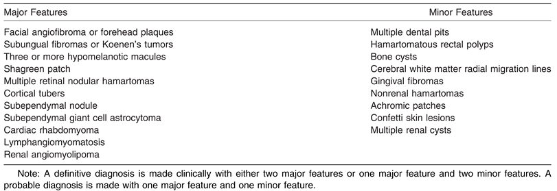
Tuberous sclerosis is a complex syndrome characterized pathologically by the presence of hamartomas in multiple organ systems [1]. There are major and minor diagnostic features to aid in the identification of tuberous sclerosis (Table 1). The most common organ systems affected by tuberous sclerosis are the brain, heart, kidneys, skin, and lungs [2]. Tuberous sclerosis causes disabling neurologic disorders, including epilepsy, autism, and intellectual disabilities. In addition to neurologic disorders, additional major features of the disease include dermatologic manifestations such as facial angiofibromas, ungual and periungual fibromas, renal angiomyolipomas, and pulmonary lymphangiomyomatosis [3]. The clinical spectrum of tuberous sclerosis is very wide, with many patients having minimal signs and symptoms with no neurologic disability.
Table 1.
Major and Minor Diagnostic Criteria of Tuberous Sclerosis Complex.
Tuberous sclerosis complex (TSC) is an autosomal dominant–inherited disease. The disease is caused by the mutation of one of two tumor suppressor genes: TSC1 and TSC2. TSC1 is found on chromosome 16 and encodes for the protein hamartin, and TSC2 is found on chromosome 9 and encodes for the protein tuberin [4]. Hamartin and tuberin are believed to have a role in the growth and differentiation of cells, and both proteins are found throughout the body and interact with each other. The function and interaction of these proteins are not yet fully understood, although they may function as tumor suppressors [2,4]. Most tumors associated with tuberous sclerosis are benign, although malignant neoplasms do occur, with most cases of malignancy occurring in the kidney [5].
Hamartomas in the brain, called cortical tubers, are composed of abnormal glial and neural cells, and the size, number, and location vary among patients [6]. It is thought that the number of tubers, which are present in more than 80% of patients, possibly correlates with the severity of seizures. Renal manifestations of TSC include angiomyolipomas and renal cysts [5]. Cardiac involvement is mainly in the form of rhabdomyomas, which cause important clinical problems in only a very small fraction of those patients. Pulmonary lesions are lymphangioleiomyomatosis and pulmonary cysts and affect women with tuberous sclerosis almost exclusively [3]. Skin lesions include ash-leaf spots, confetti lesions, facial angiofibromas, shagreen patches, fibrous plaques, and periungual fibromas. Additional acral findings include longitudinal nail grooves without visible fibromas, red comets, longitudinal leukonychia, splinter hemorrhages, and, rarely, pachydermodactyly or macrodactyly [7]. Subungual and periungual fibromas are common in adults with tuberous sclerosis and are a major feature for diagnosis. They are among the last skin lesions to appear in tuberous sclerosis, with 88% of patients older than 30 years experiencing these lesions. A study by Aldrich et al [7] has shown that periungual fibromas are more common on the toes than on the fingers. The characteristic histologic changes of these tumors include an epidermis that is acantotic with a thickened horny layer and a stroma that contains capillaries surrounded by collagen fibers [7]. Generally, ungual fibromas in patients with tuberous sclerosis are benign but may bleed and may produce pain and nail distortion [8].
With an incidence of 1 in 6,000 people, tuberous sclerosis currently affects approximately 40,000 people in the United States [7]. Tuberous sclerosis does not seem to have a sex or race predilection. Based on a meta-analysis of 380 patients with tuberous sclerosis, the frequency of TSC1 mutations was found to be twice as high in the American and British populations compared with those from Poland and the Republic of China [9]. The age of diagnosis of tuberous sclerosis occurs anywhere from birth to adulthood due to the wide array of clinical findings.
Case Report
A 63-year-old African America man presented to the clinic with pain in the right fourth toe with an associated soft-tissue mass. The patient stated that he noticed the mass near the nail approximately 2 years earlier. He admitted to the mass enlarging during the past 2 years. The patient had sharp pain in that area that became excruciating with walking. He had tried rubbing an unknown salve on the mass with no success in relief.
The patient’s medical history was significant for recurrent seizures and anemia. He was a former smoker but denied caffeine, alcohol, or illicit drug use. The patient’s surgical and family histories were unremarkable. On physical examination, the patient was further questioned regarding the diagnosis of tuberous sclerosis. He was unaware of that diagnosis but had seen a neurologist for his seizure history.
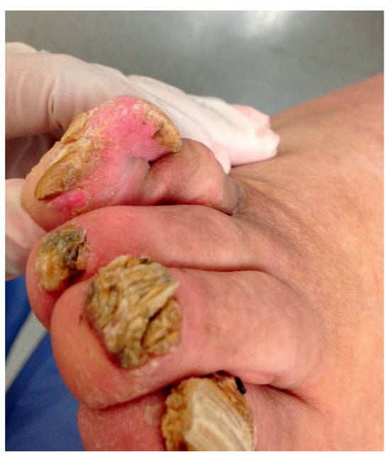
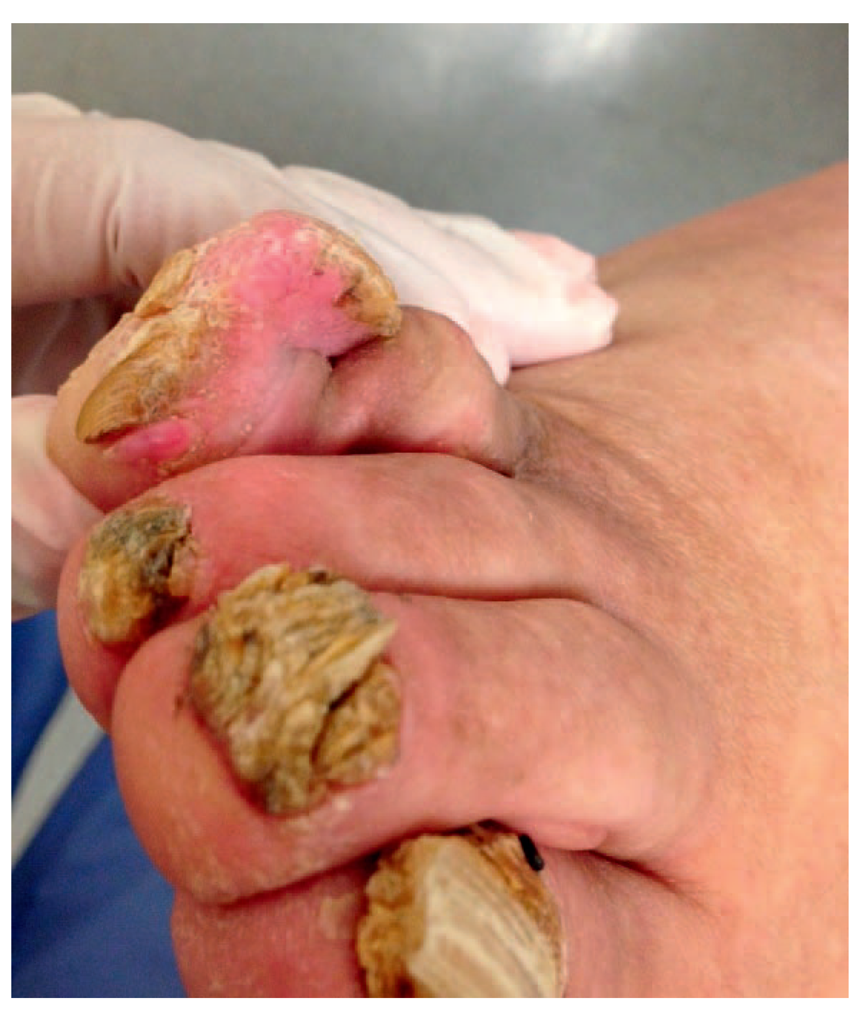
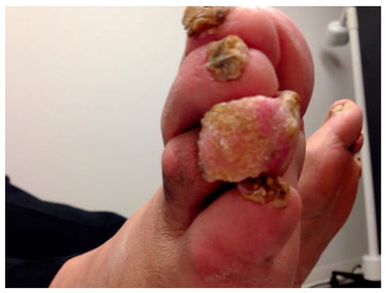
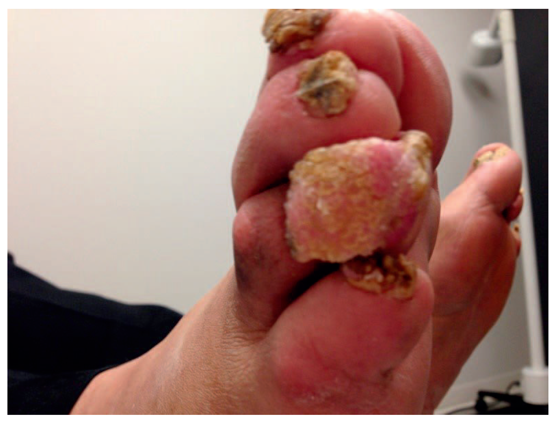
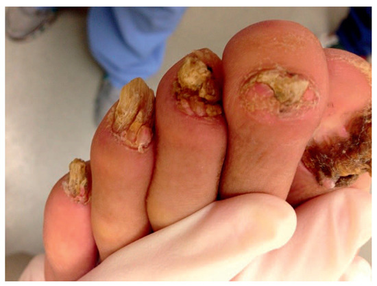
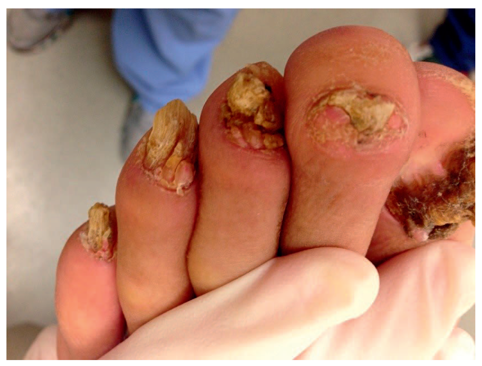
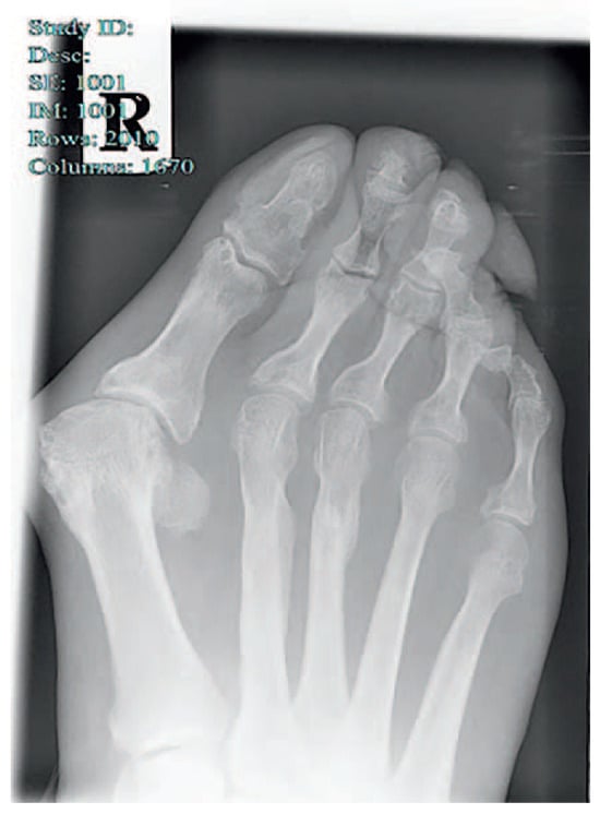
On physical examination, the patient was noted to have an intact neurovascular status. A large softtissue mass measuring 2.0 × 1.5 cm was noted on the distal aspect of the right fourth toe near the proximal nail plate (Figure 1 and Figure 2). Multiple other small lesions were noted growing near or underneath the remaining nails (Figure 3). An open lesion with serous fluid was noted on his right arm, which the patient stated was secondary to a chemical burn during a work incident.
Figure 1.
Large soft-tissue mass to the proximal fourth digit.
Figure 2.
Lateral view of a large mass near the nail plate.
Figure 3.
Contralateral foot demonstrating smaller lesions around the nail plates.
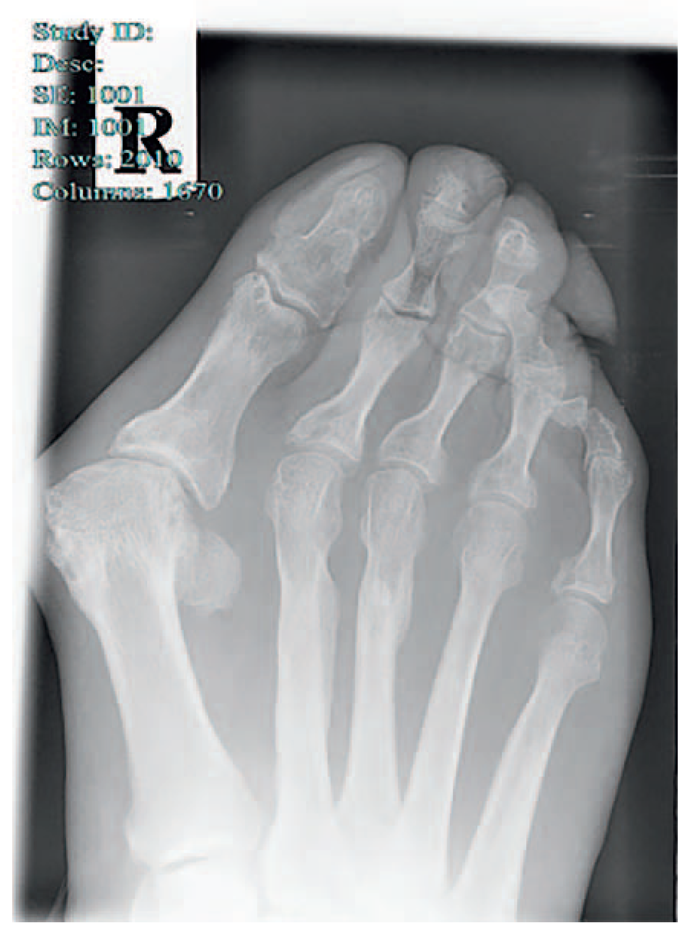
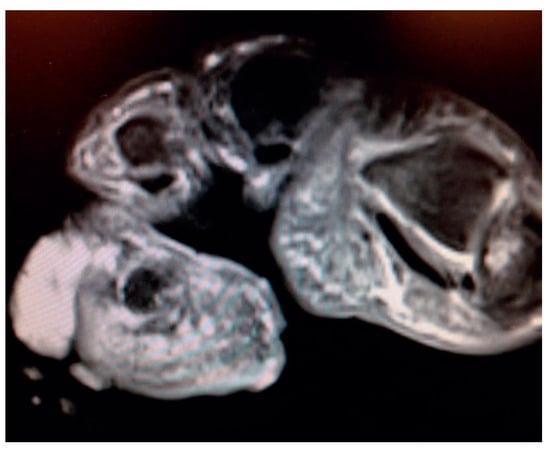
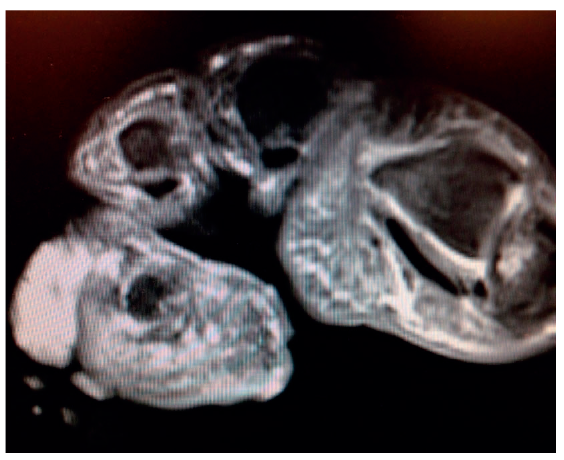
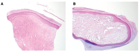
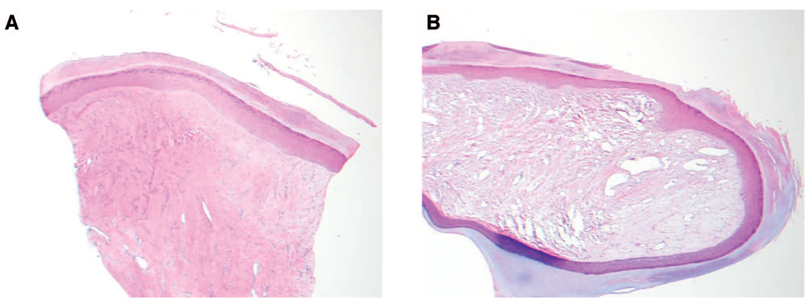
Radiographs were obtained during the patient’s initial visit (Figure 4). The fourth digit distal and middle phalanges are fused. The fused phalanx has an odd, curved appearance but without lysis of the cortex. No osteochondroma/subungual exostosis was noted. A magnetic resonance image (MRI) was obtained that demonstrated a lobulated low T1 and high T2 signal intensity mass infiltrating the skin and subcutaneous tissues around the fourth distal phalanx (Figure 5). A punch biopsy of the lesion was then performed, with the report reading as an acral fibrokeratoma with associated scar formation (Figure 6). On medical record request from his neurologist, the patient did have a diagnosis of tuberous sclerosis, with MRI of his brain indicating cortical tubers. Treatment options, both conservative and surgical, were discussed with the patient. He was seen for a surgical consult with a plan for surgical excision of the lesion but was lost to follow-up.
Figure 4.
Radiograph of the foot.
Figure 5.
Coronal plane T2 magnetic resonance image of the lesion.
Figure 6.
Pathology slides from punch biopsy reading acral fibrokeratoma with associated scar formation.
Discussion
This case report highlights the need for podiatric physicians to be aware of pedal cutaneous manifestation of systemic diseases. The podiatric medical evaluation and examination of the feet and lower extremities may lead to early diagnosis of systemic disease. As podiatric physicians, there is a need to recognize signs and symptoms of systemic disease so that proper referrals can be made to ensure that patients receive optimal work-up and treatment.
In this specific case, the patient presented with several soft-tissue lesions near the toenail plates. The most prominent and painful mass was thoroughly evaluated, including radiography, MRI, biopsy, and a plan for surgical intervention. Evaluation of the mass demonstrated a fibroma-like growth to the patient’s fourth digit. The patient’s history of a seizure disorder, mild learning disabilities, and other cutaneous lesions made us immediately suspect tuberous sclerosis as the cause of the pedal growths. The review of medical records from his neurologist confirmed this diagnosis and that he was being treated for his seizures and tuberous sclerosis diagnosis.
There are many treatment options for these pedal lesions. In the present patient, imaging and biopsies were performed to ascertain that there was no infectious or neoplastic process occurring. In some cases, observation of the lesions may be all that is necessary. With this patient, the lesion was increasing in size and causing painful ambulation, so surgical intervention was deemed necessary. There has also been evidence that excision of the lesions can be performed using a CO2 laser or phenolization. A CO2 laser provides efficient and bloodless excision of the lesion, whereas phenolization allows for a better cosmetic result [10]. Recently, it has been discovered that the use of oral rapamycin may induce the regression of tumors associated with TSC [11,12].
Conclusions
Recognizing pedal and lower-extremity cutaneous lesions as a symptom of a systemic disease is a key role of the podiatric physician. There are many treatment options for managing these cutaneous lesions, but confirming that a patient is receiving proper medical management is vital. For a patient with TSC, it is important to ascertain that the patient has a general internist, neurologist, or any other specialist who may be required depending on which specific organ system is affected. Being able to recognize systemic disease from a cutaneous manifestation will make any podiatric physician more effective in treating the whole patient and not just the lower extremity.
Financial Disclosure
None reported.
Conflicts of Interest
None reported.
References
- O’CALLAGHAN, F.J. Tuberous sclerosis: epidemiological research is needed to complement new findings in genetics. BMJ 1999, 318, 1019. [Google Scholar] [CrossRef] [Scilit] [PubMed]
- JOZWIAK, J.; JOZWIAK, S.; WLODARSKI, P. Possible mechanisms of disease development in tuberous sclerosis. Lancet Oncol 2008, 9, 73. [Google Scholar] [CrossRef] [Scilit] [PubMed]
- CRINO, P.B.; NATHANSON, K.L. PETRI HENSKE E: The tuberous sclerosis complex. N Engl J Med 2006, 355, 1345. [Google Scholar] [CrossRef] [Scilit] [PubMed]
- JOZWIAK, J. WLODARSKI P: Hamartin and tuberin modulate gene transcription via beta-catenin. J Neurooncol 2006, 79, 229. [Google Scholar] [CrossRef] [Scilit] [PubMed]
- AL-SALEEM, T.; WESSNER, L.L.; SCHEITHAUER, B.W. ET AL: Malignant tumors of the kidney, brain, and soft tissues in children and young adults with the tuberous sclerosis complex. Cancer 1998, 83, 2208. [Google Scholar] [CrossRef]
- KRISHNAN, M.L.; COMMOWICK, O.; JESTE, S.S. ET AL: Diffusion features of white matter in tuberous sclerosis with tractography. Pediatr Neurol 2010, 42, 101. [Google Scholar] [PubMed]
- ALDRICH, S.L.; CHIEN-HUI, H.; GROVES, L. ET AL: Acral lesions in tuberous sclerosis complex: insights into pathogenesis. J Am Acad Dermatol 2009, 63, 244. [Google Scholar] [CrossRef] [Scilit] [PubMed]
- MA, D.; DARLING, T.; MOSS, J. ET AL: Histologic variants of periungual fibromas in tuberous sclerosis complex. J Am Acad Dermatol 2011, 64, 442. [Google Scholar] [CrossRef] [Scilit] [PubMed]
- ZWIAK, J.; SONTOWSKA, I.; PLOSKI, R. Frequency of TSC1 and TSC2 mutations in American, British, Polish and Taiwanese populations. Mol Med Rep 2013, 8, 909. [Google Scholar]
- HAKE S: Cutaneous manifestations of tuberous sclerosis. Ochsner J 2010, 10, 200.
- LEE, N.; WOODRUM, C.L.; NOBIL, A.M.; ET, A.L. Rapamycin weekly maintenance dosing and the potential efficacy of combination sorafenib plus rapamycin but not atorvastatin or doxycycline in tuberous sclerosis preclinical models. BMC Pharmacol 2009, 9, 8. [Google Scholar] [CrossRef] [Scilit] [PubMed]
- SAMPSON, J.R. Therapeutic targeting of mTOR in tuberous sclerosis. Biochem Soc Trans 2009, 37, 259. [Google Scholar] [CrossRef] [Scilit] [PubMed]

© 2017 American Podiatric Medical Association