


Abstract
Cutaneous horns (cornu cutaneum) are chronic, dense, hyperkeratotic cutaneous lesions resembling the horn of an animal. These lesions are associated with a variety of benign, premalignant, and malignant cutaneous diseases. Cutaneous horns are often found on the upper parts of the body, such as the face, neck, and shoulders. These lesions rarely occur in areas with no sun exposure, such as the feet. We present the case of a 51-year-old man with two cutaneous horns on the lateral aspect of the third digit of the left foot. Treatment consisted of excision of the lesions and application of a full-thickness skin graft from the ipsilateral sinus tarsi.
Cutaneous horns usually present as elongated, keratinous, white or yellowish projections that are variable in size. These lesions are rare and most commonly occur in fair-skinned patients 50 years or older. The condition is thought to be more prevalent in men, but no consistent sex pattern has been demonstrated.[1]
Cutaneous horns generally appear on sun-exposed areas, such as the face, scalp, ears, chest, neck, and shoulders, but can also be found on non–sun-exposed areas, such as the lower lip, nasal vestibule, and lower extremity, on a smaller scale.[2,3] Some studies have shown that benign lesions are most frequently evident in areas less exposed to the sun, such as the lower limbs. On the other hand, areas more exposed to the sun have higher chances of presenting premalignant or malignant lesions compared with other parts of the body.[3]
The underlying condition may be benign, such as seborrheic keratosis, viral warts, histiocytoma, inverted follicular keratosis, and verrucous epidermal nevi; premalignant, such as solar keratosis, arsenical keratosis, and Bowen's disease; or malignant, such as squamous cell carcinoma, rarely, basal cell carcinoma, and Kaposi's sarcoma.[4,5] An extensive study of 643 cutaneous horns by Yu et al[6] concluded that 39% of cutaneous horns were derived from malignant or premalignant epidermal lesions and 61% from benign lesions. In general, malignant lesions seem to be harder at the base due to an inflammatory process.[3]
In 1941, Montgomery [7] classified cutaneous horns based on apparent causation, appearance, and histologic features: 1) cutaneous horn springing from an epidermoid cyst, 2) horn of the mucosa developing from mucous membrane, 3) verrucous horn resulting from a wart, 4) papillomatous horn developing from cornified epithelium, and 5) filliform horn arising from normal or hyperkeratotic skin.
Detachment, cauterization of the papillary base, and cryosurgery are among the most common treatment options for removal of these cutaneous lesions. Excisional biopsy is favored over the others because it gives us enough specimens for dermatopathologic evaluation.[2] In the case of malignant lesions at the base, excision with clean margins is necessary. These cases should be evaluated for metastasis as well.[8]
Case Report
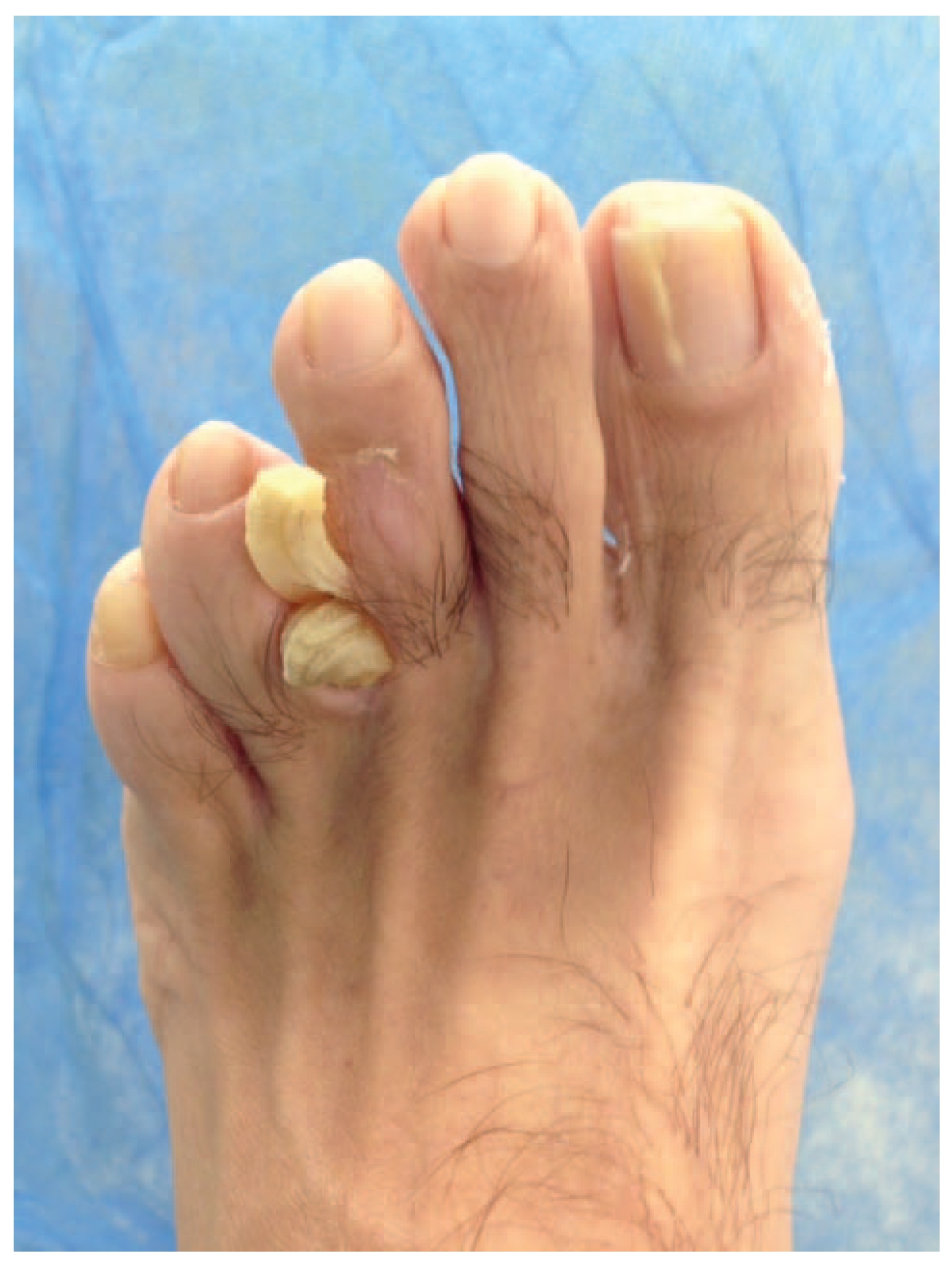
A 51-year-old male patient with a medical history of depression and colon polyps presented with two hyperkeratotic, hornlike raised lesions on the lateral aspect of the left third digit extending to the third interspace accompanied by maceration of the interspace on the left foot (Fig. 1). Each lesion measured 0.6 × 1.0 × 0.6 cm. Despite surgical resection, the lesions recurred within 2 years with the same presentation and location. The patient noted pain on ambulation, irritation to the fourth toe, and pain with all types of shoes. He stated that he had the lesions surgically removed in 2012; however, they grew back in the past 2 years. Therefore, he requested a subsequent surgical excision of the lesions.
Figure 1.
Two cutaneous horns at the lateral aspect of the left third digit.
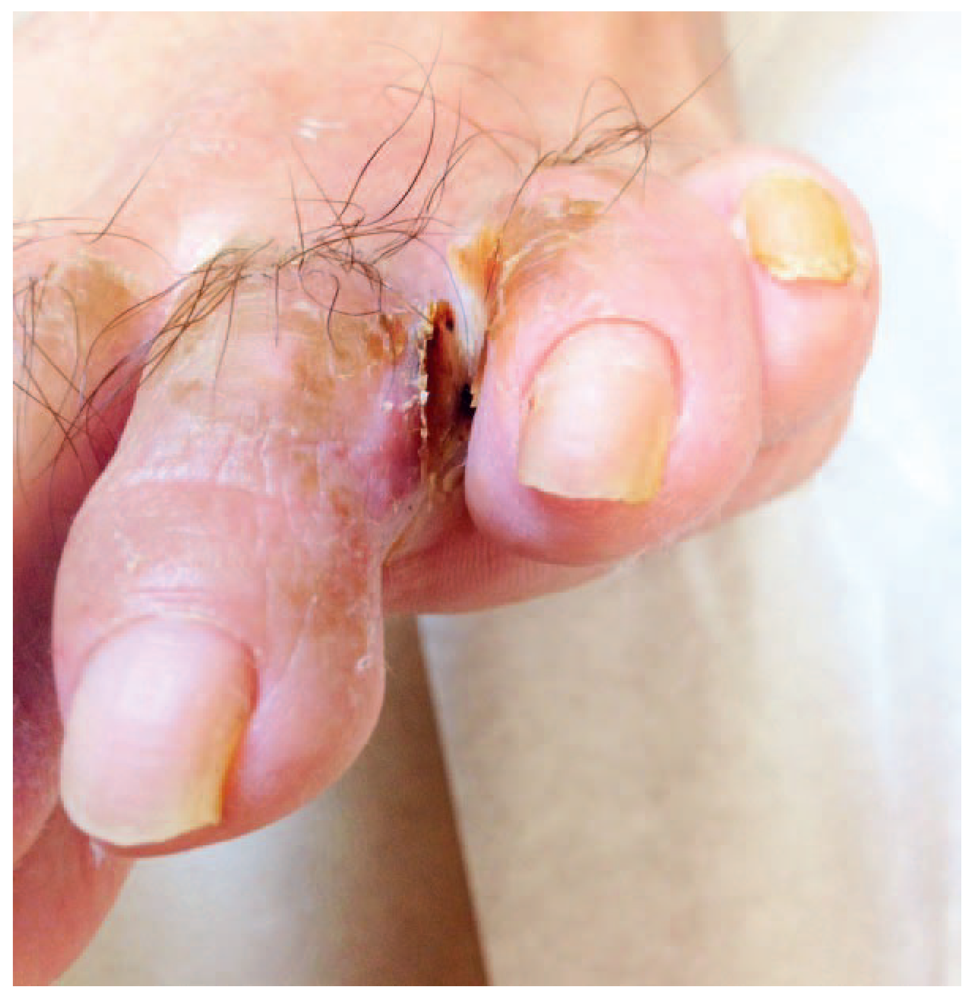
The lesions were completely excised and measured 1.4 × 1.0 × 0.6 cm; the specimens were sent for histopathologic analysis. The wound bed was inspected for abnormalities and matrices. The wound base was noted to be granular, with clear, flat margins. Due to the large soft-tissue defect, primary closure was not achievable. As a result, a full-thickness pinch graft was harvested from the sinus tarsi of the left foot and applied to the left lateral third digit and interspace. The graft was secured with 4.0 poliglecaprone 25 (Monocryl; Ethicon Inc, Somerville, New Jersey) sutures (Fig. 2). A stent dressing was applied to the surgical site. As demonstrated in Figures 3 and 4, the graft and donor sites healed completely with no recurrence in 6 months.
Figure 2.
Application of the skin graft from the sinus tarsi for primary closure following lesion removal.
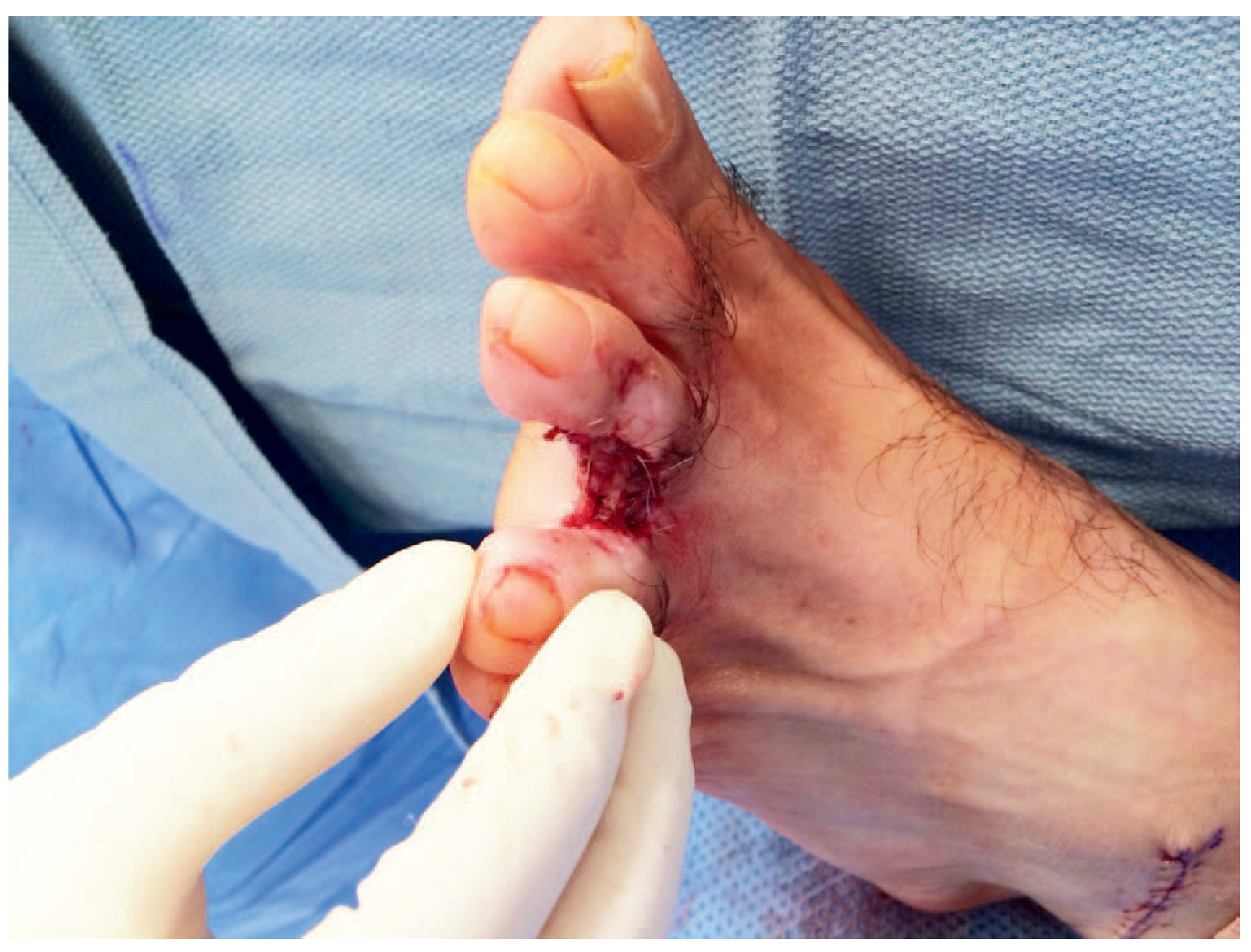
Figure 3.
Completely healed donor site.
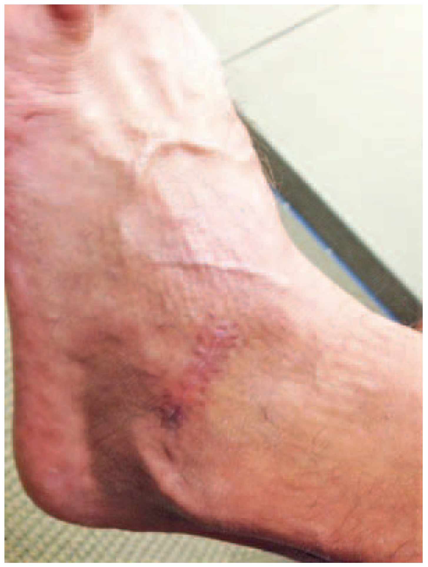
Figure 4.
Healed graft site with no signs of dehiscence. Mild hyperkeratosis is noted at the base of the graft, which was debrided later.
Results
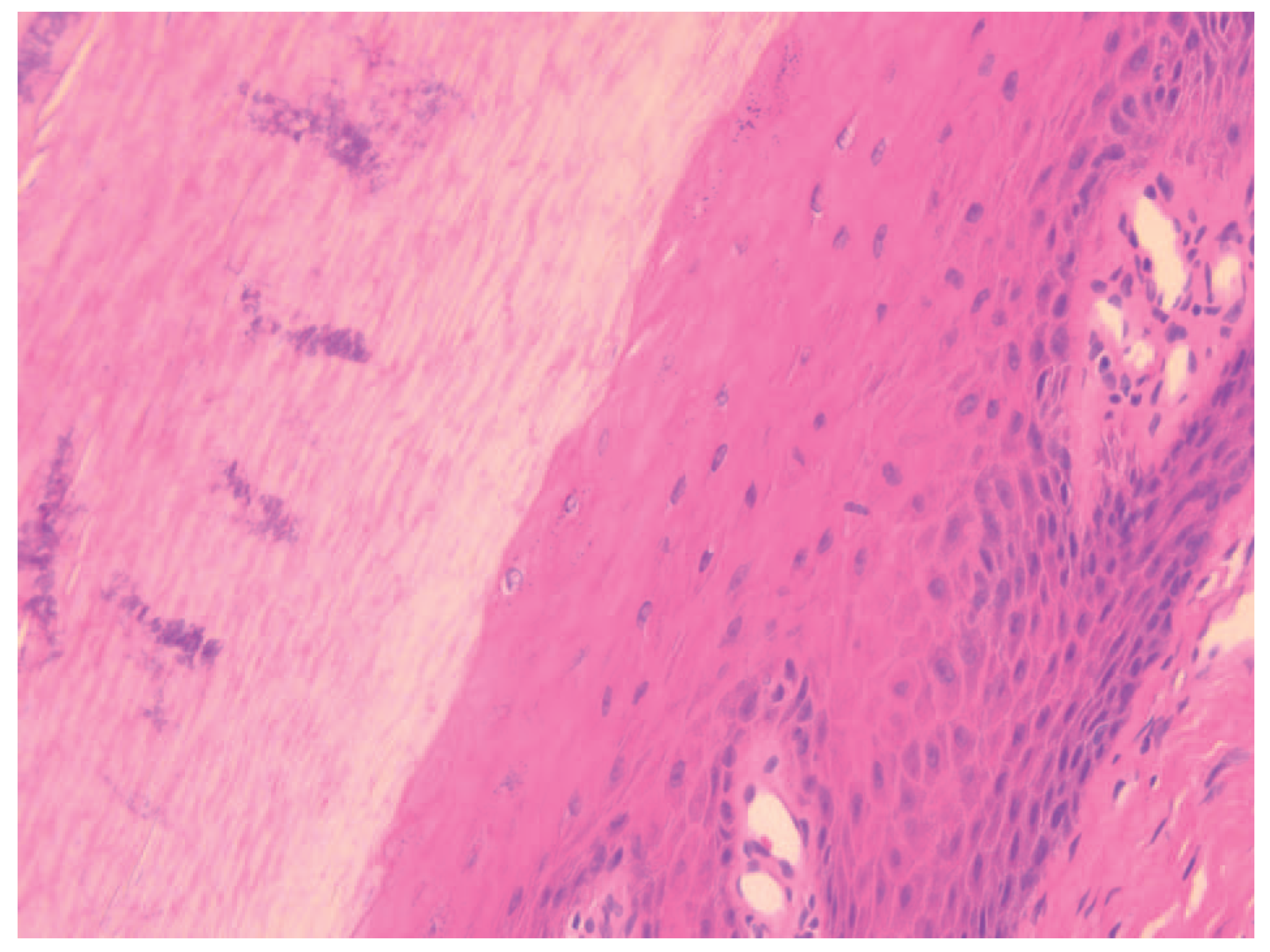
The histopathologic examination revealed yellow-tan subcutaneous nodules with marked hyperkeratosis and superficial bacterial colonization consistent with cornu cutaneum. The slides were reviewed with the pathologist to confirm the diagnosis, and no evidence of malignancy was noted (Figs. 5 and 6).
Figure 5.
Low-power view of the laminated layer of keratin consistent with cornu cutaneum showing no evidence of malignancy.
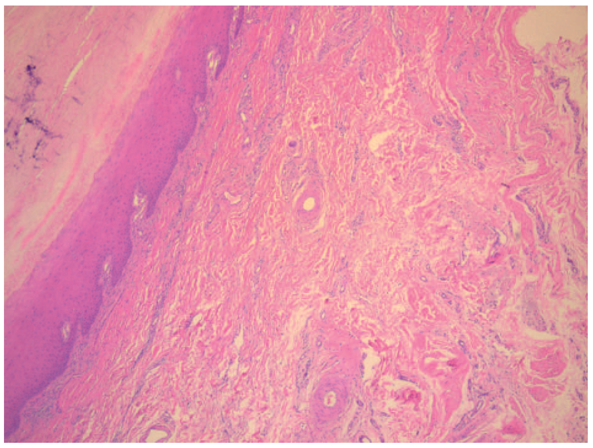
Figure 6.
High-power view of the epidermal portion of the hyperkeratotic lesion with markedly thickened stratum cornium.
Discussion
Cutaneous horns are dense, keratinized projections above the surface of the skin that are not pathologic. These lesions are generally asymptomatic, but patients may experience point tenderness or irritation depending on the location of the lesions.
The pathogenesis of these hyperkeratotic protrusions has not been fully explained. The clinical importance of these lesions is due to the underlying condition, which may be malignant. Superficial biopsies may not elucidate the definite diagnosis. Therefore, deep biopsy or total excision of lesions is recommended to confirm the dermatopathologic assessment.[1]
Depending on the size of the excised lesions, the closure of the defect may be challenging in many cases. Full-thickness or split-thickness skin grafts can be used to close the wound. We chose a full-thickness skin graft from the ipsilateral sinus tarsi for surgical site closure. Harvesting the graft from the flexor crease of sinus tarsi forms a scar parallel to relaxed skin tension lines, which results in better cosmetic outcomes. Full-thickness skin grafts have the advantages of being more durable with less contraction and color changes at the graft site compared with split-thickness skin grafts.[9]
We present a unique presentation of cutaneous horn lesions on the lower extremity. Although the presentation of cutaneous horns has been commonly associated with malignancies and occurrence on sun-exposed areas, this case is atypical in that the lesions occurred in the same location on the foot twice. In the precise histologic examination, the reports confirmed no malignancy. This case presentation includes successful surgical resection with the adjunctive aid of autologous full-thickness skin graft. No clinical recurrence was present in 6 months.
We emphasize that no dermatologic lesion has a concrete etiology, pathology, or presentation and that lesions must be treated appropriately based on histopathologic studies.
Financial Disclosure:
None reported.
Conflict of Interest:
None reported.
References
- Fernandes NF, Sinha S, Lambert WC, et al: Cutaneous horn: a potentially malignant entity. Acta Dermatovenerol Alp Pannonica Adriat18: 189, 2009.
- Sathyanarayana SA, Deutsch GB, Edelman M, et al: Cutaneous horn: a malignant lesion? A brief review of the literature. Dermatol Surg38: 285, 2012.
- Mantese SA, Diogo PM, Rocha A, et al: Cutaneous horn: a retrospective histopathological study of 222 cases. An Bras Dermatol85: 157, 2010.
- Tillo O, Chin KF, Vydia R, et al: A rare case of giant cutaneous horn. Acta Chir Plast52: 57, 2010.
- Kandemir NO, Gun BD, Barut F, et al: Cutanous horn-related Kaposi's sarcoma: a case report. Case Rep Med2010: 825949, 2010.
- Yu RC, Pryce DW, Macfarlane AW, et al: A histopathological study of 643 cutaneous horns. Br J Dermatol124: 449, 1991.
- Montgomery DW: Cornu cutaneum. Arch Dermatol Syph44: 231, 1941.
- Masic T, Babajic E, Dizdarevic D, et al: A giant cutaneous horn. Med Arh64: 375, 2010.
- Southerland JT, Boberg JS, et al: McGlamry's Comprehensive Textbook of Foot & Ankle Surgery, 4th Ed, p1205, Lippincott Williams & Wilkins, Philadelphia, PA, 2013.
© 2017 American Podiatric Medical Association