


Abstract
Os vesalianum is a rare accessory bone located proximal to the base of the fifth metatarsal in the peroneus brevis tendon. It is a radiographic diagnosis and mostly an asymptomatic incidental finding with a reported prevalence of 0.1% to 0.9%. Only 11 symptomatic cases have been described in the literature. Surgical therapy has been reported with good outcome in adults, whereas recurrence may follow excision during skeletal growth. We report a case of a 19-year-old girl with chronic weightbearing pain proximal to the base of the fifth metatarsal of her left foot. She first experienced exacerbated pain on increased loading when she started professional training as a shop assistant. Because several months of nonsurgical therapy failed, the decision was made to surgically excise the accessory bone from the peroneus brevis tendon via a longitudinal incision and a simple tendon-to-tendon reconstruction. Postoperative treatment consisted of using a walker to avoid weightbearing for 6 weeks, followed by a gradual return to full weightbearing as tolerated. At final follow-up, the patient was fully asymptomatic and was able to return to work. Citing this case, this article discusses differential diagnoses and treatment options for os vesalianum.
Os vesalianum is a rare accessory bone of the foot that is located in the peroneus brevis tendon proximal to the base of the fifth metatarsal. It was first described and named by Andreas Vesalius in 1543[1] and has since been reported to be observed in 0.1% to 0.9% of the population in several cross-sectional radiographic studies.[2-7] Tsuruta et al,[7] in a series of 3,460 foot radiographs of Japanese patients older than 7 years, detected os vesalianum in only four radiographs (0.1% of all patients). Coskun and coworkers[3] identified four cases of 984 foot radiographs of Turkish patients, corresponding to a frequency of 0.4%. An earlier study of 1,800 radiographs of the foot in a German cohort revealed os vesalianum pedis in 0.9%.[6] In relation to other types of accessory bones in the foot skeleton, os vesalianum has been reported to account for 5.9% of all detectable accessory ossicles in 464 analyzed radiographs of the Turkish population.[2]
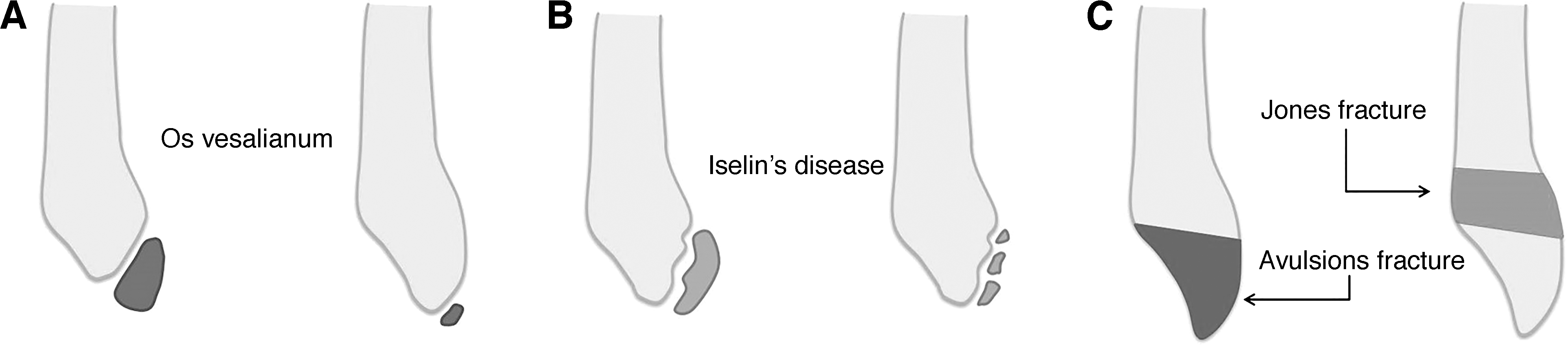
Os vesalianum is usually an incidental diagnosis in asymptomatic patients who undergo imaging studies of the foot for unrelated reasons. Only 11 symptomatic cases are described in the literature,[5,8-18] most of which have been treated by simple surgical excision. It is important to recognize these rare cases because the adequate treatment of symptomatic os vesalianum differs from that of the relevant differential diagnoses, which are by far more common: acute extra-articular avulsion fractures of the base of the fifth metatarsal and delayed healing and nonunions after fractures of the proximal fifth metatarsal, which—unlike os vesalianum—more often require open reduction and internal fixation of the proximal fragment of the fifth metatarsal and occur at an incidence of approximately 67 per 100,000 inhabitants per year.[19] Radiographically, these entities are easily distinguishable. Os vesalianum is a truly accessory bone that is found proximal to a normal-appearing base of the fifth metatarsal in the peroneus brevis tendon and is surrounded by a cortical shell, whereas fractures of the proximal fifth metatarsal and its fracture sequelae always reveal a fracture line in the base of the fifth metatarsal that runs transversely or obliquely to the long axis of the metatarsal shaft and interrupts the cortical integrity (Fig. 1). A further differential diagnosis in the growing skeleton is apophysitis of the tuberosity of the fifth metatarsal (Iselin's disease), for which nonoperative treatment is indicated and which is a self-limiting disease once ossification of the apophysis is completed.[3,10,14] In contrast to os vesalianum and fractures of the proximal fifth metatarsal, the characteristic radiographic appearance of Iselin's disease is a small flake of bone oriented obliquely to the long axis of the metatarsal shaft on the lateral plantar aspect of the base of the fifth metatarsal (Fig. 1). There are no reported epidemiologic data in the literature about the prevalence of Iselin's disease.
Figure 1.
Schematic of the characteristic differential radiologic presentation of the os vesalianum (A), Iselin's disease (B), and Jones' fracture (C).
We present a case of a 19-year-old girl with a severely symptomatic os vesalianum of her left foot that was successfully treated by simple surgical excision followed by 6 weeks of partial weightbearing using a walker.
Case Report
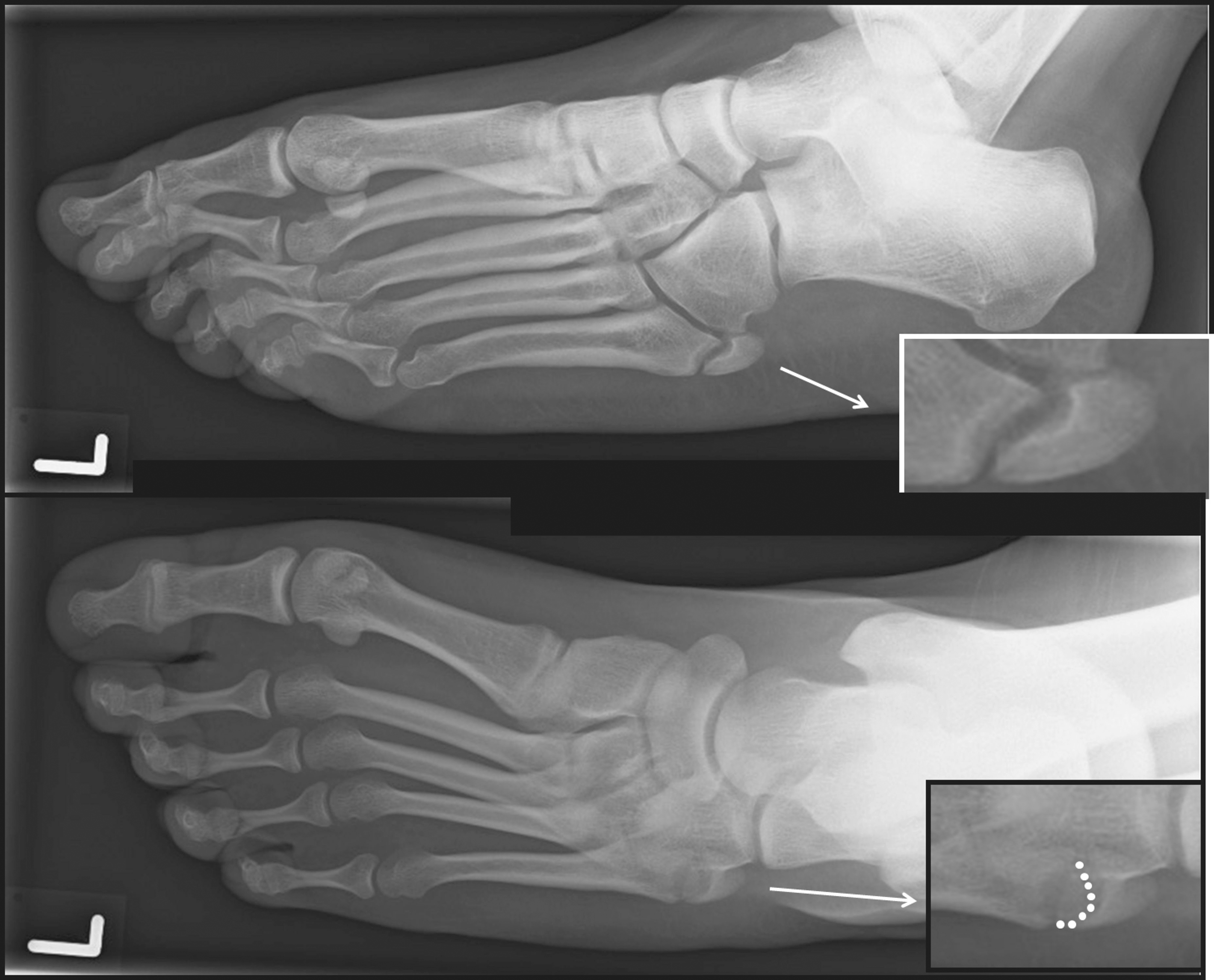
A 19-year-old girl presented to the outpatient clinic with pain over the lateral aspect of her left foot. She reported weightbearing pain that had already persisted for several years and that recently, during her professional training as a shop assistant, had increased considerably to a degree that rendered her unable to work. Her medical history was unremarkable, and she had no history of trauma to the foot or ankle. She did not report any intense athletic overuse during childhood or adolescence. On physical examination, the skin and soft tissues appeared unremarkable, without visible swelling or signs of irritation. Longitudinal and transversal foot arches appeared physiologic on standing. Barefooted gait pattern revealed that the patient was putting less weight on the lateral aspect of her left foot, resulting in a slight but visible limp. There was a distinct tenderness on palpation just proximal to the base of the fifth metatarsal. Pain was provoked by resisted eversion and dorsiflexion of the foot and by forced combined inversion and plantarflexion of the foot. The range of motion of the ankle was normal in all planes in the tibiotalar and subtalar joints, and there was no hindfoot, midfoot, or forefoot deformity. Standard radiographs demonstrated an accessory ossicle just proximal to the base of the fifth metatarsal, surrounded by a complete cortical shell and separated from the metatarsal base by a thin radiolucent line of constant width as if there was a normal joint covered with articular cartilage (Fig. 2). Based on the characteristic radiographic appearance, the ossicle was diagnosed as an os vesalianum. We initiated an intensified conservative treatment course of 24 weeks with individually made arch support orthotic devices with rearfoot support, physical antiphlogistic therapy, and partial nonweightbearing. Despite full compliance with the prescribed treatment modalities, there was no substantial pain relief. Given the favorable reports in the literature regarding the outcome of simple excision of symptomatic os vesalianum, a decision was made for surgical resection.
Figure 2.
Preoperative dorsoplantar (top) and oblique (bottom) radiographs of the reported case.
With the patient in a supine position with a tourniquet applied to the thigh, a 4-cm longitudinal skin incision centered over the tuberosity of the fifth metatarsal base was used for a lateral approach to the peroneus brevis tendon insertion and the metatarsal base. With the tendon exposed, the ossicle was not easily palpable. The tendon was incised longitudinally, and the ossicle became apparent and was easily mobilized toward the base of the metatarsal and from the tendon to which it was partially adherent. The distal aspect facing the metatarsal base was covered by cartilage, creating a joint-like formation (Fig. 3). After enucleation of the ossicle, the peroneus brevis tendon insertion to the metatarsal base appeared not to be damaged so that refixation of the tendon was not necessary. The tendon was reconstructed by a simple tendon-to-tendon resorbable polyglactin-910 (Vicryl; Ethicon, Inc, Somerville, New Jersey) suture. The postoperative radiographic appearance of the base of the fifth metatarsal proved complete excision of the os vesalianum (Fig. 4). The foot was elevated for 48 hours postoperatively. The ankle was immobilized in a walker, and partial weightbearing with 20 kg was permitted for 6 weeks. Use of the walker was then discontinued, and weightbearing was allowed as tolerated during the following 2 weeks in normal footwear. At 8 weeks postoperatively, the patient was fully asymptomatic and returned to work without any restrictions. At her last clinical follow-up visit 6 months after surgery she was fully mobile, had free range of motion, and was pain free even after long working days (Fig. 5).
Figure 3.
The resected os vesalianum contains an articular cartilage-like distal facet toward the proximal aspect of the base of the fifth metatarsal.
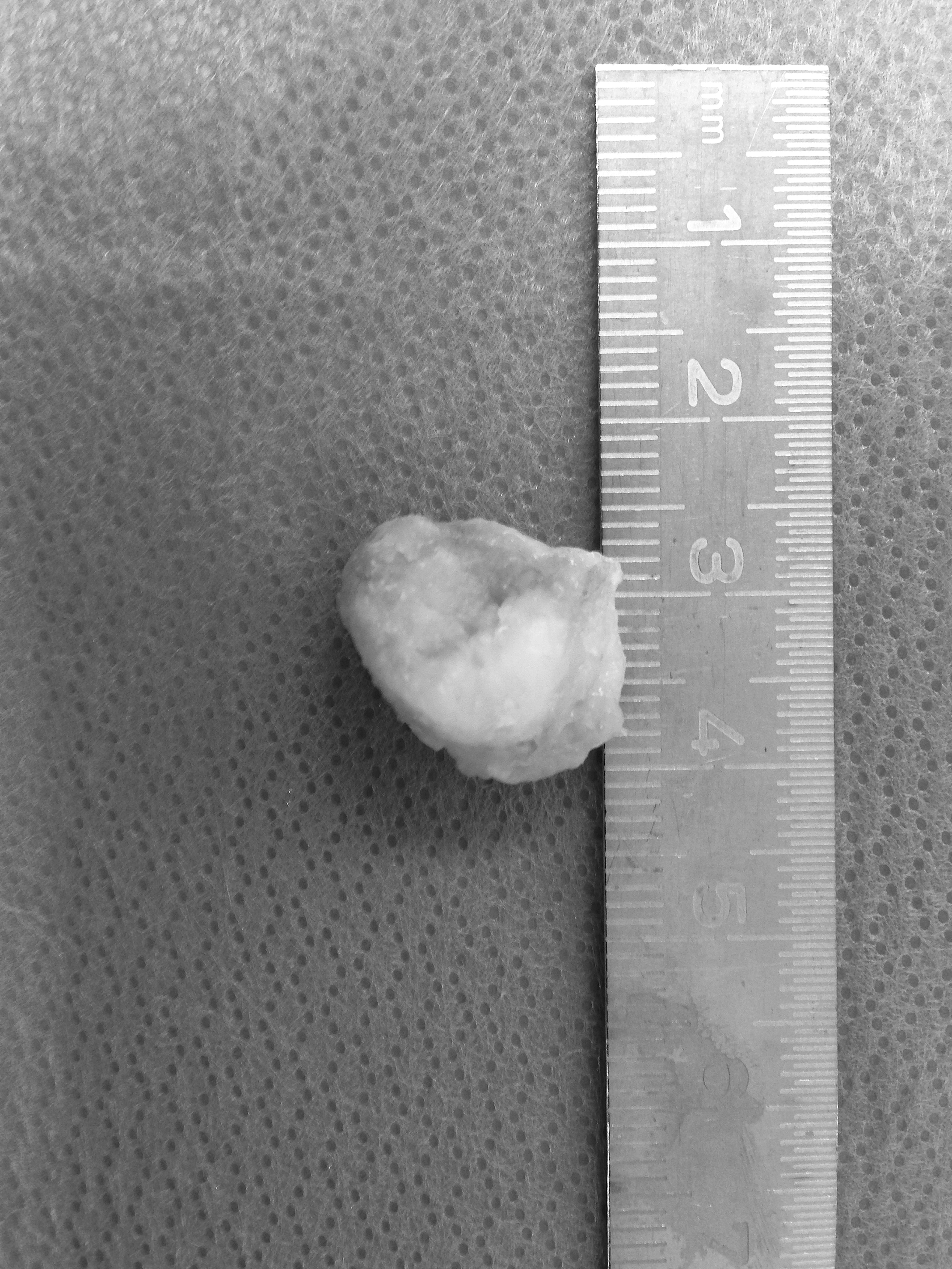
Figure 4.
Postoperative anterior posterior radiograph of the forefoot and midfoot after complete resection of the os vesalianum.
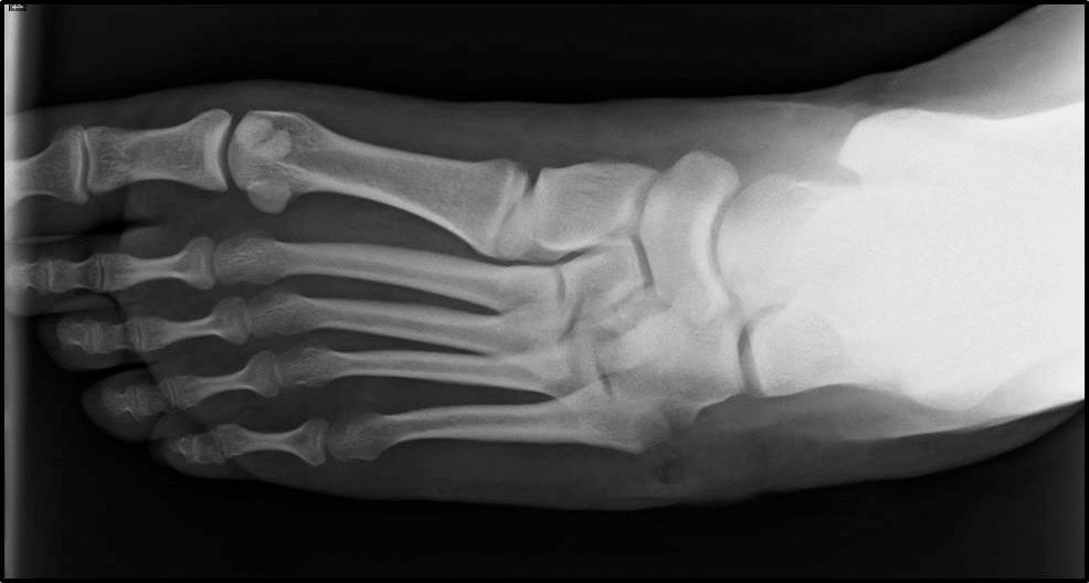
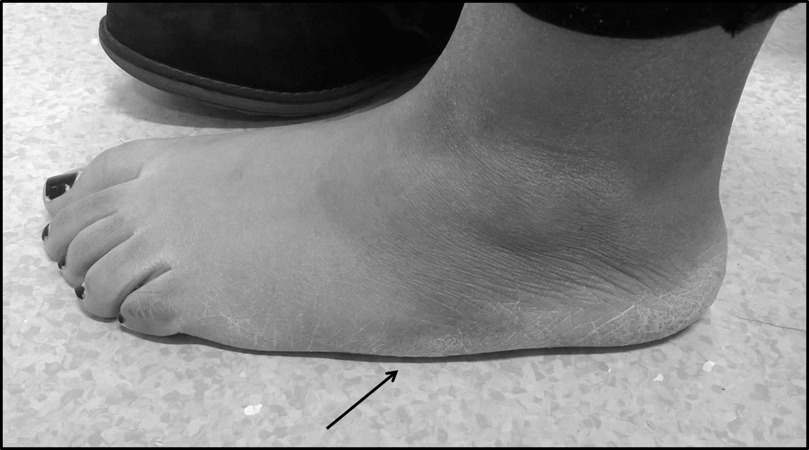
Figure 5.
Clinical presentation 6 months after surgery. The patient was fully mobile, had unlimited range of motion, and was pain free. The arrow shows the area of the surgical scar after resection of the os vesalianum.
Discussion
Os vesalianum represents 5% to 6% of all accessory bones of the foot skeleton[4] and is estimated to occur with a prevalence of 0.1% to 0.9%.[2-7] Bilateral cases as well as familial clustering have been described,[5,10,13] suggesting a potential genetic predisposition. Usually, os vesalianum is asymptomatic and is diagnosed as an incidental finding; symptomatic os vesalianum is an extremely rare entity. Usually plain midfoot radiographs in two or three planes are diagnostic and sufficient. In rare circumstances, such as clinical suspicion of a local tumor or infection, an additional magnetic resonance image may be helpful in the differential diagnosis. If os vesalianum is chronically resistant to conservative therapy, surgery represents a reliable treatment option. Various surgical procedures have been described in the literature. Eleven cases of symptomatic os vesalianum have been described to date (including two bilateral cases[5,17]), nine of which have been treated surgically. In one bilateral case, symptoms dissipated spontaneously after the contralateral side had been operated on.[10] Initial treatment is nonsurgical, with immobilization and rest.[5,10] Surgically, two treatment options have been described: resection[5,13] and osteosynthesis,[10] with most reports favoring resection. In case the peroneus brevis tendon insertion is violated by the excision to a considerable degree, refixation of the tendon, for example with a suture anchor, may be indicated.[13] There is only one report of osteosynthesis with bone grafting from the iliac crest.[10]
Given the nature of the ossicle as a truly accessory bone proximal to a normally shaped base of the fifth metatarsal, we think that excision is the natural surgical approach and should be favored over osteosynthesis. This case report demonstrates that the treatment of simple longitudinal excision of the ossicle from the peroneus brevis tendon plus tendon-to-tendon reconstruction has led to full recovery, and we, therefore, propose that this should be considered to be the standard care for this rare entity.
Conclusions
Os vesalianum should be recognized and distinguished from its differential diagnoses of fractures of the proximal fifth metatarsal and from Iselin's disease because adequate treatment of these entities differs substantially. Symptomatic os vesalianum resistant to conservative therapy can be successfully treated by simple longitudinal excision of the ossicle from the peroneus brevis tendon and tendon-to-tendon reconstruction.
Financial Disclosure:
None reported.
Conflict of Interest:
None reported.
References
- Vesalius A: “De Pedis Ossibvs,”inDe Humani Corporis Fabrica, p 142,1543.
- Cilli F, Akçaoğlu M: The incidence of accessory bones of the foot and their clinical significance [in Turkish]. Acta Orthop Traumatol Turc39: 243, 2005.
- Coskun N, Yuksel M, Cevener M, et al: Incidence of accessory ossicles and sesamoid bones in the feet: a radiographic study of the Turkish subjects. Surg Radiol Anat31: 19, 2009.
- Dameron TBJr: Fractures and anatomical variations of the proximal portion of the fifth metatarsal. J Bone Joint Surg Am57: 788, 1975.
- Dorrestijn O, Brouwer RW: Bilateral symptomatic os vesalianum pedis: a case report. J Foot Ankle Surg50: 473, 2011.
- Heimerzheim A: Über einige akzessorische Fußwurzelknochen nebst ihrer chirurgischen Bedeutung. Langenbecks Arch Surg (formerly DtschZChir)190: 96, 1925.
- Tsuruta T, Shiokawa Y, Kato A, et al: Radiological study of the accessory skeletal elements in the foot and ankle (author's transl) [in Japanese]. Nihon Seikeigeka Gakkai Zasshi55: 357, 1981.
- Boya H, Oztekin HH, Ozcan O: Abnormal proximal fifth metatarsal and os vesalianum pedis. JAPMA97: 428, 2007.
- Boya H, Ozcan O, Tandoğan R, et al: Os vesalianum pedis. JAPMA95: 583, 2005.
- Iselin H: Wachstumsbeschwerden zur Zeit der knöchernen Entwicklung der Tuberositas metatarsi quinti. Langenbecks Arch Surg (formerly DtschZChir)117: 529, 1912.
- Kim MH, Kim WH, Kim CG, et al: Os vesalianum pedis detected with bone SPECT/CT. Clin Nucl Med39: e190, 2014.
- Kose O: Os vesalianum pedis misdiagnosed as fifth metatarsal avulsion fracture. Emerg Med Australas21: 426, 2009.
- Petrera M, Dwyer T, Ogilvie-Harris DJ: A rare cause of foot pain with golf swing: symptomatic os vesalianum pedis: a case report. Sports Health5: 357, 2013.
- Smith AD, Carter JR, Marcus RE: The os vesalianum: an unusual cause of lateral foot pain: a case report and review of the literature. Orthopedics7: 86, 1984.
- Wilson TC, Wilson RC, Ouzounov KG: The symptomatic os vesalianum as an uncommon cause of lateral foot pain: a case report. JAPMA101: 356, 2011.
- Canale ST, Williams KD: Iselin's disease. J Pediatr Orthop12: 90;1992.
- Inoue T, Yoshimura I, Ogata K, et al: Os vesalianum as a cause of lateral foot pain: a familial case and its treatment. J Pediatr Orthop B8: 56, 1999.
- Ralph BG, Barrett J, Kenyhercz C, et al: Iselin's disease: a case presentation of nonunion and review of the differential diagnosis. J Foot Ankle Surg38: 409, 1999.
- Petrisor BA, Ekrol I, Court-Brown C: The epidemiology of metatarsal fractures. Foot Ankle Int27: 172, 2006.
© 2017 American Podiatric Medical Association