


Abstract
Hereditary hemorrhagic telangiectasia (HHT), which is also known as Osler-Weber-Rendu syndrome, is a group of related disorders characterized by the development of arteriovenous malformations. These malformations occur in almost all organs but predominantly in the skin, intestines, liver, lungs, and brain. This is a case report of a patient with cutaneous manifestations of HHT in the lower extremities as diagnosed by his podiatric physician. To our knowledge, the literature does not present any case reports in which cutaneous manifestations of the lower extremities followed by a further work-up allowed a diagnosis of HHT.
Hereditary hemorrhagic telangiectasia (HHT), which is also known as Osler-Weber-Rendu syndrome, is a group of disorders with clinical subtypes of HHT genetically inherited in an autosomal dominant manner. It is characterized by the development of arteriovenous malformations (AVMs) that can occur anywhere in the human body. Most of these manifestations occur in the skin, mucus membrane, and internal organs, including the brain, liver, lungs, and heart. The prevalence of the disease is approximately one in 5,000 to 8,000, with no real preference by race [1]. Similar to any other systemic disease, HHT can cause many complications in those affected. The disease is characterized by its lack of intervening capillaries between arteries and veins. As a result, small lesions ranging from pink to red and measuring approximately 1.0 mm in diameter can be seen. These lesions can be seen on nearly any part of the human body, but they are more commonly found on the lips, face, gastrointestinal mucosa, buccal, nasal, and tongue. Larger AVMs can also be seen, which often are more threatening to the patient's well-being. They may be as large as several centimeters and are mostly found in the lungs, liver, and brain. The shunts that are created in these internal organs can easily cause strokes and brain abscesses [2]. The most common manifestation of HHT is epistaxis, which approximately 50% of patients have by age 10 years and nearly 90% will have by the third decade of life. Epistaxis regularly precedes cutaneous manifestations; however, 100% of patients will develop cutaneous manifestations in later adulthood. Other symptoms of HHT can involve the lungs and usually occur in approximately 30% of patients due to AVMs, causing dyspnea, fatigue, and cyanosis. Brain, liver, gastrointestinal, and genitourinary involvement can occur as well, but it is less likely [3].
Although HHT is a clinical diagnosis, the genetic factor in the disease should be discussed because molecular diagnostics has more recently become a routine aspect of medical management. Hemorrhagic telangiectasia is inherited in an autosomal dominant manner, with considerable intrafamilial variability. Most individuals have an affected parent. It is caused by mutations in a variety of genes, including ENG, ACVRL1, SMAD4, and GDF2. Mutations in these genes are found in approximately 85% of individuals who meet the unequivocal clinical diagnosis criteria for HHT [4]. It is crucial to perform a genetic work-up, which should include genetic counseling, and risk assessment in families with this disorder. Genetic information from this patient may be used for his children, who have a 50% chance of receiving the disease in their lifetime [4,5]. This case report involves a 43-year-old man who was being treated for plantar warts for several years. It was only recently that he presented his symptoms to another podiatric physician, who conducted a further work-up that later led to a diagnosis of HHT.
Case Report
A 43-year-old man presented to a clinic with painful bleeding lesions on his feet. He was originally being treated for plantar warts for several months by another podiatric physician before his arrival. After finding no relief for some time, he decided to get a second opinion for the lesions. Like any new patient, a thorough patient history was taken. It was noted that he had a history of epistaxis as a child. The patient has had a history of these lesions for several years. It was revealed that 3 to 4 years earlier he first developed these growths on his feet that bled excessively. He currently reports them to be sporadic. He denies any history of anemia but has recently had rectal bleeding. He had a previous skin lesion located on his forehead that was diagnosed as melanoma more than 10 years ago. The patient takes atorvastatin for his high cholesterol level and otherwise is a healthy individual. When considering his family history, his father died of lung cancer. The maternal ancestry is from England and Germany. The paternal ancestry is from Germany. He has twin daughters, and one has shown symptoms of epistaxis. On physical examination, the patient had palpable pedal pulses, adequate protective sensation, and excellent muscle strength. No foot deformity was noted. The only abnormal findings were red lesions that were visible on the plantar aspects of his feet. The lesions themselves were flat, red, circular, and 0.1 cm in diameter (Fig. 1). There was no apparent pattern of distribution on the plantar aspects of the feet, and the lesions were painful on palpation. The lesions themselves did not represent the characteristics of plantar warts, and the lesions were not itchy.
Figure 1.
Telangiectasia noted at the plantar aspect of the foot.
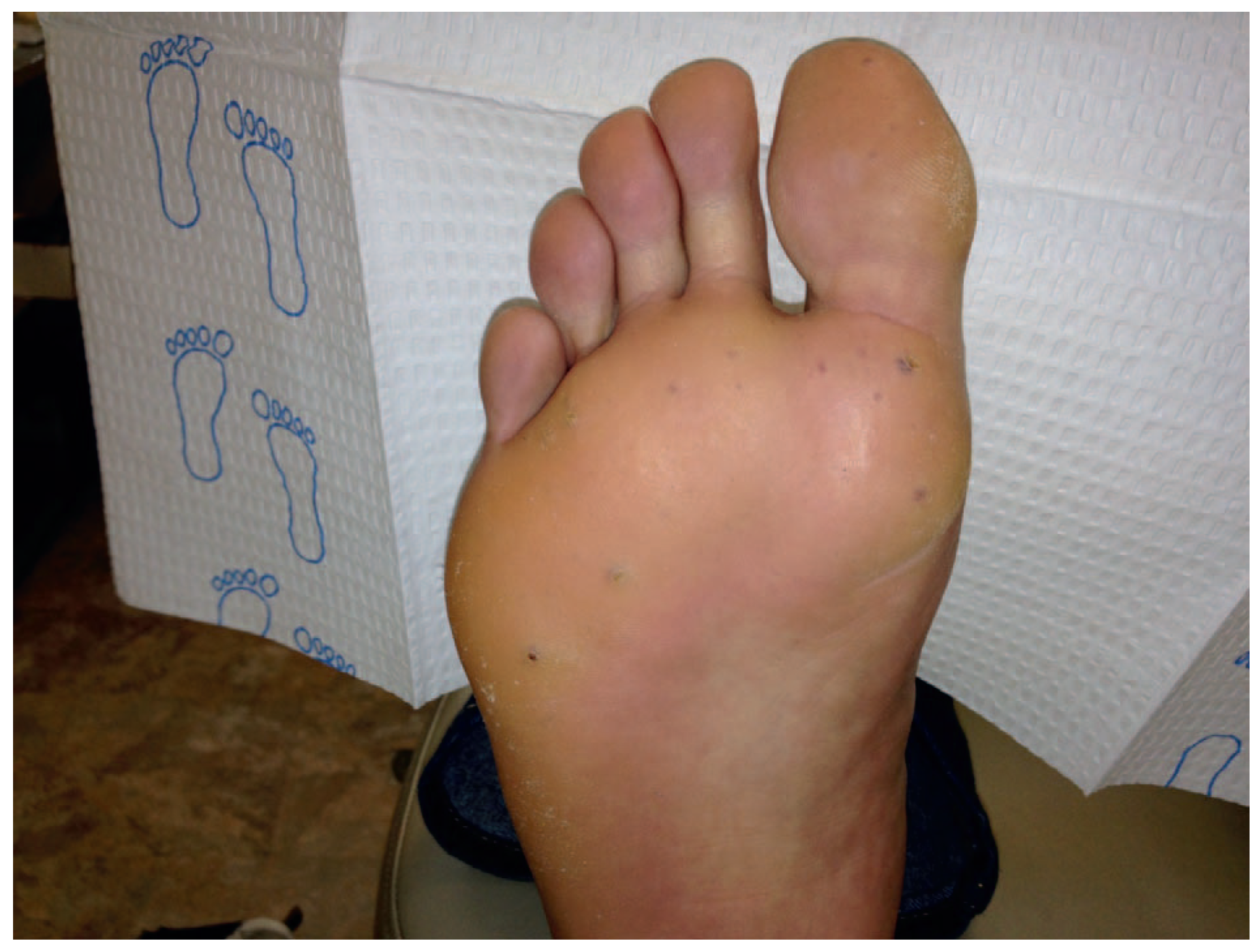
With a lesion as such, many differential diagnoses should be considered. Differential diagnosis of these cutaneous manifestations in the lower extremity may include petechiae; bug bites; trauma; chicken pox; metastatic melanoma; and hand, mouth, and foot disease, which can make it difficult to treat the patient without delaying care [6]. However, when putting great emphasis on the subjective information, the diagnosis became apparent and was confirmed clinically on the basis of the Curacao criteria: 1) epistaxis (spontaneous and recurrent); 2) telangiectases (multiple characteristic sites [lips, oral cavity, fingers, or nose]); 3) visceral lesions (gastrointestinal telangiectasia [with or without bleeding], AVMs [hepatic, cerebral, and spinal]); and 4) family history (a first-degree relative who has HHT [according to these same criteria]).
Three of the four criteria are needed to make the diagnosis of HHT. In this case report, the patient had expressed gastrointestinal bleeding, epistaxis, telangiectasia, and an alarming suspicion of HHT in one of his daughters [3]. With these criteria, the podiatric physician was able to diagnose the patient as having HHT. Owing to the genetic quality of the disease, genetic counseling was recommended for the patient and his family. His genetic testing results revealed a c.313+_2T>C in the ACVRL1 gene. The patient was also referred to a specialist who recommended brain magnetic resonance imaging, echocardiography for pulmonary shunting/AVM, and computed tomography or ultrasonography for hepatic AVM [7]. Although results are pending for the diagnostic imaging tests, the patient has been receiving weekly cauterizations of his lower-extremity lesions that have helped with the pain and bleeding. He has also been advised to refrain from anticoagulants to help with the excessive bleeding [8]. Further treatments will be decided once his diagnostic imaging is completed.
Discussion
To our knowledge, there is no report of a podiatric physician who has diagnosed a patient as having HHT. The scope of podiatric medicine generally includes the lower extremity from the knee down. However, this should not be an excuse to ignore the patient as a whole. Systematic diseases such as HHT may show clinically in the lower extremity, but it is primarily a disease that involves much more. Therefore, it is important for providers of all types to be more aware of systematic diseases that can be otherwise difficult to diagnose. Because HHT does not involve one medical specialty, it becomes crucial for all types of providers to be educated on proper care and diagnosis of the disease. The patient spent most of his life undiagnosed. Although he had seen multiple specialists throughout his life for various conditions, none of them were able to make the connections. For example, his previous podiatric physician was treating this patient for plantar warts for several months with no improvements, and this should have warranted the possibility of a different reason behind the patient's symptoms. It was not until he visited the clinic that the pieces of the puzzle were put together by simply listening to the patient and conducting a thorough history. The answer had always been in the patient's history.
After diagnosing the patient with HHT, proper follow-up was crucial. He was referred to a specialist and a genetic counselor. His cutaneous manifestations of HHT are not as severe as the other manifestations that may be present. For example, if this patient has larger AVMs present in major organs, including the brain, his condition will prove to be much more serious. Although diagnostic test results are still pending, it is difficult to say how advanced his condition may be. This is why it is important for all types of physicians practicing medicine not to delay proper treatment. In this case report, it simply was a matter of taking a proper history of the patient. In conclusion, providers should not ignore the fundamentals of medicine and should appreciate the patient's subjective as well as objective information to prevent delay of care.
Financial Disclosure:
None reported.
Conflict of Interest:
None reported.
References
- Dittus C, Streiff M, Ansell J: Bleeding and clotting in hereditary hemorrhagic telangiectasia. World J Clin Cases3: 330, 2015.
- Garg N, Khunger M, Gupta A, et al: Optimal management of hereditary hemorrhagic telangiectasia. J Blood Med5: 206, 2014.
- Govani F, Shovlin C: Hereditary haemorrhagic telangiectasia: a clinical and scientific review. Eur J Hum Genet17: 860, 2009.
- Shovlin CL, Guttmacher AE, Buscarini E, et al: Diagnostic criteria for hereditary hemorrhagic telangiectasia (Rendu-Osler-Weber syndrome). Am J Med Genet91: 66, 2000.
- Botella L, Albinana V, Ojeda-Fernandez L, et al: Research on potential biomarkers in hereditary hemorrhagic telangiectasia. Front Genet6: 115, 2015.
- Hunt K, Kissel R, Vidal L: Progressive widespread cutaneous telangiectasias. AMA Dermatol150: 1103, 2014.
- McDonald J, Wooderchak W, Whitehead K, et al: Hereditary hemorrhagic telangiectasia: genetics and molecular diagnostics in a new era. Front Genet6: 8, 2015.
- Boza JC, Dorn T, Oliveira FB, et al: Case for diagnosis: hereditary hemorrhagic telangiectasia. An Bras Dermatol89: 999, 2014.
© 2017 American Podiatric Medical Association