


Abstract
Background: We investigated the effectiveness and durability of two types of plantar padding, the plantar metatarsal pad and the single wing plantar cover, which are commonly used for reducing forefoot plantar pressures. Methods: Mean peak plantar pressure and impulse at the hallux and at the first, second, third, and fourth metatarsophalangeal joints across both feet were recorded using the two-step method in 18 individuals with normal asymptomatic feet. Plantar paddings were retained for 5 days, and their durability and effectiveness were assessed by repeating the foot plantar measurement at baseline and after 3 and 5 days. Results: The single wing plantar cover devised from 5-mm felt adhesive padding was effective and durable in reducing peak plantar pressure and impulse at the first metatarsophalangeal joint (P = .001 and P = .015, respectively); however, it was not found to be effective in reducing peak plantar pressure and impulse at the hallux (P = .782 and P = .845, respectively). The plantar metatarsal pad was not effective in reducing plantar forefoot pressure and impulse at the second, third, and fourth metatarsophalangeal joints (P = .310 and P = .174, respectively). Conclusions: These results imply limited applicability of the single wing plantar cover and the plantar metatarsal pad in reducing hallux pressure and second through fourth metatarsophalangeal joint pressure, respectively. However, the single wing plantar cover remained durable for the 5 days of the trial and was effective in reducing the peak plantar pressure and impulse underneath the first metatarsophalangeal joint.
The soft tissue between the epidermis and the bone is known to provide a cushioning effect on the plantar aspect of the foot, protecting it from severe mechanical stresses.[1] Certain chronic conditions, such as rheumatoid arthritis and diabetes, can result in fat pad atrophy, causing increasing dynamic pressures, which may lead to pain and plantar wounds.[2]
It has been well documented that peak plantar pressure is among the high-risk factors for foot ulceration.[1,3-5] Plantar forefoot abnormalities are often treated by off-loading with felt paddings, which are common chairside appliances that provide a cushioning effect and are thought to relieve pain and off-load the afflicted area by reducing plantar pressures.[6] Felt padding provides a first-line, short-term method of accommodating and off-loading the foot, depending on the design of the device used. A crucial property of off-loading devices is to reduce any abnormal mechanical forces to which the foot is subjected during walking.[7] For long-term management, a longer-lasting and more convenient method of padding is devised as an addition to total-contact insoles that, when inserted into footwear, are then used as part of a clinical management plan to prevent or treat ulceration.[8]
It is common practice for felt paddings to be used during diabetic foot care as a method of off-loading the forefoot.[1,9,10] However, there are several inconsistencies and conflicting evidence regarding their effectiveness in reducing plantar pressures,[9,11] making their use uncertain[12,13] in the high-risk foot. To date, no data are available to help in suggesting appropriate clinical guidelines on the effectiveness and durability of different plantar forefoot paddings when used as chairside appliances in clinical practice. The literature suggests that more studies should be conducted to evaluate the durability of plantar felt paddings to add to the body of existing knowledge on the importance and effectiveness of off-loading devices.[14] Traditional practices involving the use of forefoot paddings have scarcely been challenged, notwithstanding the fact that these devices are being used extensively on a daily basis for off-loading purposes. This has prompted the need for this study, which sought to determine the applicability of two commonly used plantar forefoot paddings fashioned out of semicompressed felt, namely, the single wing plantar cover (SWPC) and the plantar metatarsal pad (PMP), by evaluating their effectiveness and durability in reducing plantar forefoot pressure. However, one possible dilemma facing the researcher is that although these paddings are frequently used as off-loading devices for the diabetic or high-risk foot, the use of self-adhesive devices on actual high-risk patients could compromise their skin integrity, possibly resulting in maceration or even ulceration. Therefore, there is a clear need for a pilot study that uses a normative healthy adult population to investigate the effectiveness of these padding devices without endangering actual high-risk patients. This pilot study will then inform and has the potential to contribute to a wider, strictly controlled clinical study involving high-risk patients, such as those living with diabetes or rheumatoid arthritis.
Methods
This study was approved by the University of Malta Research Ethics Committee and was performed in accordance with the principles of the Declaration of Helsinki. A single-center, prospective, experimental, same-subject design on a healthy adult population was used, with a convenience sampling method used to recruit individuals into this study. Data collection was conducted at a clinical biomechanics laboratory for 5 days per participant. Written informed consent was obtained from all of the participants before the study. Participants had to visit the laboratory three times during this study: at the start of the study (time 0), on day 3 (time 1), and on day 5 (time 2), at all times ensuring that the felt padding applied was not removed during the study period.
Eighteen healthy volunteers without any known history of diseases and foot abnormalities who were able to walk independently were included in this pilot study. Individuals with walking impairment and those unable to follow the whole study protocol were excluded. The appropriate paddings were fashioned from 5-mm-thick semicompressed felt (Cellona; Lohmann & Rauscher GmbH & Co KG, Neuwied, Germany). This padding material is composed of 50% polyester, 30% polypropylene, and 20% viscose. It is air permeable and has self-adhesive properties through the use of adhesive strips on the underside of the material. For this study, the SWPC and the PMP (Fig. 1) were tested for effectiveness and durability for 5 days.
Figure 1.
Single wing plantar cover (left foot) and plantar metatarsal pad (right foot).
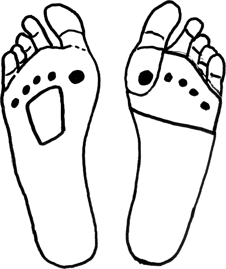
The SWPC is used to redistribute plantar pressure from the first metatarsal head, to provide shock absorption, and to reduce plantar friction. A felt pad with an aperture cutout at the first metatarsophangeal joint was devised and placed on the left foot of each participant. This was done to simulate an active ulcer location or a painful hyperkeratotic lesion underneath these areas of interest.
A PMP is thought to deflect pressure from the second, third, and fourth metatarsophalangeal joints, transferring the pressure to the metatarsal shafts, with the added benefit of also straightening the central three toes. Usually, the PMP is rhomboid shaped because the pad is wider distally and becomes narrower proximally. For this study, the PMPs were devised and placed on the right foot of each participant, 5 mm proximal to the second, third, and fourth metatarsal heads as recommended in the literature.[11]
Pressure Mapping Protocol
Peak plantar pressure beneath the foot was recorded using a pressure platform (HR Mat; Tekscan, Boston, Massachusetts). Initially, participants were asked to stand on the mat to calibrate the system according to their body mass as per manufacturer instructions. The study protocol was discussed with the participants before data collection. A period of acclimatization was included to help participants understand the two-step walking protocol that was adopted. This protocol has been reported to be reliable and not very time-consuming to implement,[15] requiring the participants to make contact with the platform at the second step.[16] Each participant was instructed to stand on both feet exactly two steps from stepping on the pressure platform. The participants started walking by stepping first with one foot on the ground and the next step on the platform and continued walking for the next three steps.[17] Several practice trials were performed to familiarize the participants with the procedure and encourage natural gait, avoiding targeting. When the participants felt confident with their walking pattern, the dynamic plantar pressures under each foot were recorded.
This procedure was repeated five times for each foot, and an average reading was calculated regarding forefoot peak plantar pressures and impulse at the areas of interest, which included the first metatarsophalangeal joint and the hallux for the SWPC and the second through fourth metatarsophalangeal joints for the PMP. Each trial produced five stance phases for each foot. The first and last stance phases of each trial were discarded; thus, the mean value of the three remaining stance phases for each foot were used for evaluation.
Study Protocol
Initially, all of the 18 participants had both feet tested barefoot without any padding (time = 0). Thereafter they were fitted with the two types of padding. To ensure secure positioning for the padding, hypoallergenic adhesive tape was placed on top. After this, the participants were asked to walk once again on the pressure platform for a set of three trials: day 1 (time 0), day 3 (time 1), and day 5 (time 2) using the same two-step walking protocol. Participants were instructed not to remove the paddings from their original positions and to keep them dry at all times.[18]
For analysis of pressure data, both forefeet were then masked according to the areas of interest, where it is traditionally hypothesized that each pad deviates pressure, ie, the first metatarsophalangeal joint and the hallux for the left foot and the second, third, and fourth metatarsophalangeal joints for the right foot. The mean peak contact pressures and impulses were consequently compared with barefoot measurements.
The one-sample Kolmogorov-Smirnov test was used to test normality of data. Repeated-measures analysis of variance with Tukey post-hoc test was used to test for any statistical significance at the 95% confidence level for all pressure data.
Results
A total of 18 participants without significant foot abnormalities, including eight men with a mean body mass of 72.5 kg (range, 45–88 kg) and ten women with a mean body mass of 64.7 kg (range, 45–85 kg), participated in this study. The mean age of the study group was 22 years (range, 21–25 years).
Effect of the SWPC at the First Metatarsophalangeal Joint and Hallux
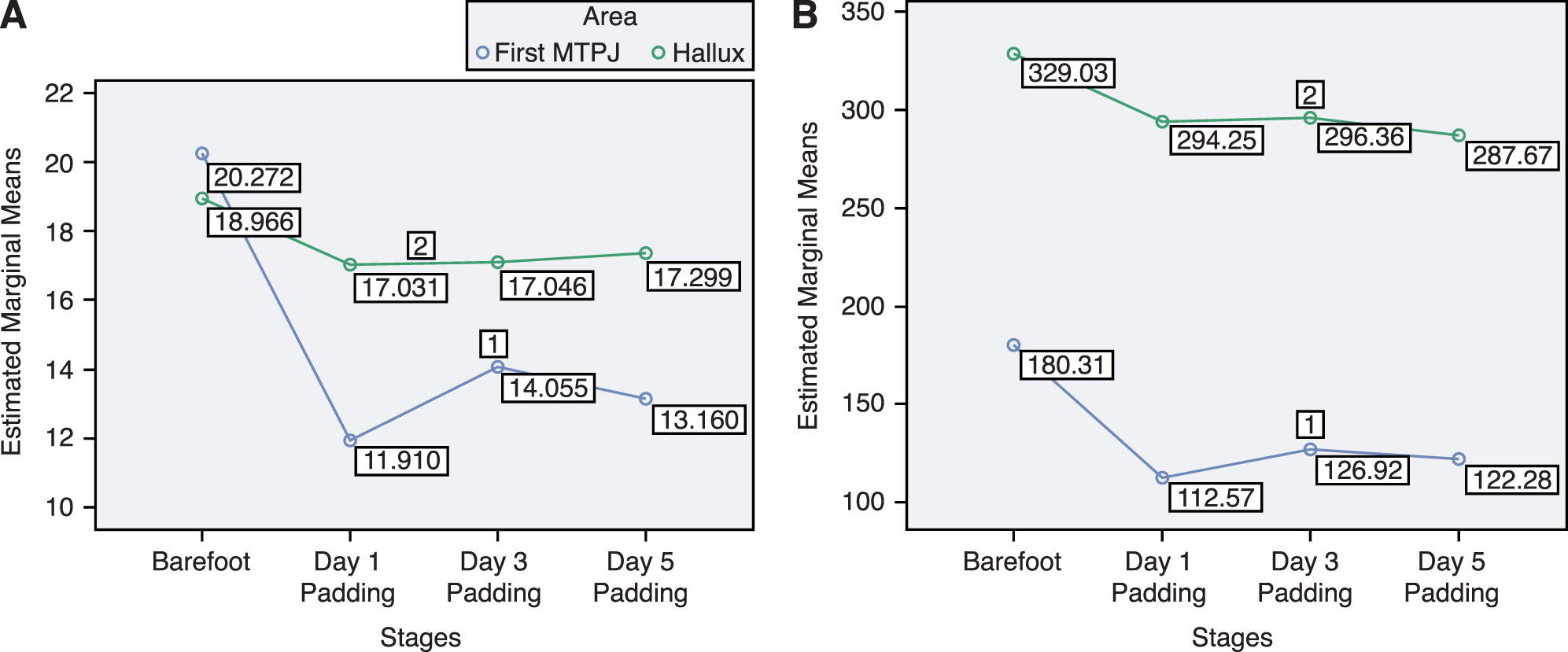
One-way analysis of variance was used to analyze the effectiveness of the SWPC on mean peak plantar pressures and impulse among the different intervals of the experiment at the region of the first metatarsophalangeal joint. With the available values, significant differences in peak plantar pressures among the four different intervals were found (P = .001). The results also indicate that there were significant differences in impulse among these intervals (P = .015). Consequently, the Tukey post hoc significant difference was used to further analyze the results, which indicate that comparing the barefoot stage with day 1 (time = 0), there is a significant reduction in pressure (P = .001) and impulse (P = .014). Comparing the barefoot stage with day 3 (time = 1), there is also a significant difference in peak plantar pressure reduction (P = .017); however, there was no significant difference in impulse (P = .105). Ultimately, comparing the barefoot stage with day 5 (time = 2), there was also a significant reduction in pressure (P = .008). Impulse exhibited a smiliar pattern (P = .049).
When analyzing the effect of the SWPC at the hallux, with the values available, no significant differences in peak plantar pressure (P = .782) and impulse (P = .845) among the four different intervals were found. Figure 2 illustrates the mean peak plantar pressure and mean impulse at the first metatarsophalangeal joint and hallux, respectively, at each stage.
Figure 2.
A, Means plot for impulse at the first metatarsophalangeal joint (MTPJ) (1) and hallux (2) according to different intervals. B, Means peak plantar pressure of the single wing plantar cover at the first MTPJ (1) and the hallux (2) according to different intervals.
Effect of the PMP at the Second Through Fourth Metatarsophalangeal Joints
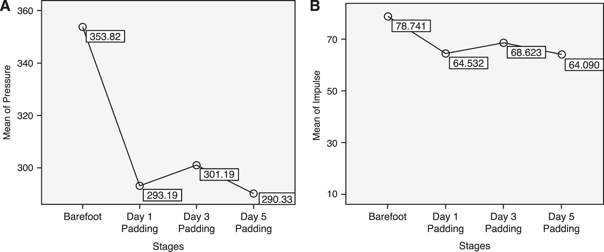
One-way analysis of variance was used to analyze the effectiveness of the PMP on the means of peak plantar pressures and impulse between the different intervals at the region of the second to fourth metatarsophalangeal joints. Although there was a slight reduction in peak plantar pressure and impulse (Fig. 3), no significant differences were found (P = .310 and P = .174, respectively).
Figure 3.
A, Means plot for peak plantar pressure on second through fourth metatarsophalangeal joints (MTPJ) according to different intervals. B, Means plot for impulse of the plantar metatarsal pad on the second through fourth metatarsophalangeal joints according to different intervals.
Discussion
The results of this study show that the SWPC devised from 5-mm adhesive felt padding was effective and durable in reducing plantar forefoot pressure at the region of the first metatarsophalangeal joint. These results are congruent with a study conducted by Tong and Ng[19] that used thicker, 7-mm, semicompressed felt for plantar forefoot padding to successfully reduce pressure at the first metatarsophalangeal joint by 37%. In addition, however, in the present study, the SWPC was not found to be significantly effective in reducing peak plantar pressure and impulse at the hallux. These results clearly indicate that the SWPC, while effective in reducing peak plantar pressure underneath the first metatarsophalangeal joint, is not effective in doing so underneath the hallux.
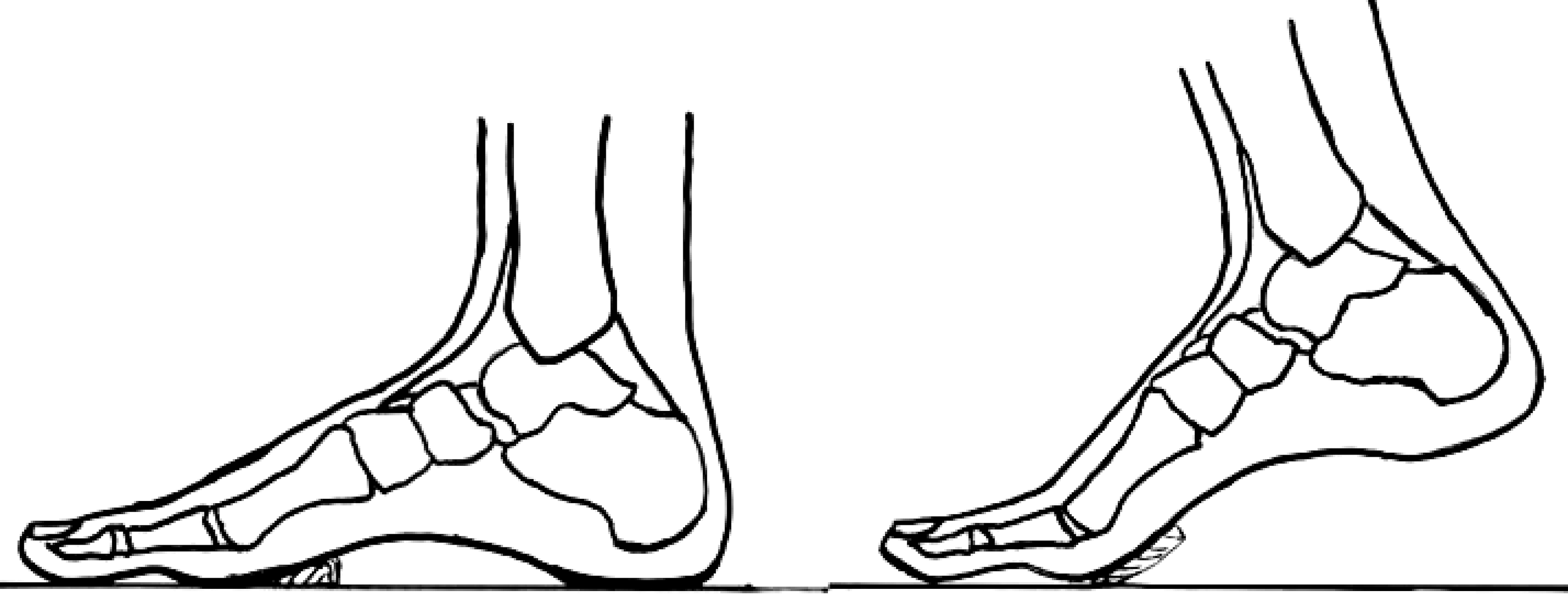
The revelation that the PMP was also not significantly effective in reducing peak plantar pressure (P = .310) and impulse (P = .174) at the region of the second, third, and fourth metatarsophalangeal joints could be surprising, initially, considering that this device has been widely used for decades in clinical practice for this purpose. The reason for nonsignificance in results could be attributed to the position of the PMP during the propulsive phase of the stance phase of gait, at a time when only the forefoot is in contact with the ground. As seen in Figure 4, at this point, the PMP is totally off the ground and, consequently, ineffective. One could hypothesize that, as a result, a PMP could be effective only during standing and apropulsive gait, such as when shuffling is present, because in these instances the foot is maintained in a plantigrade position.
Figure 4.
Plantar metatarsal pad during midstance or standing (left) and propulsion (right).
Regarding clinical application, the results of this study support the use of the SWPC as an effective method of reducing peak plantar pressure underneath the first metatarsophalangeal joint during walking but not underneath the hallux.
Furthermore, the effect of a felt PMP may be limited only to standing and a shuffling type of gait. During dynamic function, the PMP does not have the desired effect; thus, one should be careful when implementing this type of padding, either as a chairside appliance or as an orthotic intervention, when treating forefoot ulcerations or other conditions, such as Morton's neuroma and intermetatarsal bursitis, when off-loading the metatarsophalangeal joint area is crucial to treatment. This has important ramifications because these two types of padding have been used, and are still being used, during the clinical management of conditions requiring off-loading of the central metatarsophalangeal joints in the case of the PMP and the first metatarsophalangeal joint and hallux when using the SWPC. This latter type of padding is effective in reducing peak plantar pressure underneath the first metatarsophalangeal joint only.
There are a few limiting factors to consider when comparing this study with previous studies of forefoot plantar padding. The material used in each study varies in density, and the ability of each material to absorb shock may negate the ability to compare them across studies. The studied population also varies across studies, thus making comparison of results difficult. This study has included a young healthy population to evaluate the effectiveness and durability of plantar padding using synthetic felt padding. Further research, using a larger cohort to increase statistical power, should be conducted on the applicability of plantar forefoot padding to the high-risk foot with alternative off-loading scenarios.
This study showed that two frequently used designs of plantar padding using 5-mm-thick semicompressed felt are not as effective in reducing forefoot pressure as traditionally hypothesized by clinicians. The clinical implications of these findings should guide prospective clinicians who habitually attempt to off-load the forefoot during their clinical practice as to the limitations on the use of these plantar padding devices. In addition, this research should also inspire the design of alternative padding devices that may be more effective and durable; however, learning from this study, these devices should be properly researched and studied before their application to clinical practice.
Conclusions
The felt SWPC was effective in reducing peak plantar pressure and impulse underneath the first metatarsophalangeal joint and remained durable after 5 days of application. On the other hand, it was ineffective in reducing these parameters underneath the hallux. The PMP was also not effective in reducing peak plantar pressure and impulse underneath the second, third, and fourth metatarsophalangeal joints. These results cast doubts as to the effectiveness of both the SWPC and the PMP incorporated into both accommodative and functional insoles when designed to off-load the high-risk foot as a means of prevention or treatment of diabetic ulceration.
Funding
None reported.
Conflicts of Interest
None reported.
References
- Boulton AJ: Pressure and the diabetic foot: clinical science and offloading techniques. Am J Surg187: 17S, 2004.
- Foley L: Pressure point offloading in the diabetic foot. Primary Intention7: 102, 1999.
- Pham H, Armstrong DG, Harvey C, et al: Screening techniques to identify people at high risk for diabetic foot ulceration: a prospective multicenter trial. Diabetes Care23: 606, 2000.
- Caselli A, Pham H, Giurini JM, et al: The forefoot-to-rearfoot plantar pressure ratio is increased in severe diabetic neuropathy and can predict foot ulceration. Diabetes Care25: 1066, 2002.
- Lavery LA, Armstrong DG, Boulton AJ; Diabetex Research Group: Ankle equinus deformity and its relationship to high plantar pressure in a large population with diabetes mellitus. JAPMA92: 479, 2002.
- Kang JH, Chen MD, Chen SC, et al: Correlations between subjective treatment responses and plantar pressure parameters of metatarsal pad treatment in metatarsalgia patients: a prospective study. BMC Musculoskelet Disord7: 95, 2006.
- Wu SC, Jensen JL, Weber AK, et al: Use of pressure offloading devices in diabetic foot ulcers: do we practice what we preach?Diabetes Care31: 2118, 2008.
- Paton J, Bruce G, Jones R, et al: Effectiveness of insoles used for the prevention of ulceration in the neuropathic diabetic foot: a systematic review. J Diabetes Complications25: 52, 2011.
- Nubé VL, Molyneaux L, Bolton T, et al: The use of felt deflective padding in the management of plantar hallux and forefoot ulcers in patients with diabetes. The Foot16: 38, 2006.
- Birke JA, Pavich MA, Patout CAJr, et al: Comparison of forefoot ulcer healing using alternative off-loading methods in patients with diabetes mellitus. Adv Skin Wound Care15: 210, 2002.
- Lee PY, Landorf KB, Bonanno DR, et al: Comparison of the pressure-relieving properties of various types of forefoot pads in older people with forefoot pain. J Foot Ankle Res7: 18, 2014.
- Fleischli JG, Lavery LA, Vela SA, et al: 1997 William J. Stickel Bronze Award: comparison of strategies for reducing pressure at the site of neuropathic ulcers. JAPMA87: 466, 1997.
- Zimny S, Schatz H, Pfohl U: The effects of applied felted foam on wound healing and healing times in the therapy of neuropathic diabetic foot ulcers. Diabet Med20: 622, 2003.
- Nordsiden L, Van Lunen BL, Walker ML, et al: The effect of 3 foot pads on plantar pressure of pes planus foot type. J Sport Rehabil19: 71, 2010.
- Oladeji O, Stackhouse C, Gracely E, et al: Comparison of the two-step and midgait methods of plantar pressure measurement in children. JAPMA98: 268, 2008.
- Hafer JF, Lenhoff MW, Song J, et al: Reliability of plantar pressure platforms. Gait Posture38: 544, 2013.
- Bus SA, de Lange A: A comparison of the 1-step, 2-step, and 3-step protocols for obtaining barefoot plantar pressure data in the diabetic neuropathic foot. Clin Biomech (Bristol, Avon). 20: 892, 2005.
- Zimny S, Reinsch B, Schatz H: Effects of felted foam on plantar pressures in the treatment of neuropathic diabetic foot ulcers. Diabetes Care24: 2153, 2001.
- Tong JW, Ng EY: Preliminary investigation on the reduction of plantar loading pressure with different insole materials (SRP–Slow Recovery Poron, P–Poron, PPF–Poron+Plastazote, firm and PPS–Poron+Plastazote, soft). Foot20: 1, 2010.
© 2016 American Podiatric Medical Association