


Abstract
Simultaneous pancreas-kidney transplant (SPKT) is an accepted approach and the treatment of choice in patients with type 1 diabetes with accompanying end-stage renal disease. Charcot's neuroarthropathy of the foot (CN) is a fairly common and devastating complication found in patients with long-standing, mostly uncontrolled, diabetes. However, CN has also been identified as a posttransplant consequence of SPKT. Traditional postoperative immunosuppressive therapy, particularly the use of corticosteroids, is acknowledged as an additional risk factor for the development of de novo CN after SPKT. This article describes an unusual case of a patient who presented with full-blown CN deformity after SPKT.
Simultaneous pancreas-kidney transplant (SPKT) is an accepted therapy for patients with type 1 diabetes and end-stage renal disease (ESRD). In successfully restoring normoglycemic levels and correcting uremia associated with end-stage diabetic nephropathy, SPKT is considered to be a lifesaving measure for these patients. However, SPKT has been linked to the development of Charcot's neuroarthropathy (CN), a complex pathologic bone and joint destructive process normally associated with diabetes. [1-3]
Pancreas and kidney transplants are usually performed together for patients with type 1 diabetes and concomitant renal failure. In one study, quality of life significantly improved for patients receiving SPKT compared with those receiving a kidney transplant alone. [4] Besides restoration of a normoglycemic state, a functioning pancreas-kidney transplant usually succeeds in halting the progression of diabetic polyneuropathy. [5] However, a significant consequence of SPKT is the development of neuroarthropathy. [1-3]
In the case presented, a diabetic patient developed de novo CN after SPKT was performed (this pathology was not present before SPKT). In a study by Rangel et al, [1] CN was diagnosed in 4.6% of patients who underwent SPKT without a history of CN before transplant, and all of the patients were diagnosed during the first year after transplant. Matricali et al [3] reports that eight of 66 patients (12%) developed CN, four within the first year after transplant. Del Vecchio et al [2] reported Charcot-related “rapid and fulminant bone and joint destruction without a leading infectious pattern” in two cases after SPKT. Caldera et al [6] reported a patient who developed acute CN despite 11 years of tight metabolic control after successful SPKT. They concluded that in patients already affected by severe diabetic complications, tight metabolic control achieved by pancreas transplant did not prevent the development of CN. [6] The patient highlighted in this study demonstrated clinical and radiographic signs of CN 6 months after SPKT.
The goal of immunosuppressive therapy after SPKT is to minimize graft rejection. However, the use of relatively high-dose corticosteroids contributes to bone loss. [7] Preexisting osteopenia due to diabetes and persistent hyperparathyroidism due to impaired renal function or vitamin D insufficiency/deficiency also plays a significant role in the pathogenesis of bone loss. Osteoporosis is prevalent after successful SPKT, and studies show that this procedure is associated with nonvertebral fractures in up to 45% of these patients, [7] with most of these fractures involving the feet and ankles. [1] It has been further reported that five of six patients diagnosed as having CN received higher glucocorticoid doses compared with the regular dose, confirming glucocorticoid dose as the main factor associated with de novo CN after SPKT. [1]
Corticosteroids, in particular, are the mainstay of posttransplant immunosuppressive treatment, with more than 95% of transplant recipients treated with corticosteroids. [8] A requirement for larger doses of corticosteroids for immunosuppression has been reported after SPKT than after kidney transplant alone, a more frequent pulse of methylprednisone being required after SPKT, mainly because the rate of organ rejection is increased after a multiorgan transplant. [7] Significant bone loss has also been reported in liver and heart transplant recipients, with a high percentage of vertebral fractures. [7] A decline in trabecular bone mass, accepted as corticosteroid induced, is also well-documented in kidney transplant recipients. [9]
The aim of this article is to report the occurrence of acute CN in a patient in the short term after SPKT; this pathologic condition was nonexistent before this procedure.
Case Report
A 47-year-old white man presented in September 2009 with a callus on the sole of his left foot that was causing minor discomfort. His medical history included type 2 diabetes (first diagnosed in 1993), anemia, ESRD, and hypertension. The patient admits to receiving insulin 6 months after his initial diagnosis. His surgical history included only left second digit amputation.
On examination of the lower extremities, all of the pedal pulses were palpable; skin temperature was warm, with no appreciable trophic changes. Neurologic evaluation revealed intact deep tendon reflexes, proprioception, and vibratory senses, bilaterally. Light touch sensation was within normal limits via Semmes-Weinstein 5.07 monofilament testing. Examination of the left foot revealed a superficial plantar keratotic lesion extending from the second to the fifth metatarsal heads. The stump area of the second digit remaining after amputation healed without complication. A radiograph of the left foot did not reveal any pathologic osseous changes. The keratotic lesion was mechanically debrided to patient comfort. A brochure with information describing proper diabetic foot care was given to the patient. He did not request a follow-up appointment.
The patient returned 3 years later stating that he noticed that his right foot had suddenly become swollen and that he had soon afterward developed a large wound on the sole of this foot. He stated that the wound, first starting as a blister, was present for at least 30 days, but he had not sought treatment earlier because the wound was painless. He claimed that he had not sustained a recent injury to this foot.
In querying the patient regarding his medical and surgical history, he revealed that he had undergone SPKT 6 months earlier. He stated that his diabetes was now well controlled, and he no longer required insulin. Posttransplant relevant clinical laboratory data showed glycemic levels averaging 92.2 mg/dL and blood urea nitrogen levels of 9.3 mg/dL. The postoperative immunosuppresant regimen included tacrolimus (2.0 mg/d, adjusted according to blood tacrolimus levels), mycophenolate (500 mg twice daily), and prednisone (10 mg/d).
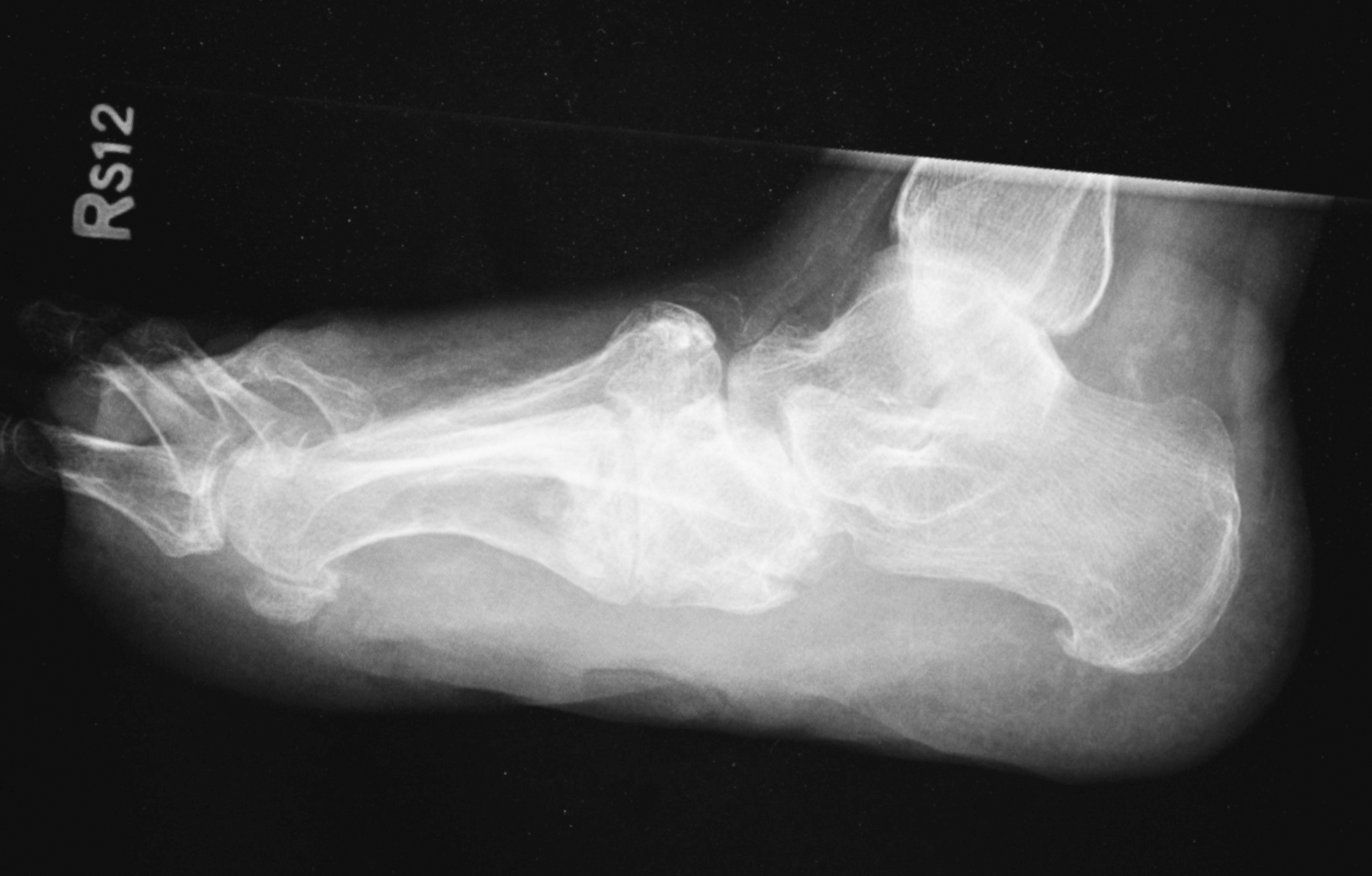
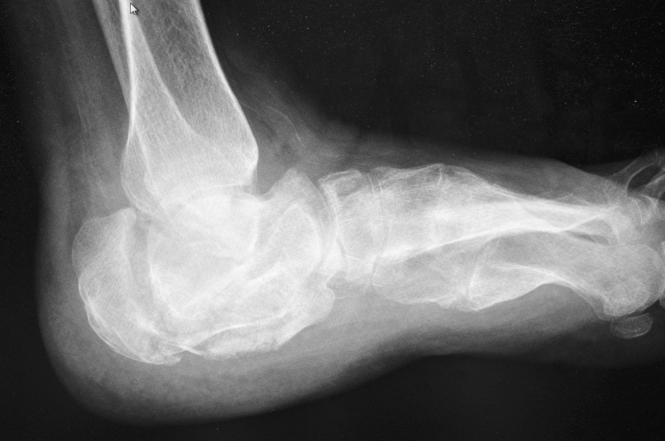
Examination of the patient's right foot displayed moderate edema of the midtarsal joint area, with an increase in skin temperature and erythyma compared with the contralateral extremity. There was a loss of protective sensation as demonstrated by Semmes-Weinstein 5.07 microfilament bilaterally. Deep tendon reflexes were absent bilaterally. There was a noticeable lack of response to sharp-dull sensory testing, particularly to the right foot. Physical examination of the right foot demonstrated painless ranges of motion of the subtalar and midtarsal joints. The patient denied any pain on normal weightbearing. The right midfoot (arch) was completely collapsed with rocker-bottom deformity, demonstrating a large neurotrophic ulcer (4.0 cm) in the center of the sole area (Fig. 1). A right anteroposterior view radiograph revealed bony demineralization with fragmentation and sclerosis of the bases of the second and third metatarsals with transverse fractures involving the navicular; first, second, and third cuneiforms; and bony bridging across the midfoot joints (Fig. 2). A right lateral view radiograph revealed significant soft-tissue swelling, multiple fractures and dislocations of the midtarsal joints, and dorsal subluxation at Chopart's joint (Fig. 3). All of the osseous degenerative changes were consistent with CN.
Figure 1.
Rocker-bottom deformity, with neurotrophic ulceration of Charcot foot.
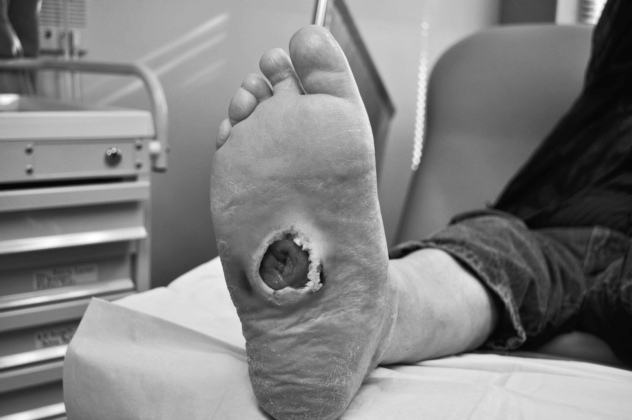
Figure 2.
Bony demineralization with fragmentation and sclerosis of the bases of the second and third metatarsals with transverse fractures involving the navicular and cuneiforms of Charcot foot.
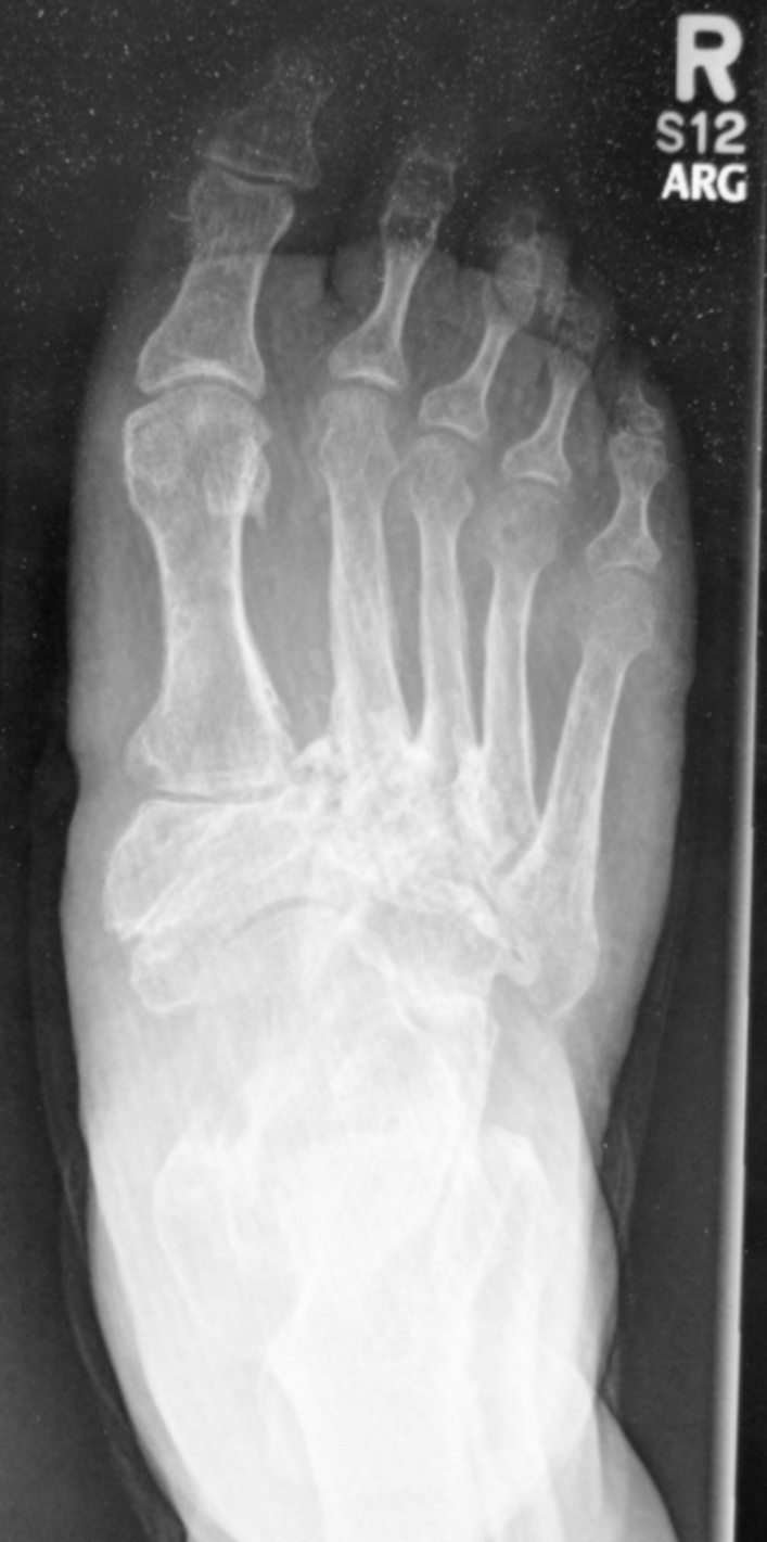
Figure 3.
Nonweightbearing lateral view of the right (Charcot) foot demonstrating dorsal subluxation of Chopart's joint, midfoot collapse, and a skin defect showing the plantar ulcer location.
The initial treatment strategy included serial total-contact casting to the affected right foot. However, the patient refused this therapy. Instead, he was placed in a right Charcot restraint orthotic walker and was advised to practice as little weightbearing as possible on the right foot. However, on the patient's return visit 1 month later, the contralateral (left) heel demonstrated significant warmth and edema. A radiograph revealed a contralateral (left) calcaneal fracture (Fig. 4), progressing to a comminuted fracture 3 weeks later (Fig. 5). After then immobilizing the left foot, the patient was transferred to an assisted living facility, where his post-CN rehabilitation could be more closely monitored.
Figure 4.
Early calcaneal fracture of the contralateral (left) foot.
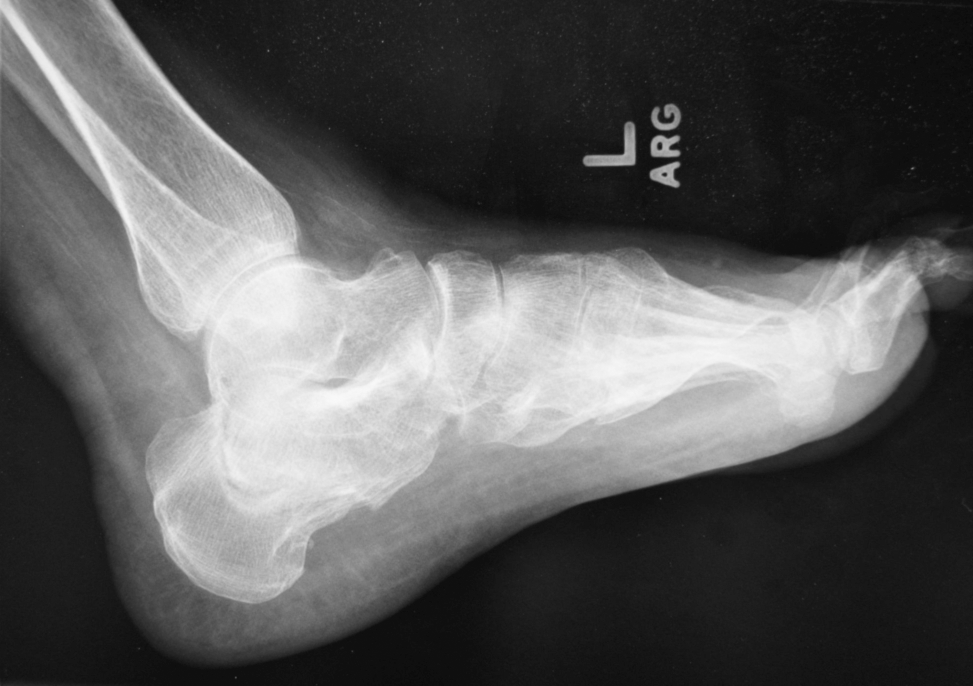
Figure 5.
Progression to comminuted fracture and collapse of the contralateral (left) calcaneus 3 weeks after the radiograph in Figure 4.
Discussion
In the reported case, the patient presented with full-blown CN deformity 6 months after SPKT was performed. There was no clinical/radiographic evidence of CN before transplant, but the patient clearly demonstrated signs of bone damage characteristic of CN 6 months after SPKT. Radiographic presentation demonstrated clear pathology characteristic of CN, with the bony bridging indicative of late-stage CN. Posttransplant immunosuppression with glucocorticoids has been identified as a main pathologic factor increasing the risk of fracture. [1] Despite his degenerative pedal condition, the patient continues with the same posttransplant immunosuppressive regimen, including predisone (10 mg/d).
Reduced-dose corticosteroid withdrawal or avoidance after transplant is encouraged. [10] This practice can aid transplant recipients in avoiding loss of bone mass because it is confirmed that SPKT patients present with an increased risk of bone fracture of CN after transplant. One useful strategy is the gradual (monthly) tapering down of the posttransplant corticosteroid dose. Substituting bisphosphonates and vitamin D after transplant has been attempted for reducing the risk of Charcot-related bone damage. Rangel et al [1] noted that short-term administration of pamidronate seemed to stabilize the progression of CN.
During this patient's convalescence, with total off-loading of the affected foot, this strict nonweightbearing approach for the affected foot led to increased mechanical forces on the contralateral nonaffected foot, ultimately also progressing into a Charcot deformity. Development of contralateral Charcot's arthropathy has been reported in up to 40% of patients due to abnormal stress patterns causing fractures on the contralateral limb. [11] In the case studied, the patient returned with a comminuted calcaneal fracture of the contralateral foot while undergoing off-loading therapy of the affected Charcot right foot.
Summary
In this case report, a patient developed full-blown CN soon after SPKT was performed, neuroarthropathy not being present before transplant. This posttransplant phenomenon has established that patients who undergo SPKT run the risk of de novo CN as a comorbidity.
The SPKT is a commonly used treatment option for type 1 diabetic patients with ESRD. However, besides autonomic neuropathy, trauma, peripheral vascular disease, and prolonged corticosteroid therapy, SPKT is now identified as an additional risk factor for CN. Glucocorticoid use in posttransplant immunosuppressant therapy has been implicated as the main factor in the development of CN. It is well accepted that corticosteroid-induced osteoporosis puts posttransplant patients at risk for fractures.
A posttransplant patient suddenly presenting with a hot, red, and swollen foot, in the presence of autonomic neuropathy (reduced pain or painless foot), should be highly suspicious for CN. If possible, minimization or avoidance of post-SPKT corticosteroid use should be considered whenever possible to avoid pedal bone and joint damage characteristic of CN. Bisphosphonates have been documented as a short-term substitute.
Before SPKT is performed, it is important for transplant surgeons to recognize the risk of the patient developing CN postoperatively. It is suggested that both feet be monitored monthly by visual and radiographic observation for the first year after transplant. Afterward, regular podiatric medical visits are recommended, inspecting the feet for preemptive signs of arthropathic changes that would warrant immediate steps to prevent the development of CN.
Acknowledgment
Dr. Alberto Orosco, Dr. John Mueller, and Elizabeth Roach, NP, for their review of the initial manuscript.
Financial Disclosure
None reported.
References
- Rangel EB, Sa JR, Gomes SA, et al: Charcot neuroarthropathy after simultaneous pancreas-kidney transplant. Transplantation94: 642, 2012.
- Del Vecchio JJ, Raimondi N, Rivarola H, et al: Charcot neuroarthropathy in simultaneous kidney-pancreas transplantation: report of two cases. Diabet Foot Ankle 4: 21819, 2013. Available at: http://dx.doi.org/10.3402/dfa.v4i0.21819. Accessed December 20,2014.
- Matricali GA, Bammens B, Kuypers D, et al: High rate of Charcot foot attacks after early simultaneous pancreas-kidney transplantation. Transplantation83: 245, 2007.
- Navarro X, Sutherland DER, Kennedy W: Long term effects of pancreatic transplantation on diabetic neuropathy. Ann Neurol42: 727, 1997.
- Recasens MJ, Ricart J, Valls-Sole A, et al: Long-term follow-up of diabetic polyneuropathy after simultaneous pancreas and kidney transplantation in type I diabetic patients. Transplant Proc34: 200, 2002.
- Caldera R, Grispigni C, La Rocca E, et al: Acute Charcot's arthropathy despite 11 years of normoglycemia after successful kidney and pancreas transplant. Diabetes Care24: 1690, 2001.
- Smets YF, van der Pijl JW, de Fijter JW, et al: Low bone mass and high incidence of fractures after successful simultaneous pancreas-kidney transplantation. Nephrol Dial Transplant13: 1250, 1998.
- Montero N, Webster AC, Royuela A, et al: Steroid avoidance or withdrawal for pancreas with kidney transplant recipients. Cochrane Database Syst Rev doi:10.1002/14651858.CD007669.pub2. http://onlinelibrary.wiley.com/doi/10.1002/14651858.CD007669.pub2/full. Accessed January 20,2015.
- O'Shaughnessy EA, Dahl DC, Smith CL, et al: Risk factors for fractures in kidney transplantation. Transplantation74: 362, 2002.
- Pascual J, Zamora J, Galeano C, et al: Steroid avoidance or withdrawal for kidney transplant recipients. Cochrane Database Syst Rev1: CD005632, 2009.
- Wilson M: Charcot foot osteoarthropathy in diabetes mellitus. Mil Med156: 563, 1991.
© 2016 American Podiatric Medical Association