


Abstract
Lipomas are the most common benign soft-tissue tumors found in the body. They tend to occur in areas of abundant adipose tissue, which explains why they are rarely found in the sole. Very few published cases of lipomas in the sole exist. When lipomas cannot be excised for histologic confirmation, ultrasonography has been shown to be a valuable assessment tool with high diagnostic capabilities. We present the case of 100-year-old patient with a long-standing plantar mass with late-stage clinical and sonographic features of a calcified lipoma.
Lipomas are the most common benign soft-tissue neoplasms found in the body. They predominantly occur in areas of abundant adipose tissue. The overall incidence is estimated to be 2.1 per 100 individuals [1], and they are mostly observed in the fifth and seventh decades of life. Plantar lipomas are rare and are characterized by their slow growth and asymptomatic presentation, which delays their diagnosis. Reports of plantar lipoma do exist in the published literature, but there are very few [2,3]. These slow-growing masses often require excision when located plantarly because of the cosmetic deformity and symptoms. Lipomas are usually relatively small, with diameters of approximately 1 to 3 cm, but in rare cases they can grow to large masses that are 10 to 20 cm in diameter and weigh up to 4 to 5 kg. Soft-tissue lipomas are classified by location into superficial and deep lesions. Superficial lipomas occur in the subcutaneous tissues, and deep lipomas occur deep to the investing fascia (ie, subfascial). Deep-seated soft-tissue lipomas are less common than superficial lipomas and may be located above muscle (supramuscular), between muscle (intermuscular), within muscle (intramuscular), or below muscle (submuscular) [4]. Superficial lipomas are commonly found in the posterior trunk, neck, and proximal extremities. Calcified cases of subcutaneous lipomas are thought to be extremely rare [5] and have been reported in up to 11% of benign fatty tumors [6].
The histologic structure of lipomas is characterized by mature adipose fat tissue with a fine capsule. The classic lipoma is composed entirely of fat, without areas of nodularity or thickened septations [7]. A substantial percentage of benign lipomas, however, show nonadipose content, attributed to fat necrosis and associated calcification, fibrosis, inflammation, and myxoid change [7]. Lipomas are typically slow-growing tumors, and calcification can occur in lipomas of long duration, probably due to degeneration of fat [5]. When subcutaneous giant lipomas are excised, histologic analysis usually reports mature adipocytes surrounded by fibrous stroma and calcified areas, which is confirmed by von Kossa staining [5]. Because histologic analysis was not performed in this case, the diagnosis was made by clinical assessment and ultrasonography. Ultrasonography, as suggested by the literature [8], is an important diagnostic tool for the examination of subcutaneous benign lesions. In particular, for lipomas it shows a high sensitivity of 88.1% (P < .01) [8]. Although, the diagnosis can be made clinically, ultrasound can be used to further assess the lesion and if there is doubt regarding the clinical diagnosis. Ultrasound examination is usually performed using a 7- to 18-MHz linear array transducer by a radiologist experienced in this field. Higher megahertz (ie, 18 MHz) yields better resolution but less penetration ability. The features specifically evaluated are the site of the tumor, location in the muscle compartment, size, shape, marginal definition, internal echogenicity (including the presence of linear internal echoes), acoustic attenuation, and vascularity. Because pure fatty tumors have few interfaces and less of an acoustic impedance mismatch, they appear echo free, whereas lipomas with mixed cellularity have an increased number of interfaces and appear echogenic. Altered acoustic transmission resulting in either posterior enhancement or attenuation is not a feature of subcutaneous lipomas. This is to be expected because the tissue is comparable with surrounding fat. Thick septa (>2 mm) and nodular or globular regions of nonadipose tissue are reported in 28% to 31% of patients with soft-tissue lipomas [4]. Such lipomas cannot be distinguished from well-differentiated liposarcomas with imaging alone, and biopsy is mandatory to exclude well-differentiated liposarcoma and the possibility of dedifferentiation.
Lipoma variants, such as angiolipoma and myolipoma, are another group of tumors that predominantly contain fat but that demonstrate nonadipose features that may be difficult to dismiss as a benign lipoma [6]. The important differential diagnosis for a benign lipoma is a well-differentiated liposarcoma, which may also demonstrate a large fat component. Features found to favor a diagnosis of well-differentiated liposarcoma include lesion size greater than 10 cm (>3.94 inches), thickness greater than 2 mm (>0.08 inches), septa (diffuse or focal), presence of globular or nodular nonadipose areas or masses, older than 60 years of age, male sex, and lesion composition of less than 75% fat [7]. Changes in the mass size can help with diagnosis. Although rapid growth is certainly of concern for malignancy, a benign mass may grow rapidly owing to hemorrhage [9]. We present a case of giant calcified lipoma of very long duration left to its natural evolution demonstrating late-stage clinical and ultrasonographic features.
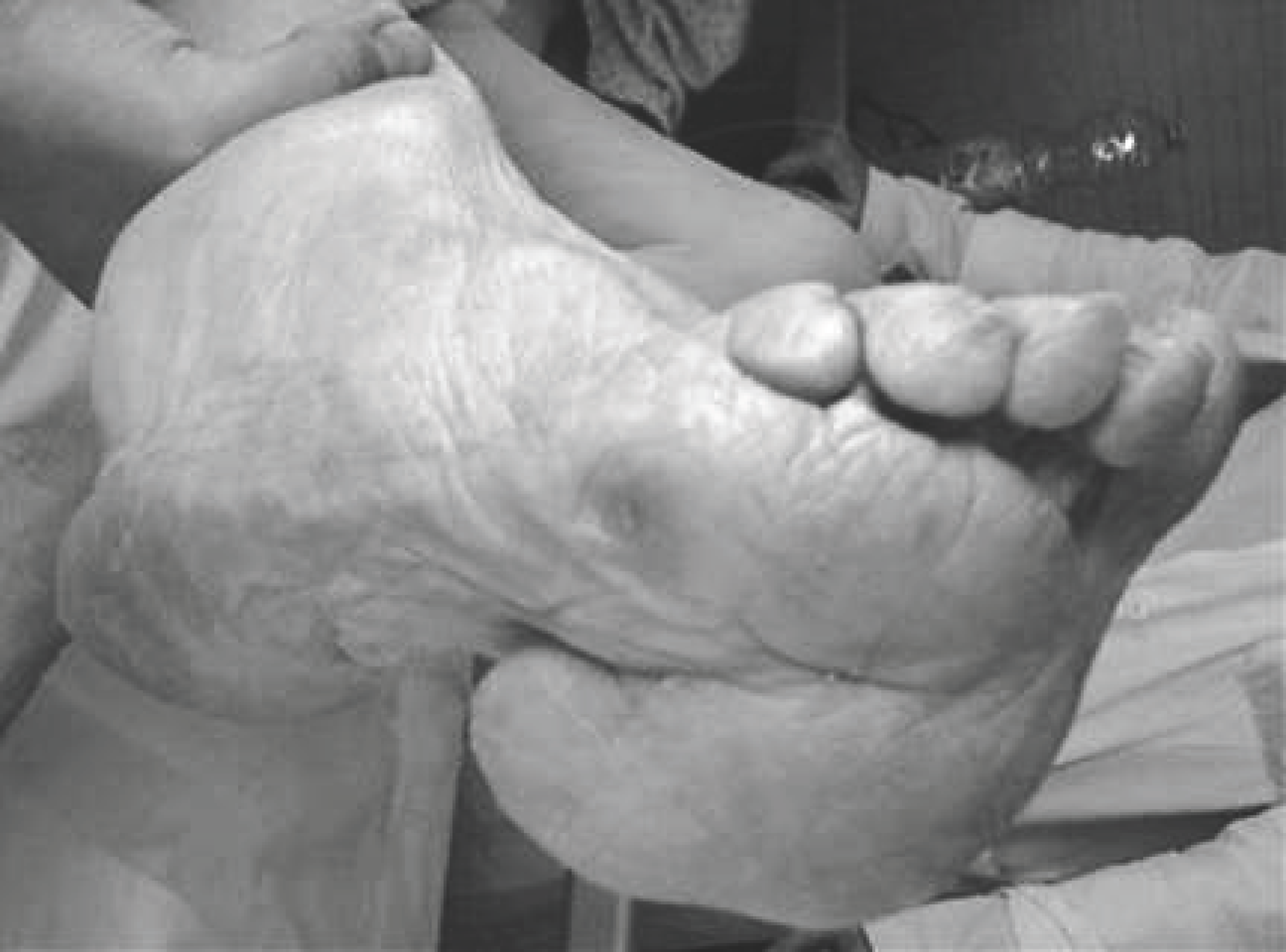
A 100-year-old woman was admitted to the geriatric ward with dyspnea and generalized edema determined by acute congestive heart failure in ischemic-hypertensive-valvular (mitro-aortic) heart disease. The physical examination revealed a voluminous, palpable, movable, painless soft-tissue mass measuring approximately 8.0 × 7.0 cm (3.15 × 2.75 inches) at the base of the first metatarsal in the plantar right foot. There were no ulcers or skin hyperkeratosis (Fig. 1).
Figure 1.
Asymptomatic, voluminous, palpable, movable, soft-tissue mass measuring approximately 8.0 × 7.0 cm at the base of the first metatarsal in the plantar right foot.
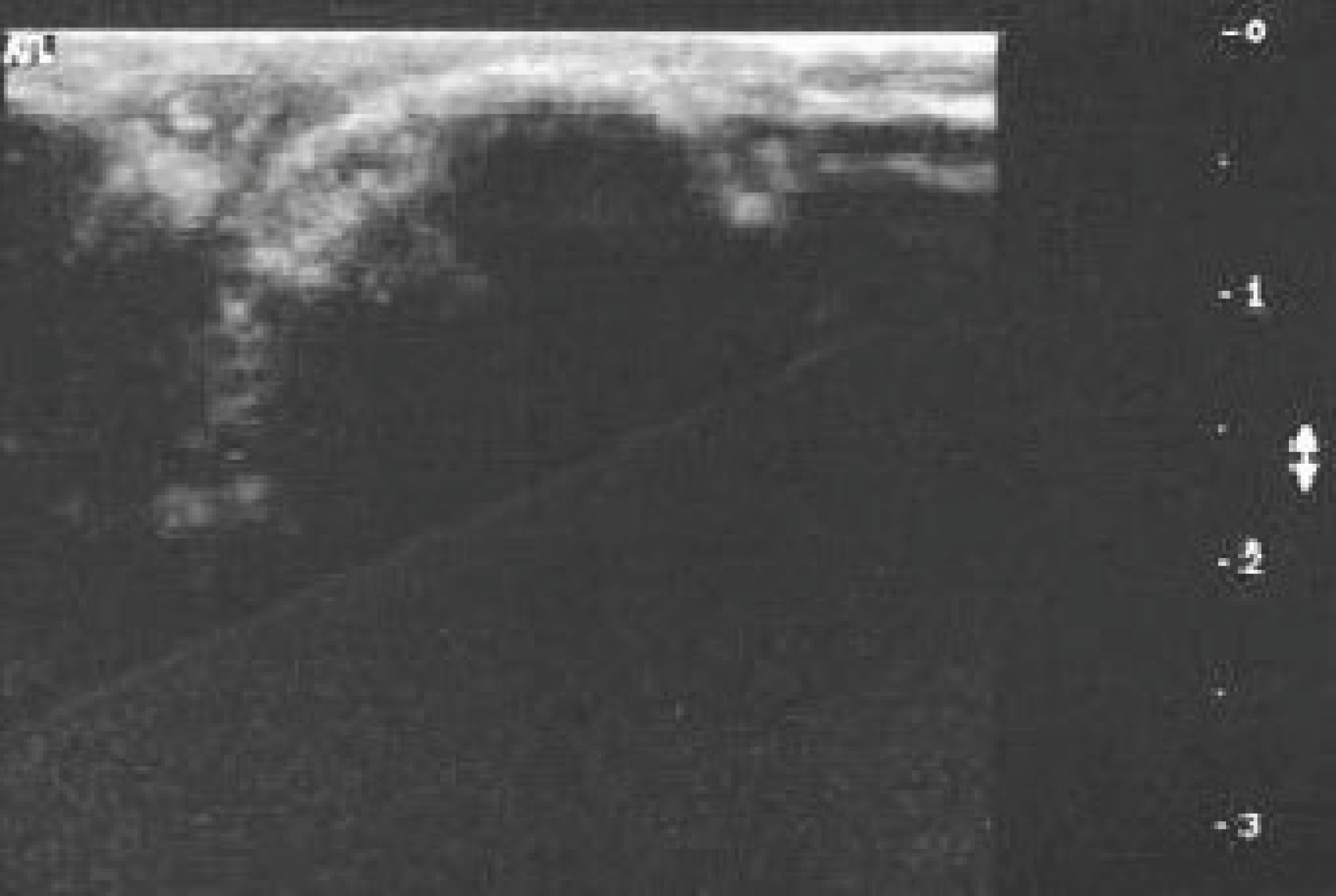
The patient and her daughter confirmed that the mass had been present for 20 years and had progressively increased in size. However, it did not affect the gait and was asymptomatic. This subcutaneous suprafascial lesion was examined by ultrasound using a 7.5-MHz linear array transducer (HDI 5000; Philips ATL, Bothell, Washington). The longitudinal and transverse planes were assessed. Ultrasound showed a multilobulated and almost calcified fatty mass underneath the plantar aponeurosis measuring 8.9 × 7.1 cm (3.50 × 2.80 inches), establishing the primary diagnosis of a calcified lipoma (Fig. 2). Radiographs of the lipoma were not acquired. The patient opposed a surgical approach, thus histologic confirmation was not obtained. Few cases of plantar lipoma have been described in the literature. In the present patient, who would refuse any invasive procedure, ultrasound proved to be very valuable and confirmed the clinical diagnosis of the lipoma. Ultrasound determined its location, its size, its relationship to the investing fascia, and the associated calcification.
Figure 2.
Ultrasound examination performed with a 7.5-MHz linear array transducer demonstrated an elongated, multilobulated, almost calcified mass (8.9 × 7.1 cm) in the subcutaneous tissue underneath the plantar aponeurosis. There are striated echoes in the mass corresponding to the septa in the subcutaneous tissues.
Although this was a rare presentation of a lipoma, and the long duration and progressing size were suggestive of malignancy, our clinical impression and the sonographic findings were enough to lead us to believe that this was a benign process that did not require an invasive procedure.
Financial Disclosure
None reported.
Conflict of Interest
None reported.
References
- MurpheyMD, CarrollJF, FlemmingDJ, et al: From the archives of the AFIP: benign musculoskeletal lipomatous lesions. RadioGraphics24: 1433, 2004.
- El KhatibK, LakouichmiM, AbouchadiA, et al: Plantar forefoot lipoma: an uncommon case report. Ann Chir Plast Esthet54: 71, 2009.
- PontiousJ, ZielaskowskiLA, KingG: Extensive lipoma of the sole. JAPMA93: 402, 2003.
- PaunipagarBK, GriffithJF, RasalkarDD, et al: Ultrasound features of deep-seated lipomas. Insights Imaging1: 149, 2010.
- KinoshitaR, YasudaH, YamamotoO, et al: Giant suspensory lipoma with calcification. J Dermatol31: 133, 2004.
- BancroftLW, KransdorfMJ, PetersonJJ, et al: Benign fatty tumors: classification, clinical course, imaging appearance, and treatment. Skeletal Radiol35: 719, 2006.
- KransdorfMJ, BancroftLW, PetersonJJ, et al: Imaging of fatty tumors: distinction of lipoma and well-differentiated liposarcoma. Radiology224: 99, 2002.
- KuwanoY, IshizakiK, WatanabeR, et al: Efficacy of diagnostic ultrasonography of lipomas, epidermal cysts, and ganglions. Arch Dermatol145: 761, 2009.
- SimonMA, FinnHA: Diagnostic strategy for bone and soft-tissue tumors. J Bone Joint Surg Am75: 622, 1993.
© 2015 American Podiatric Medical Association