


Abstract
Squamous cell carcinomas are often seen on the sun-exposed areas of the skin and are rarely observed on the digits of the foot. However, there have been incidences of squamous cell carcinoma developing in the presence of chronic wounds with osteomyelitis, thus complicating the treatment. We present a patient with osteomyelitis who developed invasive squamous cell carcinoma of the third digit. We conclude that wounds with osteomyelitis may have underlying pathologic abnormalities that are not obvious on initial presentation.
Squamous cell carcinoma (SCC) is a malignant tumor arising from epithelial keratinocytes. It is commonly seen on sun-exposed surfaces of the skin, especially of the face and the back of the hands. Approximately 1% to 2% of SCCs arise on the foot and are usually observed on the plantar surface [1]. It rarely occurs on the digits, but when seen, it is often in the presence of previously damaged skin, such as infection, premalignant lesions, actinic keratosis, chronic ulceration, etc [2,3]. We report an atypical presentation of SCC in the presence of a chronic wound with osteomyelitis of the third digit, which had been previously treated conservatively. We reinforce that chronic wounds with osteomyelitis must be treated initially aggressively to prevent possible further debilitating pathologic abnormalities.
Case Report
An 89-year-old man with a medical history significant for Parkinson's disease, Alzheimer's disease, jaundice, hip fracture, and cataract, with no known history of cancers, initially presented to our medical center for consultation of an ulceration on the distal aspect of the left foot third digit. The patient stated that he had this ulcer for 3 months and was treated with local wound care by another physician. Physical examination of the lower extremity revealed that the ulcer on the left third digit measured 0.7 × 0.4 cm and had a necrotic base with irregular borders and an exposed distal phalanx (Fig. 1A). Serous drainage and malodor were noted from the ulceration site. There was no edema; however, mild periwound erythema was present. The patient was admitted to Trinitas Regional Medical Center (Elizabeth, New Jersey) for the treatment of osteomyelitis and was immediately administered intravenous antibiotic drugs.
Figure 1.
A, Photograph depicting initial presentation of the patient with a chronic wound on the third digit of the left foot with an exposed distal phalanx. B, Anteroposterior radiographic image of the left foot depicting cortical erosion of the distal phalanx of the third digit, indicative of osteomyelitis.
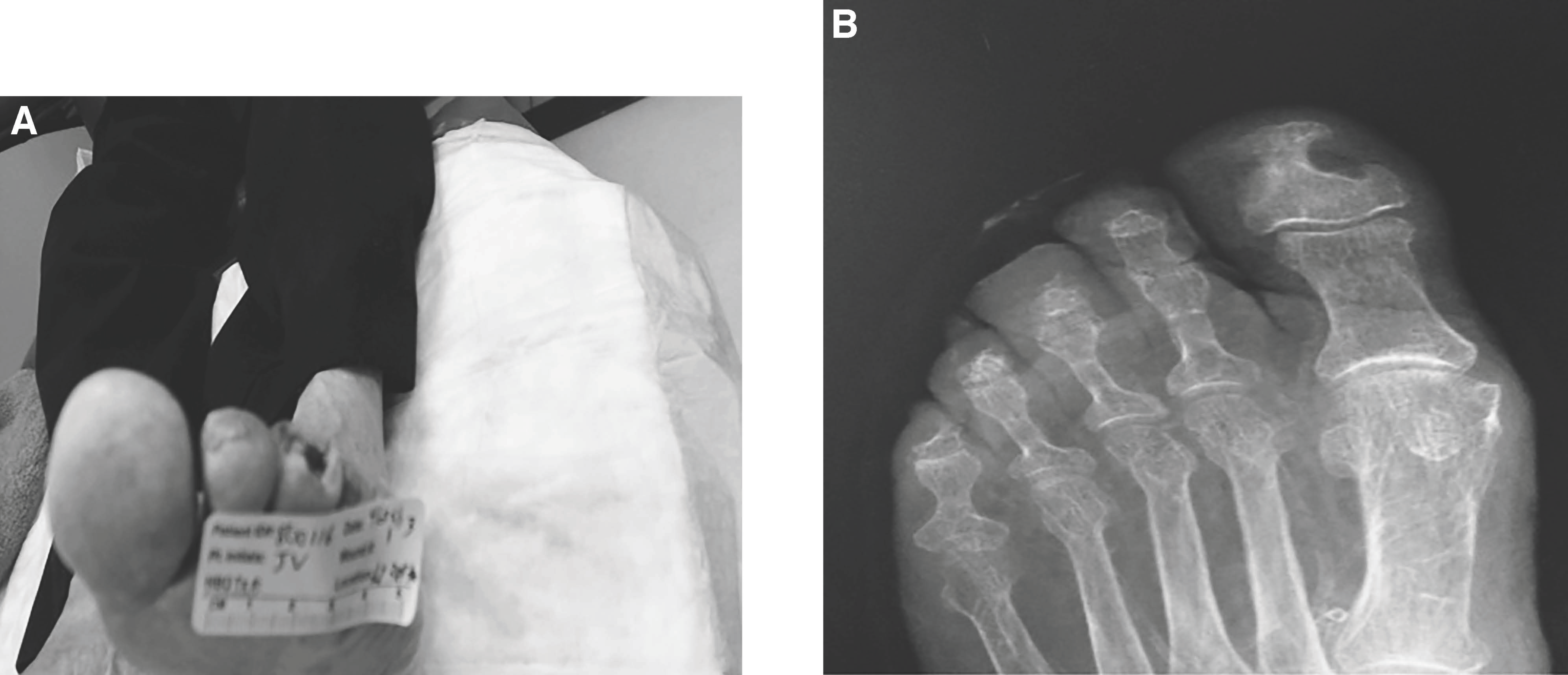
Figure 1B depicts the initial radiographic findings of bone erosion at the distal phalanx of the left third digit indicative of osteomyelitis. At this time, surgical intervention was discussed and consent was obtained for partial amputation of the left third digit. Intraoperatively, the distal phalanx and the head of the middle phalanx were noted to be eroded and were sent to the pathology laboratory along with a soft-tissue specimen. The remaining middle phalanx had no evidence of osseous destruction and, therefore, was left intact. The pathology report confirmed chronic osteomyelitis of the bone and reported that the soft-tissue specimen was positive for invasive SCC.
Figure 2 shows that the cells have loss of maturation, with an increased cytoplasm to nuclei ratio. There is increased mitotic activity, abnormal mitosis, and a hyperchromatic and eosinophilic tint. These characteristics are indicative of SCC as noted in the pathology slides of this patient's specimen.
Figure 2.
A, Low-power view showing invasive squamous cell carcinoma in the upper left corner (x40). B, Medium-power view with a confluent and invasive border surrounded by chronic inflammation (x100). C, High-power view revealing invasive “tongues” of malignant cells invading underlying stromal tissue (x200).
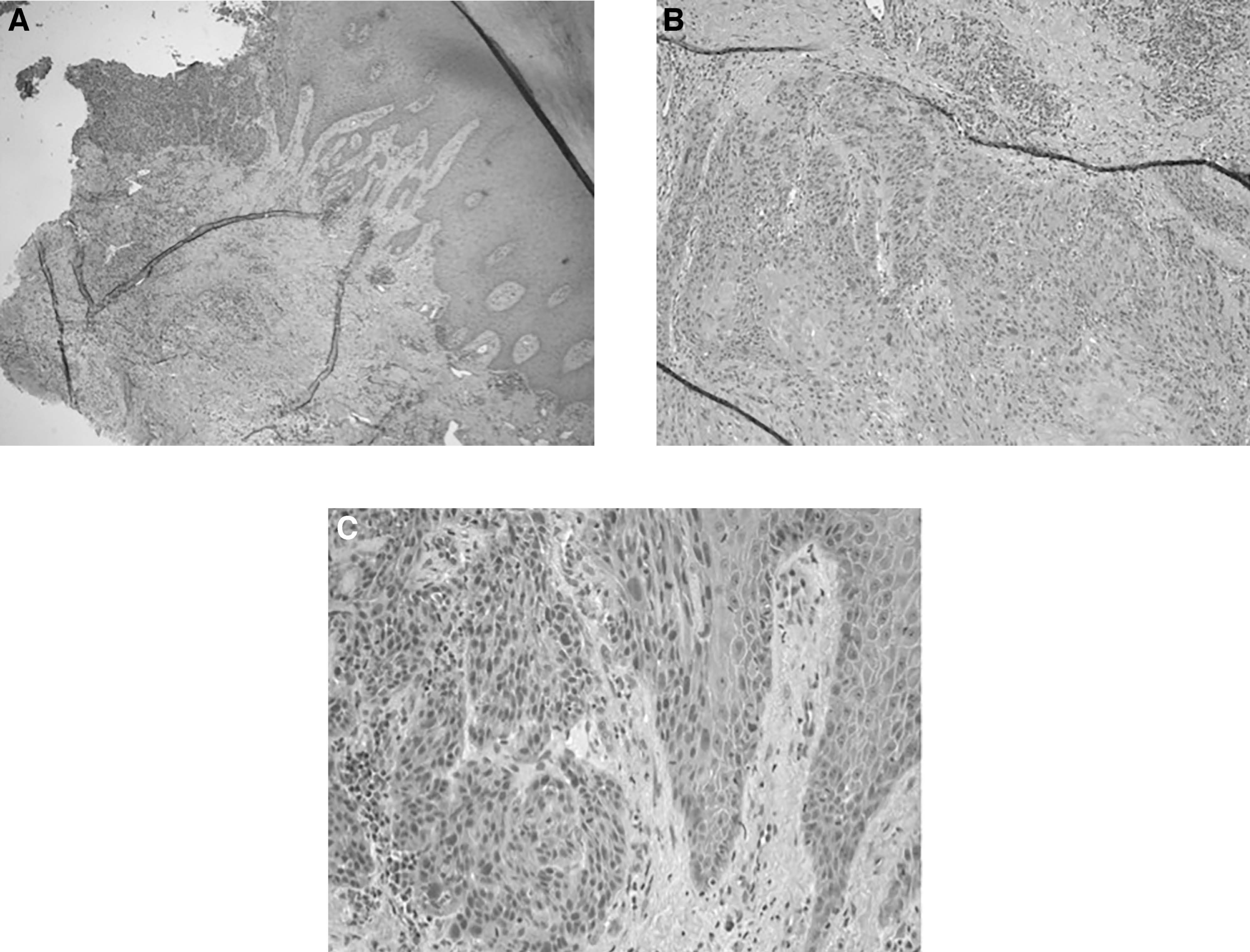
The patient was notified of the pathologic findings and was taken to the operating room for complete amputation of the left third digit with wide excision of soft tissue by excising the medial and lateral skin edges. Primary closure was obtained at the surgical site using appropriate sutures. Histopathologic analysis confirmed that the wound margins excised were clear of any pathologic abnormality. The patient was subsequently followed up in the wound care center until the surgical site completely healed, and he was also referred to a dermatologist for further work up of the invasive SCC.
Discussion
After basal cell carcinoma, SCC is the second most common type of skin cancer in the United States and accounts for approximately 700,000 new cases each year [4]. Commonly, SCC presents at areas that have been exposed to sun for prolonged periods and can cause local damage to surrounding skin and soft tissue. In addition, in some cases, SCC can even be fatal. In fact, the 5-year rate of recurrence of the primary cutaneous lesion is 8%, and the 5-year rate of metastasis is 5% [5,6]. Squamous cell carcinoma has been documented as a rare complication of osteomyelitis and chronic ulcers, especially in the lower extremity.
Squamous cell carcinoma can be confirmed based on histopathologic findings. In general, key features include abnormal pleomorphic tumor cells from the epidermis that invade into the dermis. These abnormal cells have eosinophilic cytoplasm with a central nucleus. Well-differentiated tumor cells form round nodules of intraepithelial keratinization, or “pearls.”
We present a case of SCC that developed in the presence of osteomyelitis. There have been only a limited number of these specific cases, especially in the lower extremity. Although rare, SCC has been documented in 0.2% to 1.7% of chronic wounds with osteomyelitis [7,8]. Marjolin first presented the malignant transformation in chronic wounds, and later Hawkins (1825) specified the carcinoma in the presence of osteomyelitis [8]. The exact pathogenesis of transformation is not clear. However, it is known that SCC can grow beyond the basement membrane, destroying surrounding soft tissue, and bone, and infiltrates lymph nodes through the sinus tracts [7]. Once the carcinoma has invaded epithelial boundaries into bone, amputation is unavoidable [8].
On accurate diagnosis, the most common treatments for SCC include simple surgical excision and Mohs surgery. Surgical intervention should maintain a plantigrade foot and return to functional capacity. Once the tumor has been properly removed, it is imperative to take proper preventive measures by wearing appropriate clothing, using sunscreen with at least SPF 30, and avoiding intense sun exposure. In addition, it is imperative for the patient and the health-care provider to be vigilant of wounds that have not responded to treatment because they may be an atypical presentation of SCC.
In the present patient, osteomyelitis was radiographically and clinically indicated, and pathology confirmed SCC. This finding warranted aggressive treatment with surgical excision and wide debridement of soft tissue for immediate eradication of SCC. This unique presentation of SCC is an example of why it is imperative to be vigilant of wounds that have not responded to conservative treatment as they may be an atypical presentation of a more serious pathologic abnormality.
Financial Disclosure
None reported.
Conflict of Interest
None reported.
References
- PatelA: Squamous cell carcinoma of the toe masquerading as osteomyelitis. J R Soc Med81: 418, 1988.
- TrentJT, KirsnerRS: Wounds and malignancy. Adv Skin Wound Care16: 31, 2003.
- AndrewsBT, StewartJB, AllumWH: Malignant melanoma occurring in chronic venous ulceration. Dermatol Surg23: 598, 1997.
- DavisJ, BordeauxJ: Squamous cell carcinoma. JAMA Dermatol149: 1448, 2013.
- MiriglianoE, LaTourR, AbramczukJW: Squamous cell carcinoma of the foot mimicking osteomyelitis: a case report. J Foot Ankle Surg50: 480, 2011.
- AlamM, RatnerD: Cutaneous squamous cell carcinoma. N Engl J Med344: 976, 2001.
- ZietsRJ, EvanskiPM, LusskinR, et al: Squamous cell carcinoma complicating chronic osteomyelitis in a toe: a case report and review of the literature. Foot Ankle12: 178, 1991
- KirsnerRS, SpencerJ, FalangaV, et al: Squamous cell carcinoma arising in osteomyelitis and chronic wounds. Dermatol Surg22: 1015, 1996.
© 2015 American Podiatric Medical Association