


Abstract
Background: Up to 10% of people will experience heel pain. The purpose of this prospective, double-blind, randomized clinical trial was to compare custom foot orthoses (CFO), prefabricated foot orthoses (PFO), and sham insole treatment for plantar fasciitis. Methods: Seventy-seven patients with plantar fasciitis for less than 1 year were included. Outcome measures included first step and end of day pain, Revised Foot Function Index short form (FFI-R), 36-Item Short Form Health Survey (SF-36), activity monitoring, balance, and gait analysis. Results: The CFO group had significantly improved total FFI-R scores (77.4 versus 57.2; P = .03) without group differences for FFI-R pain, SF-36, and morning or evening pain. The PFO and CFO groups reported significantly lower morning and evening pain. For activity, the CFO group demonstrated significantly longer episodes of walking over the sham (P = .019) and PFO (P = .03) groups, with a 125% increase for CFOs, 22% PFOs, and 0.2% sham. Postural transition duration (P = .02) and balance (P = .05) improved for the CFO group. There were no gait differences. The CFO group reported significantly less stretching and ice use at 3 months. Conclusions: The CFO group demonstrated 5.6-fold greater improvements in spontaneous physical activity versus the PFO and sham groups. All three groups improved in morning pain after treatment that included standardized athletic shoes, stretching, and ice. The CFO changes may have been moderated by decreased stretching and ice use after 3 months. These findings suggest that more objective measures, such as spontaneous physical activity improvement, may be more sensitive and specific for detecting improved weightbearing function than traditional clinical outcome measures, such as pain and disease-specific quality of life.
It has been estimated that approximately 10% of people will experience plantar heel pain in their lifetime, resulting in approximately 600,000 outpatient visits annually.[1] The economic burden of plantar fasciitis for 2007 was estimated to be $192 million to $376 million in direct costs.[2] Recent meta-analyses have been conducted on the use of foot orthoses for the treatment of plantar fasciitis.[3,4] Lee and colleagues[3] concluded that orthoses provide short-, intermediate-, and long-term benefit for decreasing pain and improving function in plantar fasciitis; however, true control groups were needed to assess potential bias. Hawke and colleagues[4] concluded that there was silver-level evidence for custom foot orthoses (CFOs) for the treatment of plantar fasciitis as there were conflicting results with improvements in function over noncustom orthoses at 3 and 12 months. The authors also concluded that CFOs may not reduce pain more than sham insoles or prefabricated foot orthoses (PFOs) after 3 months.[4]
Objective assessment of pain and function are important outcome measures for foot-related pathology.[5] The Medical Outcomes Study 36-Item Short Form Health Survey (SF-36) is a generic health-related quality-of-life instrument that measures a patient's physical, mental, and social well-being.[6] Using the SF-36, self-reported physical function disability from plantar fasciitis[7] is one of the most disabling foot conditions, ranking it between Charcot's neuroarthropathy[8] and adult-acquired flatfoot.[9] Disease-specific measures of these domains of health-related quality of life offer promise to be sensitive enough to detect changes over time in specific disease states or in response to treatment.[10] However, the responsiveness of these measures to detect important clinical changes has recently been called into question with the use of activity monitors.[11-14] These assessments rely on self-reporting and recall and, therefore, are subjective and prone to biases. In addition, they may not be accurate in older adults or patients with cognitive impairment. Furthermore, they often fail to capture the dynamic nature of pain and its effect on activities of daily living. In recent years, body-wearable sensor technology based on electromechanical sensors, such as accelerometers, has provided a new avenue for accurately detecting and monitoring the spontaneous daily physical activities of an individual under free conditions.[15-17] These technologies allow measurement of body postures (eg, sitting, standing, and lying), locomotion (eg, walking, gait speed, and number of steps), and its dynamics (eg, postural transition, complexity, and fractality).[11-13,18-22] Results of recent studies suggest that these objective measurements may represent the functional weightbearing capacity of an individual in detecting important clinical changes due to pain management.[13]
The purpose of this prospective, double-blind, randomized clinical trial was to compare patients randomized to receive CFOs, PFOs, or sham insoles for the treatment of plantar fasciitis. The main outcome measures at baseline, 1 month, and 3 months were first step and end of day pain, Revised Foot Function Index short form (FFI-R), SF-36, balance assessment, spontaneous physical activity monitoring, and gait analysis.
Research Design and Methods
Overall Design
Seventy-seven patients were recruited from three foot and ankle centers in the greater Chicago metropolitan area. Patients were enrolled in a prospective, double-blind, randomized clinical trial to compare three groups for the treatment of plantar fasciitis. Patients were block randomized (n = 6 in each center; created, concealed, and assigned by the research coordinator [R.T.C.]) to receive CFOs, PFOs, or sham insoles. The study was conducted in accordance with the CONSORT (Consolidated Standards of Reporting Trials) criteria for conducting and publishing clinical trials[23,24] and was registered at ClinicalTrials.gov (NCT00765843). In an effort to minimize loss to follow-up,[25] we permitted a pain rescue management allowance 3 months after all of the study evaluations were completed.[26,27] The treatment permitted a combination of nonsteroidal anti-inflammatory drugs and corticosteroid injections for patients with pain scores greater than 7 of 10 not responding to acetaminophen treatment.
Inclusion and Exclusion Criteria
Patients were included if they had 1) pain at the plantar fascial attachment to the calcaneal tubercle or pain distal from the tubercle along the plantar fascial band and 2) either pain with first steps in the morning or when getting up after being seated for a time. Patients were ambulatory, were aged 18 to 75 years, and had plantar heel pain for no more than 1 year. They could not have previous injections within 6 months or current use of CFOs. Patient history, examination, radiographs, and ultrasound ruled out other etiologies of heel pain, including proximal or local nerve entrapment, arthritis, bone cyst or tumor, and stress fracture. Because most patients likely self-treated (over-the-counter analgesics, pads, stretching, ice, etc) this condition before seeking care, we had a washout period, and acetaminophen was used as rescue analgesia. Patients were excluded if they 1) had proximal musculoskeletal pathology (ie, knee or hip arthritis, sciatica secondary to back pathology, or significant limb-length discrepancy), 2) used gait assistive devices (crutches, canes, or walkers), 3) could not wear supportive closed-toe shoes, or 4) lacked range of motion at the first metatarsophalangeal joint or subtalar joint. Willing patients meeting these criteria signed an informed consent form, and the study was conducted in accordance with the participating sites' institutional review board approval.
Imaging
Screening imaging examination included an anteroposterior and lateral radiograph and ultrasound testing to measure the thickness of the plantar fascia. Radiographs were evaluated for the presence or absence of plantar calcaneal exostoses and to rule out other causes of pain (bone cyst, tumor, etc). The single imaging examiner (A.E.F.), blinded to treatment status, performed a standardized ultrasound heel examination using an 8.0-MHz linear array transducer (Biosound LA 30C/40 MM; Aquila Esaote, Genoa, Italy).
Main Outcome Measures
The main outcome measures included overall pain as determined by the FFI-R.[28] The FFI was selected owing to its extensive reliability and validity testing.[28-32] The recent Rasch analysis of the FFI-R shortened the instrument to 34 items and improved the Cronbach alpha for scales to 0.95, making it appropriate for individual analysis.[33] Additional outcome measures at screening, baseline, 1 month, and 3 months were first step and end of day pain as assessed by a visual analog scale, SF-36, balance assessment, physical activity monitoring, and gait analysis.[6,34,35] To avoid biasing patient response owing to the influence of a clinician, patients completed the survey instruments in the examination room before each visit. This procedure was performed in an attempt to minimize influence from the examination results and the presence of the examiner.[36] Other measures included patient-reported time standing and recreational activities. Occupation was assessed for percentage of time standing, sitting, squatting, ladder or staging use, and stair climbing.
Balance Assessment
In a subset of 30 patients (randomized ten per group), activity, balance, and gait were measured. Balance and postural control was examined using the BalanSens sensor system (BioSensics LLC, Cambridge, Massachusetts) 1) to calculate the range of motion of the hip and ankle in the anteroposterior and mediolateral directions and 2) to measure variations in the center of mass. The validity and reliability of the BalanSens system have been reported previously.[37] Static balance and postural compensatory strategy was assessed in quiet upright standing for 30 sec with eyes open and eyes closed under dual- and single-leg conditions. Testing occurred in base of gait, with arms relaxed at the sides.
Physical Activity Monitoring
We used a body-worn sensor[21,38] (PAMSys; BioSensics LLC) embedded in a comfortable t-shirt at the sternal level. The sensor uses triaxial accelerometers to detect body posture (sitting, standing, and lying) and periods of walking. The validity of this approach for human motion analysis has been established by various clinical studies[21,22,39] and demonstrated overall sensitivity of 99% for detecting the time of various postural transitions (ie, sit-to-stand and stand-to-sit) and more than 87% sensitivity and specificity for identifying the transition type. Activity was monitored during 48 hours using wearable shirt-based sensors according to previous studies in which we demonstrated that two continuous weekdays of activity monitoring yields acceptable test-retest reliability (intraclass correlation coefficient >0.7)[40] and high adherence (>98% per day) to continuous wearing of the shirt-based sensor during activities of daily living.[20] This approach was also chosen to evaluate standing time and sit-to-stand transition time,[20] factors deemed important in patients with plantar fasciitis.
Gait Analysis
Spatial temporal gait was measured using a five-sensor system attached to the anterior shins, anterior thighs, and posterior lumbar region using elastic bands. The lower-limb sensors were uniaxial gyroscopes (ADXRS 250; Analog Devices Inc, Norwood, Massachusetts) that allow measurement of the angular velocity in flexion-extension. The lumbar sensors included a triaxial accelerometer and a triaxial gyroscope that permitted measurement of the range of motion of center of mass for each gait stride in the anteroposterior and mediolateral directions. Signals from sensors were digitized (16 bit) at a sampling rate of 200 Hz by a light portable data logger (LEGSys; BioSensics LLC) and were stored for off-line analysis on a Secure Digital memory card (2 MB). The methods have been described in detail and validated in previous publications.[41,42] We used this method to assess gait initiation, intercycle variability, stance time, double support, gait speed, and mediolateral and anteroposterior rotation of center of mass in previous foot orthosis studies.[43,44] Tests were conducted indoors in a well-lit, level hallway with tile flooring. Participants were instructed to walk at their habitual speed down the hallway and to stop once they reached a predetermined stopping point of 60 feet.
CFO Casting, Fabrication, and Standard Shoes
A senior experienced biomechanics instructor (B.J.) with more than 20 years of teaching and practice experience performed all of the biomechanical examinations, neutral suspension castings, and CFO prescriptions. All of the patients in the CFO group received a standard prescription for functional CFOs posted with the forefoot balanced to the rearfoot deformity (neutral calcaneal stance position) with the following exceptions: 1) valgus rearfoot (neutral calcaneal stance position) received a flat rearfoot post and 2) rearfoot varus of greater than 6° was posted to a maximum of 6° varus. Medium arch fill was used for patients who could not dorsiflex past 0° and for those who were obese (>30% over ideal body weight); minimum arch fill was used for all of the other patients. Additional accommodations to the prescription process were made depending on body stature, foot type, first-ray function, and ankle function.
A standard biomechanical examination was performed in addition to foot type assessment using the Foot Posture Index. This index has had numerous accuracy and validation studies.[45-48] The examiner evaluated the casts in a standard manner. Casts were remade if they failed one of the parameters. The examiner was not involved in the outcome or radiologic evaluation. The neutral suspension casts were sent to a laboratory accredited by the Board for Accreditation of Prescription Foot Orthotic Laboratories (Paris Orthotics Ltd, Vancouver, British Columbia, Canada) via next-day mail. The same technician evaluated all of the orthoses to ensure quality and that the device was made to the prescribed specifications. Quality assurance was a five-step process, including inspections: 1) receiving and scanning; 2) evaluation and digital cast dressing; 3) pressing, trimming, and heel posting; 4) grinding; and 5) a final quality assurance inspection to ensure accuracy, completeness, and conformity to laboratory standards. The orthoses were sent via next-day mail to ensure a maximum 14-day turnaround time. To aid in the blinding process, the CFOs, PFOs, and sham orthoses all had the same 3-mm neoprene (Spenco Medical; Waco, Texas) top cover.
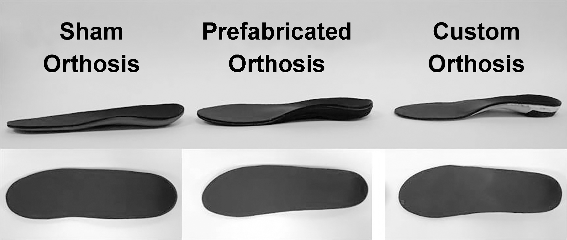
The PFO (Orthaheel Workforce; Vionic Group LLC, San Rafael, California) contained a full foot–length, Shore C, 45-durometer, ethyl vinyl acetate, triplanar orthotic footbed with a 15-mm heel cup. The device had an intrinsic 4° rearfoot medial post and was balanced through the forefoot. The manufacturer's brushed nylon top cloth was covered by the study's standard 3-mm neoprene top cover.
The sham device consisted of a full foot–length, Shore A, 35-durometer, 3-mm-thick base layer of ethyl vinyl acetate, covered by the standard neoprene top cover. The sham devices were heated for 2 min in a convection oven at 177°C and then were vacuum pressed onto a positive mold of the respective patients' feet. Thus, the sham devices had the shaped appearance of a customized device but lacked the structural integrity and support of an actual orthotic device. All sham orthoses were fabricated by the same certified pedorthist.
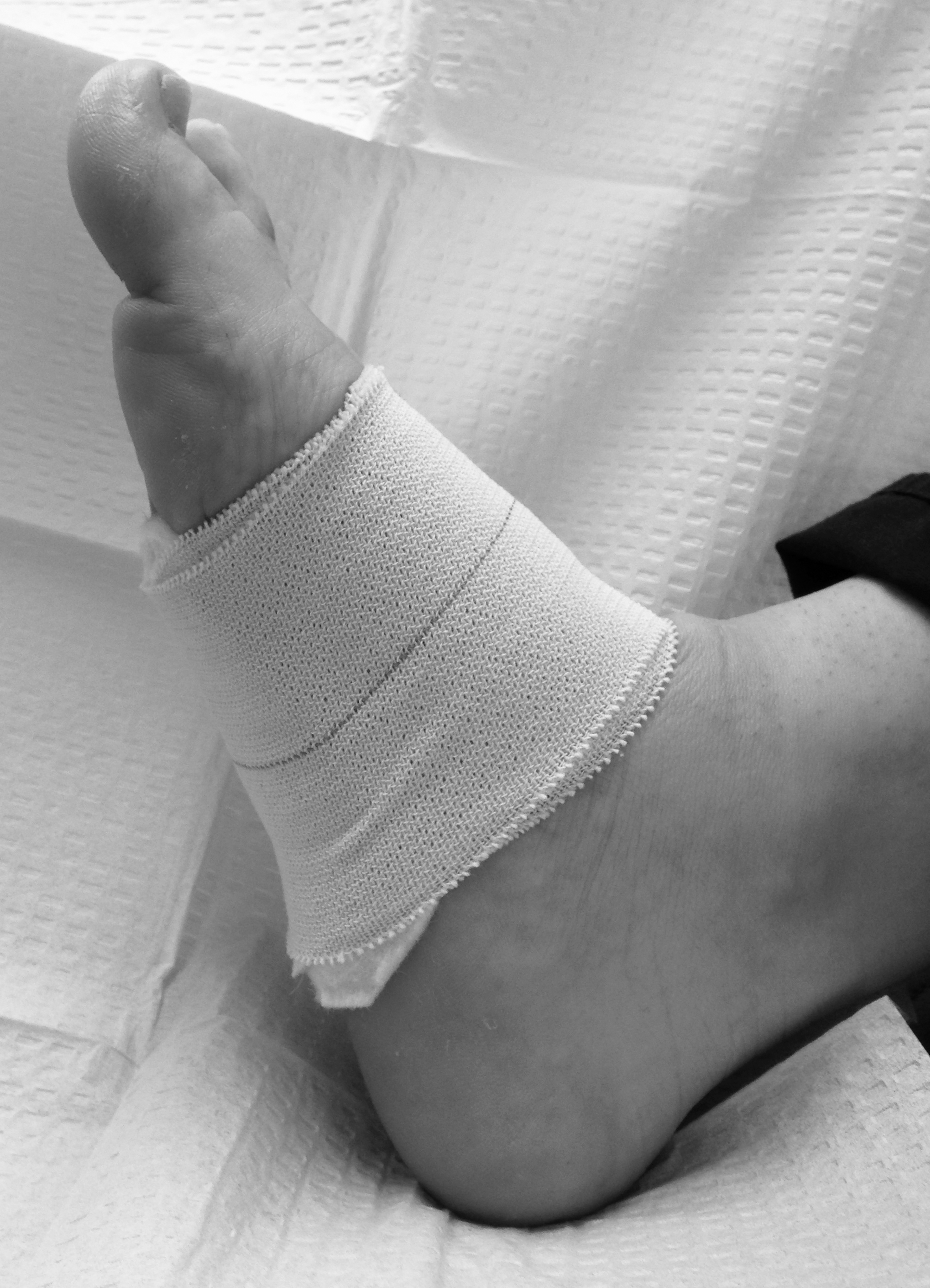
All of the patients were treated with removable longitudinal and metatarsal pads (Figs. 1 and 2) for the 7- to 14- (±3) day period before orthosis arrival. This standardized treatment avoided complications from patients having tape allergies for low-dye taping. Patients were given standardized athletic shoes (Brooks Dyad; Brooks Sports Inc, Bothell, Washington) for use with the orthoses. All of the patients were given standardized written instructions on the proper break-in for orthoses at the baseline visit. They were also instructed to wear the shoes and orthoses at all times (indoors and outdoors) when walking; however, participants were instructed that the use of slippers in the house was better than going barefoot. The same examiner (B.J.) measured and dispensed the shoes.
Figure 1.
Longitudinal and metatarsal pad.
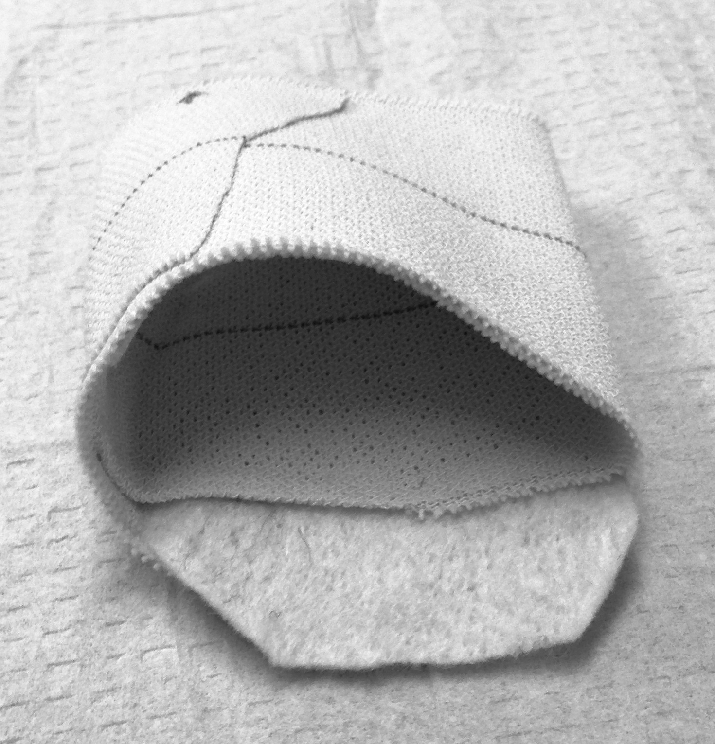
Figure 2.
Longitudinal and metatarsal pad.
Standardized Foot Self-care
All of the patients received the same self-care instructions at screening: 1) avoid using ladders or going barefoot, 2) perform stair stretching at mealtimes, and 3) use ice massage in the evening. Patients were given a demonstration of the proper stair stretching technique to be performed around mealtimes three times daily. Each stretch should total 1 min and should include a 30-sec initial period of static stretch followed by a deeper static stretch for 30 sec more. They were also given a standardized approach to ice massage using dispensed large (4-oz) paper cups. Last, they were asked to moderate their recreational activities until their pain improved.
Blinded Outcome Assessment
Casting and radiology examiners were blinded to treatment group status. Follow-up evaluations were given by the coordinator before the patient removed their shoes and saw the treating podiatric physician. Patients were blinded to the treatment condition as they all had standard biomechanical examination, neutral suspension casting, and fitting of orthoses to their shoes. The sham device was heat molded and had a similar top cover configuration as the PFO and CFO (Fig. 3). Shoes with foot orthoses were hidden by the coordinator before examination.
Figure 3.
The three study foot orthoses, with similar top covers.
Statistical Analysis and Sample Size Estimation
Parametric tests for survey data were used for the larger sample. Comparisons across the three orthosis conditions were made using analysis of variance and a Scheffé post hoc test. Comparisons between each paired condition were made using a paired two-tailed Student t test. For the activity, gait, and balance data, owing to the small sample size, the Mann-Whitney U test was used. For all of the tests, an alpha level of 0.05 was considered statistically significant. P values were adjusted for familywise error using the Holm-Bonferroni sequential approach when assessing for within-group differences for each family of outcomes. All of the calculations were performed using IBM SPSS Statistics for Windows, version 20.0 (IBM Corp, Armonk, New York) or Matlab version 7.14 (R2012a) (The MathWorks Inc, Natick, Massachuestts).
We estimated the sample size based on literature addressing the same target population. In a 2010 study,[43] we explored the impact of type of footwear on balance during walking and on gait initiation. The results suggest that the participant's dynamic balance is significantly improved on average by 22% by wearing foot orthoses compared with other footwear conditions (barefoot and regular shoes). In addition, wearing foot orthoses helped participants significantly reduce their gait initiation phase by 48% and 34% compared with barefoot and regular shoes, respectively. We assumed that a similar improvement could be observed in gait initiation and dynamic balance after using CFOs compared with other footwear conditions. Considering power = 0.80, P < .05, 19 patients are needed to observe a significant difference. We also estimated the sample size based on a previous study regarding the improvement of physical activity measured by activity monitoring in patients treated for chronic pain by spinal cord stimulation.[14] According to this study, with power = 0.80, P < .05 and an estimated population effect of d = 1.16 for the increase in the time spent walking or standing after treatment, 23 patients will be needed to have an 80% chance of finding significant changes within treatment epochs. On the same note, if we consider the improvement observed in duration of lying down during the daytime, total walking distance, and maximum distance covered in a single episode of walking, the estimated population effects of d would be −1.7, 1.17, and 1.10, respectively, which suggest a sample size of 11, 23, and 26 patients, respectively, for a significant improvement after treatment in period of lying down during the daytime, walking distance, and maximum distance in an episode of walking.
Results
Figure 4 describes the patient flow throughout the study, and we found no disparate losses to follow-up. Table 1 describes the baseline characteristics of the study population. The mean age was 49.6 years, with 63% being female. The mean height was 5 foot 5 inches, with a mean weight of 196 lb, which suggests that the population was obese, with a mean body mass index (calculated as weight in kilograms divided by height in meters squared) of 31.8. The mean duration of symptoms was 5.2 months, and the mean visual analog scale score for morning pain was 4.78 and for evening pain was 4.68. The mean self-reported standing time was 3.08 hours per day. At baseline, there was a significant correlation between self-reported standing time and age (r = −0.25; P = .03) and between self-reported standing time and duration of symptoms (r = 0.24; P = .03).
Figure 4.
Patient flow throughout the study. aPatients lost to follow-up before randomization met the inclusion criteria but did not return to the clinic to complete all of the screening visit procedures. bThe patient lost to an AE had a non–study-related AE that prohibited study treatment. AE, adverse event; CFO, custom foot orthosis; PA, daily physical activity; PFO, prefabricated foot orthosis.
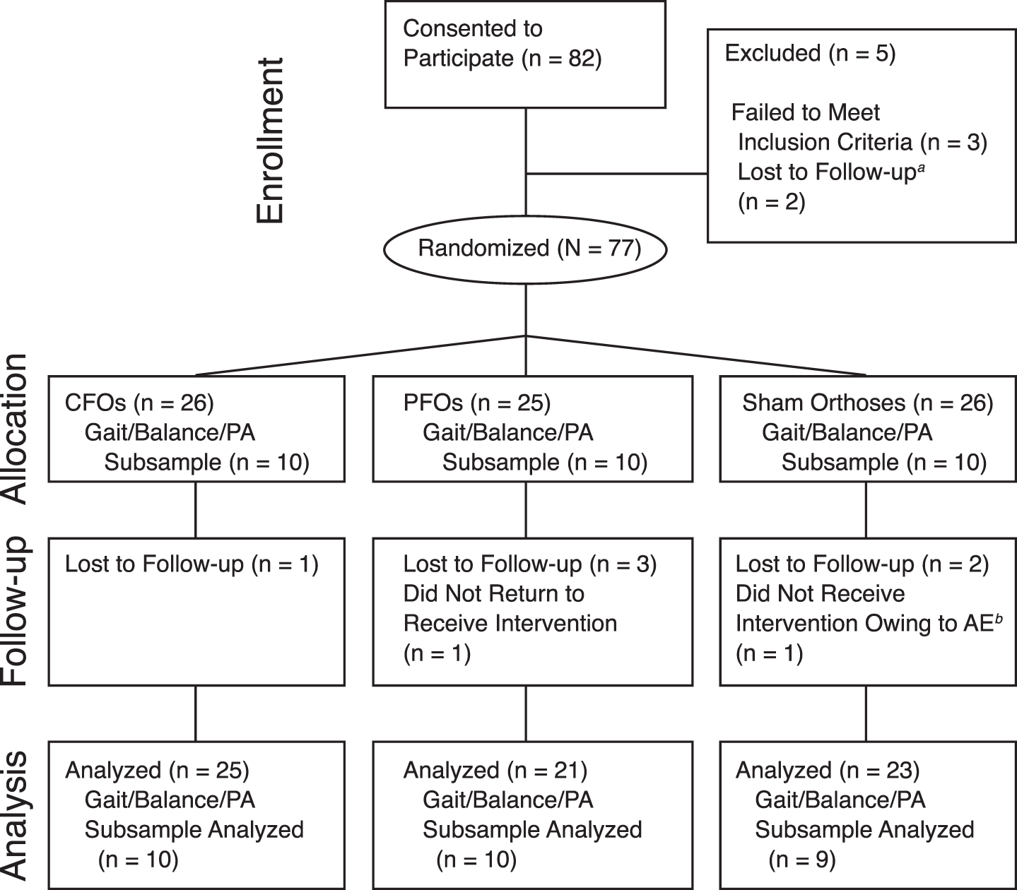
Table 1.
Baseline Characteristics of the Study Participants
Table 2 describes baseline patient characteristics, self-reported adherence, and health-related quality-of-life outcomes by treatment group. There were no significant group differences in baseline characteristics. We found no significant differences between groups at 1 or 3 months for FFI-R, FFI-R pain subscale, SF-36, or visual analog scale morning or evening pain scores. However, we did find significantly lower visual analog scale morning pain scores in all of the groups at 3 months compared with baseline. We also found significantly lower visual analog scale evening pain scores in the PFO and CFO groups over baseline. All of the groups demonstrated significantly lower pain as measured by the FFI-R from screening to baseline (P = .001–.00001; data not shown).
Table 2.
Baseline Patient Characteristics, Self-Reported Adherence, and Health-Related Quality-of-Life Outcomes by Treatment Group
A subsample of only 30 patients (ten per group) was included in activity monitoring and gait and balance assessment. Table 3 summarizes the results of spontaneous activity monitoring at baseline (before wearing orthoses) and 3 months later. No baseline between-group differences were observed for activity data for any of the assessed parameters (P > .05). Although most assessed activity parameters were improved in the CFO group compared with the other groups at 3 months, only longest walking bout (Fig. 5) and duration of sit-to-stand postural transition (Fig. 6) were significantly different between groups. Post hoc comparison suggests that the CFO group had significantly longer episodes of walking at 3 months over the sham (266%, P = .019) and PFO (169%, P = .03) groups. On the same note, postural transition was significantly lower in the CFO group compared with the sham (5%, P = .01) and PFO (5%, P = .04) groups. Although some of the activity parameters were also enhanced in the PFO group compared with the sham group, none of them achieved statistical significance in this sample.
Table 3.
Spontaneous Daily Physical Activity by Treatment Group
Figure 5.
Changes in walking bouts by number of steps from baseline to 3 months by orthosis condition. Data are given as mean ± SD. CFO, custom foot orthosis; PFO, prefabricated foot orthosis.
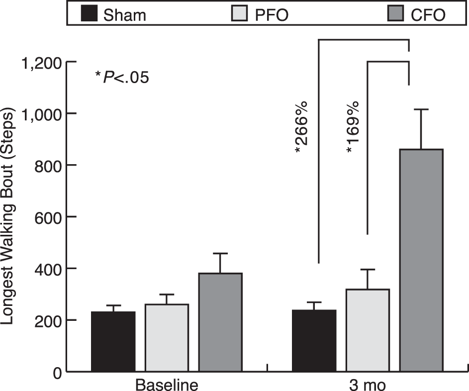
Figure 6.
Changes in sit-to-stand postural transition time from baseline to 3 months by orthosis condition. Data are given as mean ± SD. CFO, custom foot orthosis; PFO, prefabricated foot orthosis.
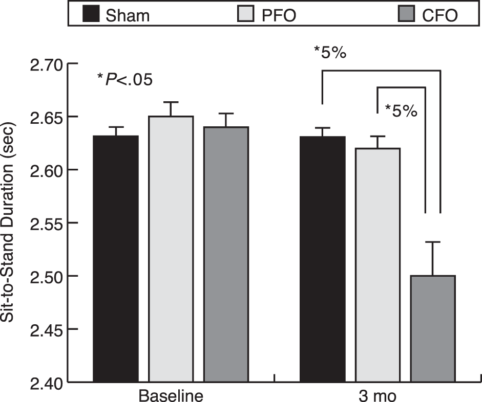
Within-group results suggest that spontaneous daily physical activities are enhanced by wearing orthoses compared with sham devices, and improvement was more pronounced with CFOs versus PFOs. The most impacted activity parameters in the CFO group compared with baseline were increases observed in the duration of standing (30%), duration of walking (18%), total number of steps (11%), number of walking bouts per day (19%), and longest walking bout, characterized by maximum number of steps per episode (125%), and a decreased sit-to-stand postural transition time (5%). The observed improvement was significant for duration of standing, duration of walking, and duration of sit-to-stand postural transition (P < .05).
For the gait parameters, moderate improvements in gait initiation speed, gait initiation steps, gait velocity, knee range of motion, and gait variability were observed in the PFO and CFO groups compared with baseline, although none reached statistical significance. On the same note, no between-group difference was observed for the tested parameters (Table 4).
Table 4.
Gait Data by Treatment Group
Similar to gait results, balance data suggest that postural control was enhanced in the CFO group after 3 months. However, results achieved statistical significance only for center of mass sway during the eyes-closed condition, with a 35% decrease compared with baseline (P = .05; 95% confidence interval, −0.10 to 0.0 cm2) (Fig. 7). No significant improvement was observed for the sham and PFO groups. Although sway improved in the CFO group during the 3 month study period, no between-group differences were observed for any of the tested balance parameters (Table 5).
Figure 7.
Changes in postural sway from baseline to 3 months by orthosis condition. Data are given as mean ± SD. CFO, custom foot orthosis; COM, center of mass; PFO, prefabricated foot orthosis.
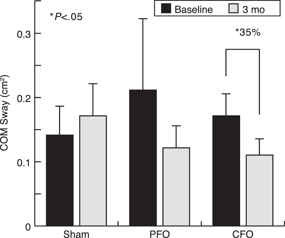
Table 5.
Balance Data by Treatment Group
The CFO group reported significantly lower use of stretching at 3 months within and among groups. The CFO group also reported significantly less ice use by 3 months, and this difference was not significant among groups. There were no differences in hours of use or comfort for shoes and foot orthoses (data not shown). There were no significant differences (P = .669) between groups regarding use of rescue pain management at the end of the study (corticosteroid injections: sham, n = 4; PFO, n = 2; CFO, n = 4).
Discussion
In this study, we found no group differences in traditional outcome measures, such as generic or disease-specific health-related quality of life, for patients with plantar fasciitis treated with sham devices, PFOs, or CFOs along with standard athletic shoes, stretching, and ice. We were not surprised by the lack of change in generic measures, such as the SF-36, because these patients were already functioning at a high level. We did find that all of the groups reported significantly lower morning pain scores after 3 months, suggesting a benefit for athletic shoe use, stretching, and ice alone. This finding is further corroborated in these data because all of the groups reported significant improvements in FFI-R pain subscale scores from screening to baseline. To our knowledge, we are the first group to describe this benefit of Achilles stretching, ice, and longitudinal and metatarsal padding for the short-term treatment of plantar fasciitis. These findings add to a growing body of literature suggesting a benefit of stretching alone[49,50] or combined with other nonorthosis treatments for the treatment of plantar fasciitis.[51-54] However, our study is the first to describe the benefit of a longitudinal and metatarsal pad in conjunction with stretching and ice. Patients were also instructed to use their athletic shoes as slippers until their pain resolved. Self-reported shoe use was 9.4 hours per day initially and diminished to 7 hours after 3 months, although this difference was not statistically significant. However, we did find that the PFO and CFO groups reported significantly lower evening pain scores at 3 months compared with baseline, although there were no group differences. The use of pain alone as a responsive outcome measure has been questioned by activity monitoring research.[14] We selected the FFI because it measures difficulties, activity limitations, and social issues in addition to pain. We found no significant improvements in function among groups at any time point. Alternatively, the CFO group demonstrated 125% increased maximal walking steps per episode over baseline and over improvements made in the PFO (22%; P = .03) and sham (0.2%; P = .019) groups. The CFO group also demonstrated improved postural transition time after 3 months compared with baseline. Another possible explanation for the CFO findings is the changes in self-reported stretching and ice use after 3 months. The CFO group demonstrated significantly less stretching over baseline and among groups after 3 months. They also reported significantly less use of ice over baseline. These two findings suggest improvement in the CFO group's condition after 3 months and may have moderated any other treatment effect measured using pain or health-related quality-of-life measures. In other words, it could be that patients with CFOs felt well enough to moderate their stretching and ice use, increase their activity, and improve their postural transition times; however, the psychometric properties of the health-related quality-of-life measures may not have been favorable enough to detect these clinically important changes.
There are strengths and limitations to this study. The study was designed and reported using the CONSORT criteria for conducting clinical trials. We designed the study to address previous criticisms from meta-analyses of plantar fasciitis trials. Namely, previous studies comparing various types of foot orthoses were underpowered, had shorter follow-up than 1 year, failed to compare with sham orthoses, did not use a validated health status measure, did not use intention-to-treat analysis, or had high levels of losses to follow-up.[26,27] The strengths of our study design included a single expert clinician for the biomechanical examination, casting, and orthosis prescription; a single orthosis laboratory accredited by the Board for Accreditation of Prescription Foot Orthotic Laboratories; patient outcome assessment before visits, with footwear hidden; a molded sham device and similar top covers to facilitate better blinding; ultrasound confirmation of plantar fasciitis; provision of standardized shoes, longitudinal and metatarsal pads, stretching, and ice; and ability of crossover into injection if needed. We believe that these strategies helped minimize to 8% the losses to follow-up and decrease the influence of missing data. The sample size estimations were made for the activity outcomes, and, thus, we may have been underpowered to detect changes in the FFI. We were also limited in the number of patients who completed follow-up greater than 3 months. We attempted a double-blind study with several precautions to ensure masking. However, we did not survey examiners and patients regarding their treatment status. Because we used objective measures, such as the FFI-R, activity monitor, and gait and balance sensors, we believe that the potential bias from unmasking is low.
Conclusions
We found that the CFO group had a significant 125% greater improvement in spontaneous physical activity after 3 months of treatment for plantar fasciitis compared with the 22% improvement with PFOs and the 0.5% improvement with sham devices. Balance, as measured by postural sway, and sit-to-stand transition time significantly improved in the CFO group by 35% and 5%, respectively. All of the groups improved in morning pain after 3 months of treatment that also included standardized athletic shoes, stretching, and ice. The CFO and PFO groups improved in evening pain over baseline, whereas the sham group did not. Changes in the CFO group may have been moderated by decreased self-reported use of stretching and ice. These findings suggest that more objective measures, such as spontaneous physical activity improvement, may be more sensitive and specific for detecting improved weightbearing function than traditional clinical outcome measures, such as pain and disease-specific quality of life.
Financial Disclosure: This study was funded by a research grant from the American Podiatric Medical Association. Brooks athletic shoes were provided at a discount price for the study. Vasyli donated the PFOs.
Conflict of Interest: None reported.
References
- RiddleDL, SchappertSM: Volume of ambulatory care visits and patterns of care for patients diagnosed with plantar fasciitis: a national study of medical doctors. Foot Ankle Int25: 303, 2004.
- TongKB, FuriaJ: Economic burden of plantar fasciitis treatment in the United States. Am J Orthop (Belle MeadNJ) 39: 227, 2010.
- LeeSY, McKeonP, HertelJ: Does the use of orthoses improve self-reported pain and function measures in patients with plantar fasciitis? a meta-analysis. Phys Ther Sport10: 12, 2009.
- HawkeF, BurnsJ, RadfordJA, et al: Custom-made foot orthoses for the treatment of foot pain. Cochrane Database Syst Rev3: CD006801, 2008.
- WrobelJS: Outcomes research in podiatric medicine. JAPMA90: 403, 2000.
- McHorneyCA, WareJEJr, LuJF, et al: The MOS 36-item Short-Form Health Survey (SF-36): III. Tests of data quality, scaling assumptions, and reliability across diverse patient groups. Med Care32: 40, 1994.
- BuchbinderR, PtasznikR, GordonJ, et al: Ultrasound-guided extracorporeal shock wave therapy for plantar fasciitis: a randomized controlled trial. JAMA288: 1364, 2002.
- GrantWP, Garcia-LavinSE, SaboRT, et al: A retrospective analysis of 50 consecutive Charcot diabetic salvage reconstructions. J Foot Ankle Surg48: 30, 2009.
- LinJL, BalbasJ, RichardsonEG: Results of non-surgical treatment of stage II posterior tibial tendon dysfunction: a 7- to 10-year followup. Foot Ankle Int29: 781, 2008.
- GuyattGH, BombardierC, TugwellPX: Measuring disease-specific quality of life in clinical trials. CMAJ134: 889, 1986.
- Paraschiv-IonescuA, PerruchoudC, BuchserE, et al: Barcoding human physical activity to assess chronic pain conditions. PLoS One7: e32239, 2012.
- Paraschiv-IonescuA, BuchserE, AminianK: Unraveling dynamics of human physical activity patterns in chronic pain conditions. Sci Rep3: 2019, 2013.
- BuchserE, Paraschiv-IonescuA, DurrerA, et al: Improved physical activity in patients treated for chronic pain by spinal cord stimulation. Neuromodulation8: 40, 2005.
- Paraschiv-IonescuA, BuchserEE, RutschmannB, et al: Ambulatory system for the quantitative and qualitative analysis of gait and posture in chronic pain patients treated with spinal cord stimulation. Gait Posture20: 113, 2004.
- AminianK, NajafiB: Capturing human motion using body-fixed sensors: outdoor measurement and clinical applications. Computer Animation Virtual Worlds15: 79, 2004.
- ZijlstraW, AminianK: Mobility assessment in older people: new possibilities and challenges. Eur J Ageing4: 3, 2007.
- NajafiB, HelbostadJL, Moe-NilssenR, et al: Does walking strategy in older people change as a function of walking distance?Gait Posture29: 261, 2009.
- NajafiB, BhararaM, TalalTK, et al: Advances in balance assessment and balance training for diabetes. Diabetes Manag2: 293, 2012.
- JeannetP-Y, AminianK, BloetzerC, et al: Continuous monitoring and quantification of multiple parameters of daily physical activity in ambulatory Duchenne muscular dystrophy patients. Eur J Paediatr Neurol15: 40, 2011.
- NajafiB, CrewsRT, WrobelJS: The importance of time spent standing for those at risk of diabetic foot ulceration. Diabetes Care33: 2448, 2010.
- NajafiB, AminianK, Paraschiv-IonescuA, et al: Ambulatory system for human motion analysis using a kinematic sensor: monitoring of daily physical activity in the elderly. IEEE Trans Biomed Eng50: 711, 2003.
- AminianK, NajafiB, BülaC, et al: Spatio-temporal parameters of gait measured by an ambulatory system using miniature gyroscopes. J Biomech35: 689, 2002.
- MoherD, SchulzKF, AltmanDG; CONSORT Group (Consolidated Standards of Reporting Trials): The CONSORT statement: revised recommendations for improving the quality of reports of parallel-group randomized trials. JAPMA91: 437, 2001.
- MoherD, SchulzKF, AltmanD: The CONSORT statement: revised recommendations for improving the quality of reports of parallel-group randomized trials. JAMA285: 1987, 2001.
- CrawfordF, AtkinsD, YoungP, et al: Steroid injection for heel pain: evidence of short-term effectiveness: a randomized controlled trial. Rheumatology (Oxford)38: 974, 1999.
- LandorfKB, KeenanAM, HerbertRD: Effectiveness of foot orthoses to treat plantar fasciitis: a randomized trial. Arch Intern Med166: 1305, 2006.
- MartinJE, HoschJC, GoforthWP, et al: Mechanical treatment of plantar fasciitis: a prospective study. JAPMA91: 55, 2001.
- Budiman-MakE, ConradK, StuckR, et al: Theoretical model and Rasch analysis to develop a revised Foot Function Index. Foot Ankle Int27: 519, 2006.
- Budiman-MakE, ConradKJ, RoachKE: The Foot Function Index: a measure of foot pain and disability. J Clin Epidemiol44: 561, 1991.
- CaselliMA, ClarkN, LazarusS, et al: Evaluation of magnetic foil and PPT Insoles in the treatment of heel pain. JAPMA87: 11, 1997.
- DiGiovanniBF, NawoczenskiDA, LintalME, et al: Tissue-specific plantar fascia-stretching exercise enhances outcomes in patients with chronic heel pain: a prospective, randomized study. J Bone Joint Surg Am85: 1270, 2003.
- BennettPJ, PattersonC, WearingS, et al: Development and validation of a questionnaire designed to measure foot-health status. JAPMA88: 419, 1998.
- Nunnally J: Psychometric Theory. McGraw-Hill, New York, 1978.
- McHorneyCA, WareJEJr, RaczekAE: The MOS 36-Item Short-Form Health Survey (SF-36): II. Psychometric and clinical tests of validity in measuring physical and mental health constructs. Med Care31: 247, 1993.
- WareJEJr: The status of health assessment 1994. Annu Rev Public Health16: 327, 1995.
- HollanderJE, ValentineSM, McCuskeyCF, et al: Long-term evaluation of cosmetic appearance of repaired lacerations: validation of telephone assessment. The Stony Brook Wound Registry Study Group. Ann Emerg Med31: 92, 1998.
- NajafiB, HornD, MarclayS, et al: Assessing postural control and postural control strategy in diabetes patients using innovative and wearable technology. J Diabetes Sci Technol4: 780, 2010.
- Najafi B: Physical Activity Monitoring and Risk of Falling Evaluation in Elderly People[PhD dissertation], Electrical Engineering Department, Ecole Polytechnique Federale de Lausanne, Lausanne, Switzerland, 2003.
- NajafiB, WrobelJ, ArmstrongD: A novel ambulatory device for continuous 24-h monitoring of physical activity in daily life. Paper presented at: North American Congress on Biomechanics; August 5-9,2008; Ann Arbor, MI.
- de BruinED, NajafiB, MurerK, et al: Quantification of everyday motor function in a geriatric population. J Rehabil Res Dev44: 417, 2007.
- AminianK, NajafiB, BülaC, et al: Spatio-temporal parameters of gait measured by an ambulatory system using miniature gyroscopes. J Biomech35: 689, 2002.
- AminianK, TrevisanC, NajafiB, et al: Evaluation of an ambulatory system for gait analysis in hip osteoarthritis and after total hip replacement. Gait Posture20: 102, 2004.
- NajafiB, e MillerD, JarrettBD, et al: Does footwear type impact the number of steps required to reach gait steady state? an innovative look at the impact of foot orthoses on gait initiation. Gait Posture32: 29, 2010.
- WrobelJS, EdgarS, CozzettoD, et al: A proof-of-concept study for measuring gait speed, steadiness, and dynamic balance under various footwear conditions outside of the gait laboratory. JAPMA100: 242, 2010.
- EvansAM, CopperAW, ScharfbilligRW, et al: Reliability of the foot posture index and traditional measures of foot position. JAPMA93: 203, 2003.
- MenzHB, MunteanuSE: Validity of 3 clinical techniques for the measurement of static foot posture in older people. J Orthop Sports Phys Ther35: 479, 2005.
- RedmondAC, CrosbieJ, OuvrierRA: Development and validation of a novel rating system for scoring standing foot posture: the Foot Posture Index. Clin Biomech (Bristol, Avon)21: 89, 2006.
- ScharfbilligR, EvansAM, CopperAW, et al: Criterion validation of four criteria of the foot posture index. JAPMA94: 31, 2004.
- RompeJD, CacchioA, WeilLJr, et al: Plantar fascia-specific stretching versus radial shock-wave therapy as initial treatment of plantar fasciopathy. J Bone Joint Surg Am92: 2514, 2010.
- DigiovanniBF, NawoczenskiDA, MalayDP, et al: Plantar fascia-specific stretching exercise improves outcomes in patients with chronic plantar fasciitis: a prospective clinical trial with two-year follow-up. J Bone Joint Surg Am88: 1775, 2006.
- KaragounisP, TsironiM, PrionasG, et al: Treatment of plantar fasciitis in recreational athletes: two different therapeutic protocols. Foot Ankle Spec4: 226, 2011.
- Renan-OrdineR, Alburquerque-SendínF, de SouzaDP, et al: Effectiveness of myofascial trigger point manual therapy combined with a self-stretching protocol for the management of plantar heel pain: a randomized controlled trial. J Orthop Sports Phys Ther41: 43, 2011.
- ClelandJA, AbbottJH, KiddMO, et al: Manual physical therapy and exercise versus electrophysical agents and exercise in the management of plantar heel pain: a multicenter randomized clinical trial. J Orthop Sports Phys Ther39: 573, 2009.
- GreveJM, GreccoMV, Santos-SilvaPR: Comparison of radial shockwaves and conventional physiotherapy for treating plantar fasciitis. Clinics (Sao Paulo)64: 97, 2009.
© 2015 American Podiatric Medical Association