


Abstract
Background: Morton’s neuroma is a perineural fibrosis of an intermetatarsal plantar nerve. Burning, numbness, paresthesia, and tingling down the interspaces of involved toes may also be experienced. Taking into account all of this information, we designed a prospective open-label study to evaluate the efficacy of pulsed radio frequency on Morton’s neuroma. Methods: Twenty patients with Morton’s neuroma were experiencing symptomatic neuroma pain in the foot not relieved by routine conservative treatment. All of the patients had been evaluated by a specialized orthopedist and were offered pulsed radio frequency as a last option before having surgery. Initially, pain level (numerical rating scale), successful pain control (a 50% pain decrease was accepted as successful pain control), comfort when walking (yes or no), and satisfaction level (satisfied or not satisfied) were evaluated. Results: We found a decrease in the pain level in 18 of 20 patients, successful pain control in 12, and wearing shoes and walking without pain in 16. Overall, satisfaction was rated as excellent or good by 12 patients with Morton’s neuroma in this series. Conclusions: This evidence indicates that ultrasound-guided pulsed radio frequency is a promising treatment modality in the management of Morton’s neuroma pain.
Morton’s neuroma is a perineural fibrosis of an intermetatarsal plantar nerve. It is a type of nerve compression syndrome that involves the common digital nerves of the second and third intermetatarsal spaces.[1] It occurs most often in middle-aged women (78% are women), and patients frequently note pain after only a short time. Burning, numbness, paresthesia, and tingling down the interspaces of the involved toes may also be experienced.[2,3]
Although the exact cause is unknown, it is believed that the interdigital nerves most commonly involving the second and third toes get pinched when walking. This trauma to the nerve causes them to swell, generating a vicious cycle of more pain, clicking, swelling, and altered sensation.[4]
Both conservative and surgical modalities are treatment options. Nonoperative treatments include nonsteroidal anti-inflammatory drugs, wider shoes with lower heels, soft metatarsal pads to reduce forefoot pressure, a metatarsal bar to shift pressure proximally, and total-contact orthoses.[2] If these management approaches fail, corticosteroid injection is widely used and can relieve inflammation in some patients; however, pain can recur after some weeks or months. Sclerosing alcohol injections, cryogenic neuroablation, and radio frequency (RF) thermoneurolysis are increasingly available treatment alternatives, but the results of these procedures are not considered to be permanent.[3] If all of these treatments fail, patients are commonly offered surgery known as “neurectomy.”[5]
Radio frequency is a treatment that uses radio waves and heat on a variety of different nerve locations to cause neural ablation or destruction of nerves, causing pain such as trigeminal neuralgia, lumbar disk hernia, and facet joint pain.[5,6] In this procedure, a constant high-frequency, high-temperature (80°–82°C) electrical current is applied to target tissue.[7,8] The high temperature applied with RF is neurodestructive and is usually characterized by a period of discomfort, including hypoanesthesia and a neuritis-like reaction. Sometimes pain may potentially worsen owing to nerve regeneration and may lead to neuroma formation.[6,8,9] Bogduk[9] subsequently discovered that the nerve did not have to be completely destroyed for patients to get relief from pain. By changing the procedure so that the temperature was decreased to 67°C and then to 40°C instead of 80° to 90°C at the tip of the probe, wherein temperatures were able to prevent total destruction of the nerve, it was demonstrated that there were no differences between patients’ levels of pain relief.[7,9,10]
The use of pulsed RF (PRF), a nonneurodestructive or minimally neurodestructive and, therefore, less painful technique, serves as an alternative to conventional RF therapy. Unlike RF, which delivers high-intensity current in pulses, allowing the heat to spread during the latent period, tissue temperatures do not exceed 38° to 42°C in this procedure, which protects the tissue from destruction.[7,9,11] The studies have demonstrated favorable outcomes in PRF compared with conventional RF.[9,10,12,13] In the Department of Anesthesiology at Gulhane Military Medical Academy (Ankara and Instanbul, Turkey), PRF has already been used as treatment for chronic orchialgia, coccygodynia, and premature ejaculation, and satisfactory outcomes have been achieved.[14–16]
Taking into account all of this information, we designed a prospective open-label study to evaluate the efficacy of PRF on Morton’s neuroma.
Patients and Methods
After receiving approval from the Gulhane Military Medical Academy ethics committee and patients’ oral and written approval, this prospective open-label study was conducted on 20 patients with Morton’s neuroma referred to the Department of Anesthesiology at Gulhane Military Medical Academy between December 1, 2010, and August 31, 2011. The patients were having symptomatic neuroma pain of the foot that was not relieved by routine conservative treatment, including changing shoes, using shoe soles and metatarsal pads, using orthoses, applying ice, taking nonsteroidal anti-inflammatory drugs, exercising the Achilles tendon, and applying local anesthetics separately or with corticosteroids or injection of alcohol. All of the patients had been evaluated by a specialized orthopedist (S.B.), and they were offered PRF as a last option before having surgery. After a thorough discussion with the patients and obtaining informed consent in accordance with institutional policies, a PRF ablation of the Morton’s neuroma was proposed. The exclusion criteria were a diabetic foot or neuropathy, coagulopathy, problems with venous drainage of the lower extremity, Burger’s disease, and not being treated by conventional treatment regimens.
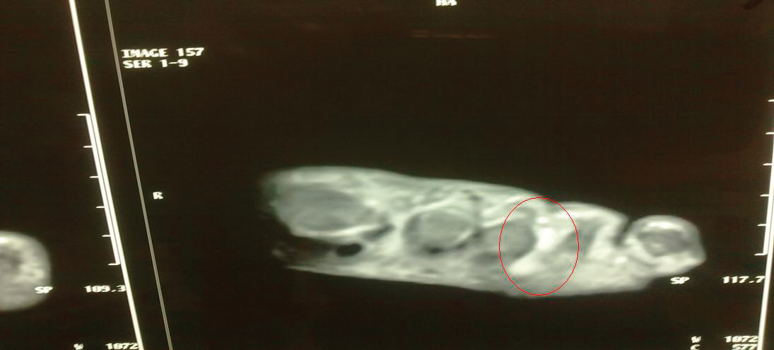
The diagnosis of neuroma is established by clinical presentation and physical examination of metatarsalgia and difficulty in walking, and the most common symptoms are localized pain, numbness, burning, and tingling while walking.[5,17–20] The pain may emerge as a crisis. The diagnosis comprises increasing pain when compressing the tender sites and neurologic signs such as hypoesthesia and hyperalgesia. If needed, ultrasonography and magnetic resonance imaging are implemented (Fig. 1).
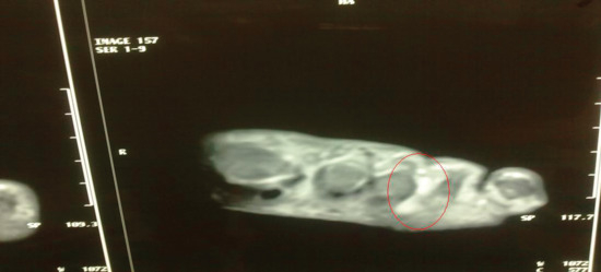
Figure 1.
Magnetic resonance imaging appearance of an hourglass-shaped Morton’s neuroma (red circle).
Initially, pain level (on a numerical rating scale from 0 to 10), successful pain control (a ≥50% pain decrease was accepted as successful pain control), comfort when walking (yes or no), and satisfaction level (satisfied or not satisfied) were questioned by an anesthesiologist, and a 6-month evaluation was made by a different anesthesiologist. The same system and technique were applied to all of the patients by the same specialized anesthesiologists (A.A. and S.T.), and a 6-month evaluation was made by different anesthesiologists (T.P. and M.Y.). The same system and technique were applied to all of the patients by the same specialized anesthesiologists (S.D. and E.K.).
Treatment Techniques
Electrocardiography, oxygen saturation, and arterial blood pressure monitoring were performed on the patients. They were sedated with 0.02 mg/kg of midazolam and were laid down in the supine position. Local anesthetic was applied by separating fingers at the two sides of the space of Morton’s neuroma.
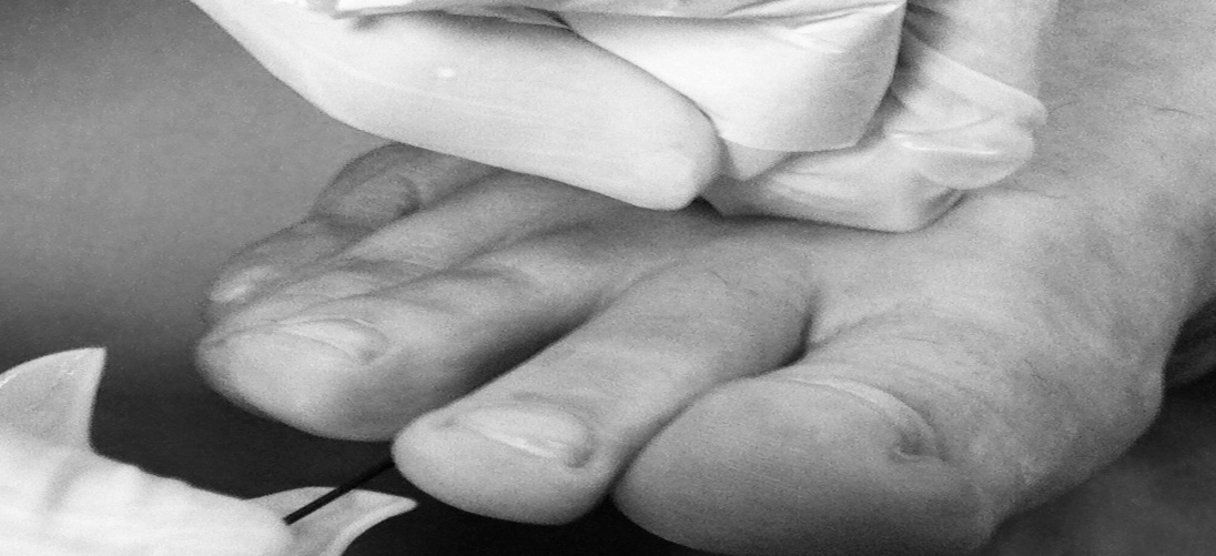
A straight 22-gauge, 5-cm, RF cannula with a 4-mm active-pinned tip (with matching electrode) that is compatible with the Cosman RFG-1A lesion generator (Cosman Medical Inc, Burlington, Massachusetts) was placed through the Morton’s neuroma with the guidance of ultrasound (the linear probe [12L-RS, 7–11 MHz] of LOGIQ Book XP ultrasound [GE Medical Systems, Solingen, Germany]) (Figs. 2 and 3). The RF electrode was placed in the cannula, and impedance measured 240 to 350 Ω. Placement of the cannula was approved neurophysiologically. Sense of difference (ie, sense of swelling, fullness, or pressure) was detected at 50 Hz with 0.4- to 0.8-V sensorial stimuli. Therapy with PRF was performed at 42°C for 300 sec. Bupivacaine, 5 mg, was injected through the cannula, and then the cannula was removed. Patients were observed for complications for 2 hours and then were discharged.
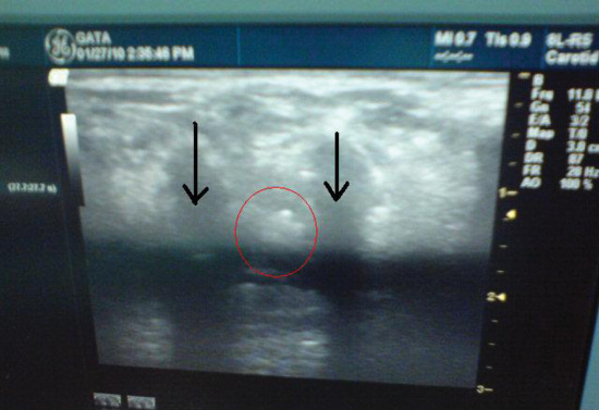
Figure 2.
Ultrasound appearance of Morton’s neuroma (red circle). Arrows indicate metatarsal bones.
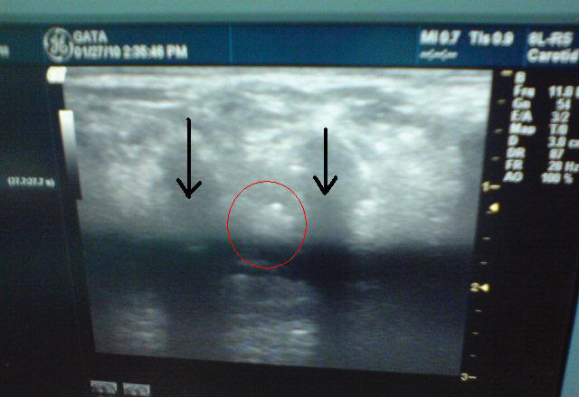
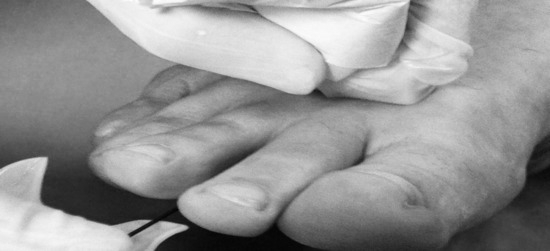
Figure 3.
Position of the cannula and ultrasound probe.
All of the analyses were performed with a statistical software program (PASW Statistics for Windows version 18.0; SPSS Inc, Chicago, Illinois). Parameters are expressed as frequency, percentile, minimum, maximum, and mean ± SE. The suitability of data to normal distribution was evaluated by the Kolmogorov-Smirnov test. Data not suitable for normal distribution were analyzed by the Wilcoxon nonparametric test. Differences between initial and 6-month numerical rating scale scores of patients were evaluated using the two-way repeated-measures analysis of variance test. Statistical significance was accepted at P < .05.
Results
Of the 20 study patients, 16 were women and four were men. The mean patient age was 48.4 years (range, 21–67 years). Two patients had two-space neuroma, so the total neuroma count was accepted to be 22. Seven of the 22 neuromas (32%) were located at the second intermetatarsal space, 13 of 22 (59%) at the third intermetatarsal space, and 2 of 22 (9%) at the fourth intermetatarsal space. Minimum follow-up was 7 months, with maximum follow-up of 15 months and mean follow-up of 9 months.
The mean ± SE numerical rating scale score of the patients was 7.1 ± 0.26 before the procedure and 3.3 ± 0.54 at 6-month follow-up (P < .0001). Of the 20 patients, 18 (90%) expressed somewhat relief of symptoms after PRF treatment, and only two patients’ (10%) symptoms had not changed. After PRF treatment, 12 patients (60%) achieved successful pain control (≥50% decrease in pain); however, the other patients also reported a decrease in pain after PRF, but this decrease was not enough to accept successful pain control. All of the patients expressed major footwear restriction and pain in walking at the initial interview, but only 4 (20%) continued to experience pain after intervention. Overall, 12 patients (60%) expressed excellent satisfaction with this procedure.
Two patients reported complications: superficial cellulitis and a moderate hematoma. Both patients were treated with antibiotic agents, nonsteroidal anti-inflammatory drugs, and elevation of the foot. None of the patients experienced permanent complications, such as numbness, tingling, or hypoanesthesia.
Discussion
The present prospective open-label study found that PRF is effective in the pain management of Morton’s neuroma. We found a decrease in the pain level in 90% of patients, successful pain control in 60%, and wearing shoes and walking without any pain in 80%. Overall, satisfaction was rated as excellent or good in 60% of patients with Morton’s neuroma in this series.
Pulsed RF is a novel therapeutic modality that offers the advantage of pain control without tissue destruction.[21,22] The mechanism of effectiveness of PRF is poorly understood, but it may involve altered signal transduction in pain pathways.[21,22] A popular theory is that the rapidly changing electric fields produced by PRF alter the transmission of pain signals via a pathway involving c-Fos, which is a hint at the inhibition of excitatory C fibers and long-term depression as a viable therapeutic mechanism in PRF.[10] Another popular theory is that serotonergic and noradrenergic stimuli with neuromodulation may have a role in the efficacy of PRF.[22]
There are few studies showing that PRF is effective in treating pain such as axial low-back pain, chronic cervical radicular pain, idiopathic trigeminal neuralgia, and chronic ilioinguinal neuralgia after inguinal herniorrhaphy.[8,13,23,24] The Department of Anesthesiology at Gulhane Military Medical Academy (Ankara, Turkey) also reported that PRF is effective in chronic orchialgia, coccygodynia, and premature ejaculation.[14–16] These studies are largely retrospective case series studies. Therefore, its clinical value needs to be explored with well-designed, randomized controlled trials with a large sample size and long-term follow-up.
Originally, Finney et al[3] reported a retrospective study in which conventional RF was effective in the treatment of Morton’s neuroma. They reported that RF provided an average of 56% improvement in symptoms, and they also reported overall satisfaction with RF in 68% of cases. Twenty-three years later, Moore et al[5] expressed a success rate of 83% in their retrospective series of patients treated with conventional RF without any significant complications. We conducted this series in a prospective manner to eliminate bias, and the success rate is also similar to that of the series by Moore et al.
In both series related to the efficacy of RF on Morton’s neuroma, it was seen that the probe was placed by means of fluoroscopy to make sure that it was in the correct intermetatarsal area. Unlike these studies, we used ultrasound to confirm whether the probe was placed in the correct spaces. Besides being used for diagnostic purposes, ultrasound guidance has also been used for diagnostic blockade and neurolytic injection of an ablative agent in chronic pain.[11] Ultrasound has a variety of advantages in the management of chronic pain, such as being a bedside test, not using radiation, and visualization and localization of neuroma. Therefore, we believe that ultrasound guidance should be used as part of placing the probe in the right spaces in the treatment of Morton’s neuroma with PRF or conventional RF.
Surgical treatment methods are usually preferred as a last choice. Achievement drive[17,18,25] in the long-term is reported to be 57% to 85%, although this success rate had complications such as localized pain, scar ache, numbness, burning, and tingling (38%–67%).[5,17–20,25] No permanent complications were observed in the present study, but two patients had transient complications (10%). Cahana et al[26] examined PRF treatment performed on more than 1,200 patients, and they found no lasting complications. The present results also coincide with the literature.
There are some limitations of the present study. This series is relatively small; 20 patients may not have been enough to support PRF in the treatment of Morton’s neuroma. In addition, prospective comparative studies are needed, and we have designed a study to compare the results of a surgical procedure and PRF in Morton’s neuroma.
In conclusion, this evidence shows that ultrasound-guided PRF is a promising treatment modality in the pain management of Morton’s neuroma. However, well-designed studies (prospective, randomized, placebo-controlled trials with a large sample size and long-term follow-up) are needed to ascertain the clinical value of this treatment modality.
Financial Disclosure
None reported.
Conflict of Interest
None reported.
References
- AddanteJB, PeicottPS, WongKY, et al: Interdigital neuromas: results of surgical excision of 152 neuromas. JAPMA76: 493, 1986.
- BartolomeiFJ, WertheimerSJ: Intermetatarsal neuromas: distribution and etiologic factors. J Foot Surg22: 279, 1983.
- FinneyW, WienerSN, CatanzaritiF: Treatment of Morton’s neuroma using percutaneous electrocoagulation. JAPMA79: 615, 1989.
- GoldmanF: Intermetatarsal neuromas: light and electron microscopic observation. JAPA70: 265, 1980.
- MooreJL, RosenR, CohenJ, et al: Radiofrequency thermoneurolysis for the treatment of Morton’s neuroma. J Foot Ankle Surg51: 20, 2012.
- SluijterME: The role of radiofrequency in failed back surgery patients. Curr Rev Pain4: 49, 2000.
- SlappendelR, CrulBJ, BraakGJ, et al: The efficacy of radiofrequency lesioning of the cervical spinal dorsal root ganglion in a double blinded randomized study: no difference between 40 degrees C and 67 degrees C treatments. Pain73: 159, 1997.
- RozenD, AhnJ: Pulsed radiofrequency for the treatment of ilioinguinal neuralgia after inguinal herniorrhaphy. Mt Sinai J Med73: 716, 2006.
- BogdukN: Pulsed radiofrequency. Pain Med7: 396, 2006.
- Van ZundertJ, de LouwAJ, JoostenEA, et al: Pulsed and continuous radiofrequency current adjacent to the cervical dorsal root ganglion of the rat induces late cellular activity in the dorsal horn. Anesthesiology102: 125, 2005.
- Restrepo-GarcesCE, MarinovA, McHardyP, et al: Pulsed radiofrequency under ultrasound guidance for persistent stump-neuroma pain. Pain Pract11: 98, 2011.
- ByrdD, MackeyS: Pulsed radiofrequency for chronic pain. Curr Pain Headache Rep12: 37, 2008.
- TekinI, MirzaiH, OkG, et al: A comparison of conventional and pulsed radiofrequency denervation in the treatment of chronic facet joint pain. Clin J Pain23: 524, 2007.
- BasalS, ErginA, YildirimI, et al: A novel treatment of chronic orchialgia. J Androl33: 22, 2012.
- BasalS, GoktasS, ErginA, et al: A novel treatment modality in patients with premature ejaculation resistant to conventional methods: the neuromodulation of dorsal penile nerves by pulsed radiofrequency. J Androl31: 126, 2010.
- AtimA, DenizS, KurtE: Re: coccygodynia. Pain Pract11: 507, 2011.
- LeeKT, KimJB, YoungKW, et al: Long-term results of neurectomy in the treatment of Morton’s neuroma: more than 10 years’ follow-up. Foot Ankle Spec4: 349, 2011.
- ValenteM, CrucilM, AlecciV: Operative treatment of interdigital Morton’s neuroma. Chir Organi Mov92: 39, 2008.
- GenonMP, ChinTY, BediHS, et al: Radio-frequency ablation for the treatment of Morton’s neuroma. ANZ J Surg80: 583, 2010.
- SchreiberK, KhodaeeM, PoddarS, et al: Clinical inquiry: what is the best way to treat Morton’s neuroma?J Fam Pract60: 157, 2011.
- CosmanERJr, CosmanERSr: Electric and thermal field effects in tissue around radiofrequency electrodes. Pain Med6: 405, 2005.
- HagiwaraS, IwasakaH, TakeshimaN, et al: Mechanisms of analgesic action of pulsed radiofrequency on adjuvant-induced pain in the rat: roles of descending adrenergic and serotonergic systems. Eur J Pain13: 249, 2009.
- Van ZundertJ, PatijnJ, KesselsA, et al: Pulsed radiofrequency adjacent to the cervical dorsal root ganglion in chronic cervical radicular pain: a double blind sham controlled randomized clinical trial. Pain127: 173, 2007.
- Van ZundertJ, BrabantS, Van de KelftE, et al: Pulsed radiofrequency treatment of the Gasserian ganglion in patients with idiopathic trigeminal neuralgia. Pain104: 449, 2003.
- CoughlinMJ, PinsonneaultT: Operative treatment of interdigital neuroma: a long-term follow-up study. J Bone Joint Surg Am83: 1321, 2001.
- CahanaA, Van ZundertJ, MacreaL, et al: Pulsed radiofrequency: current clinical and biological literature available. Pain Med7: 411, 2006.
© 2015 American Podiatric Medical Association