


Abstract
The normal radiographic anatomy of the foot and ankle, aside from my previous work, has been addressed only superficially or sparingly in the medical literature. This project correlates the detailed radiographic anatomy of the entire adult foot and ankle (two-dimensional) to osteology (three-dimensional). Each bone's position was determined after meticulous examination and correlation to an articulated skeleton relative to the image receptor and direction of the x-ray beam, with correlation to the radiograph for confirmation. Images of each foot and distal leg bone (“front” and “back” perspectives) are presented alongside a corresponding radiographic image for comparison. The normal gross and radiographic anatomy is correlated and described for each radiographic positioning technique. Foundational knowledge is provided that future researchers can use as a baseline (“normal”) and that students and practitioners can use for comparison when interpreting radiographs and distinguishing abnormal findings. The findings from the original project, owing to its broad scope, have been divided into five parts: the lower leg (the focus of this paper), the greater tarsus, the lesser tarsus, the metatarsals, and the phalanges.
A common pitfall when interpreting foot and ankle radiographs is mistaking normal anatomical shadows for pathologic abnormalities. To truly appreciate abnormalities of position, form, architecture, and density, the interpreter must have a working knowledge and appreciation of normal radiographic anatomy. In the foot and ankle, this requires correlation of normal three-dimensional (3-D) osteology (the bone specimen) to two-dimensional (2-D) radiographic anatomy (the diagnostic image).
The normal radiographic anatomy of the foot and ankle, aside from my previous works, [1,2,3,4,5,6] has been addressed only superficially or sparingly in the medical literature. Four relatively recent textbooks on radiology of the foot, for example, barely touched this subject matter. [7,8,9,10] Other textbooks that did concentrate on the radiographic anatomy of the skeleton simply named the bones, with little or no correlation to osteology. [11,12,13,14] Although osteology was described in some publications, their descriptions were either limited or were not correlated to radiographs. [15,16,17,18] The only articles that provided some detailed radiographic anatomy were limited to the tarsus and talotibial joint. [19,20,21] Rhea et al [22] were unique in their approach. A single tarsal bone was removed from an articulated foot skeleton and the standard foot views were obtained. That bone was then replaced and another bone removed, repeating the procedure. Although the radiographic appearance of each tarsal bone was identified, osteology was not described.
The comprehensive results discussed and pictorially presented in this project otherwise do not exist in the literature and are new scientific knowledge.
Methods
One set of normal-appearing radiographs demonstrating each foot and ankle radiographic positioning technique (totaling 11 views, listed later herein) was randomly chosen from the records of unidentified patients. One set of normal-appearing disarticulated foot and distal leg bones was randomly selected from a personal collection of unidentified skeletons. (The normal samples were selected based on that described in the literature and the expertise of the author.) The exclusion criteria included any abnormal osteological findings and any skeleton with open physes. The goal was then to photograph each bone from two perspectives (“front” and “back”) such that it mimicked the position in which it would be seen in each radiographic view. The equipment used to obtain the bone specimen images included a 10 × 10 × 2-inch cardboard box filled with black art sand (for positioning the bone), a professional-grade photographic copy stand with four flood lamps, a Canon SLR camera with a 50-mm f/2.8 macro lens (Canon USA Inc, Melville, New York), and Kodak Rapid Process Copy film (Eastman Kodak, Rochester, New York).
The position of each bone was determined based on meticulous examination and correlation to the normal articulated foot skeleton (which included the distal leg bones), normal radiographs of the foot and ankle, and the radiographic positioning technique of the articulated foot skeleton relative to the image receptor and the direction of the primary x-ray beam. Each foot and distal leg bone was individually examined in the articulated skeleton with the naked eye from the perspective of an x-ray tube; then the individual bone specimen was positioned on the sand in the same position as identified in the articulated skeleton. The bone's position was also correlated to the normal radiograph for confirmation, and then the bone was photographed. The same procedure was then performed with the articulated foot skeleton reversed (rotated 180°) and the bone specimen repositioned such that the “back” of the 3-D bone could be photographed.
The resultant photographs were digitized, and, when necessary, the image brightness levels were adjusted with Adobe Photoshop (Adobe Systems Inc, San Jose, California) for consistency. The “reversed” image was flipped horizontally to complement the orientation of the original image so that they both matched the radiograph orientation. This technique resulted in two images (“front” and “back”) of each bone. The corresponding 2-D radiographic images were digitized and cropped so that the individual bone under study was centered.
The resultant three images for each bone (“front” and “back” of the bone specimen and the radiograph) were placed in a collage for visual comparison, and the normal gross and radiographic anatomy was correlated and described for each radiographic positioning technique.
The following standard radiographic positioning techniques (as described by Christman23) were replicated, and images were obtained for each of the following bones:
- •
- Tibia: anteroposterior, mortise, medial oblique, lateral oblique, and lateral ankle
- •
- Fibula: anteroposterior, mortise, medial oblique, lateral oblique, and lateral ankle
- •
- Calcaneus: dorsoplantar, medial oblique, lateral oblique, lateral, and calcaneal axial foot; medial oblique and lateral oblique ankle
- •
- Talus: dorsoplantar, medial oblique, lateral oblique, and calcaneal axial foot; anteroposterior, mortise, medial oblique, lateral oblique, and lateral ankle
- •
- Medial cuneiform: dorsoplantar, medial oblique, lateral oblique, and lateral foot
- •
- Intermediate cuneiform: dorsoplantar, medial oblique, lateral oblique, and lateral foot
- •
- Lateral cuneiform: dorsoplantar, medial oblique, lateral oblique, and lateral foot
- •
- Navicular: dorsoplantar, medial oblique, lateral oblique, and lateral foot
- •
- Cuboid: dorsoplantar, medial oblique, lateral oblique, and lateral foot
- •
- First metatarsal: dorsoplantar, medial oblique, lateral oblique, lateral, and sesamoid axial foot
- •
- Second through fourth metatarsals: dorsoplantar, medial oblique, lateral oblique, lateral, and sesamoid axial foot
- •
- Fifth metatarsal: dorsoplantar, medial oblique, lateral oblique, and lateral foot
- •
- Hallux distal phalanx: dorsoplantar, medial oblique, lateral oblique, and lateral foot
- •
- Hallux proximal phalanx: dorsoplantar, medial oblique, lateral oblique, and lateral foot
- •
- Lesser toes: dorsoplantar, medial oblique, lateral oblique, and lateral foot
(All of the positioning techniques simulate the weightbearing positions except for the medial and lateral oblique foot positions, which simulate nonweightbearing.)
Results/Discussion
Osteology and its correlation to the radiograph are discussed in detail. It is strongly recommended that a loosely articulated set of foot and distal leg bones be on hand when studying this section. The contribution of soft-tissue shadows and soft-tissue radiographic anatomy is described at the end.
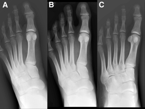
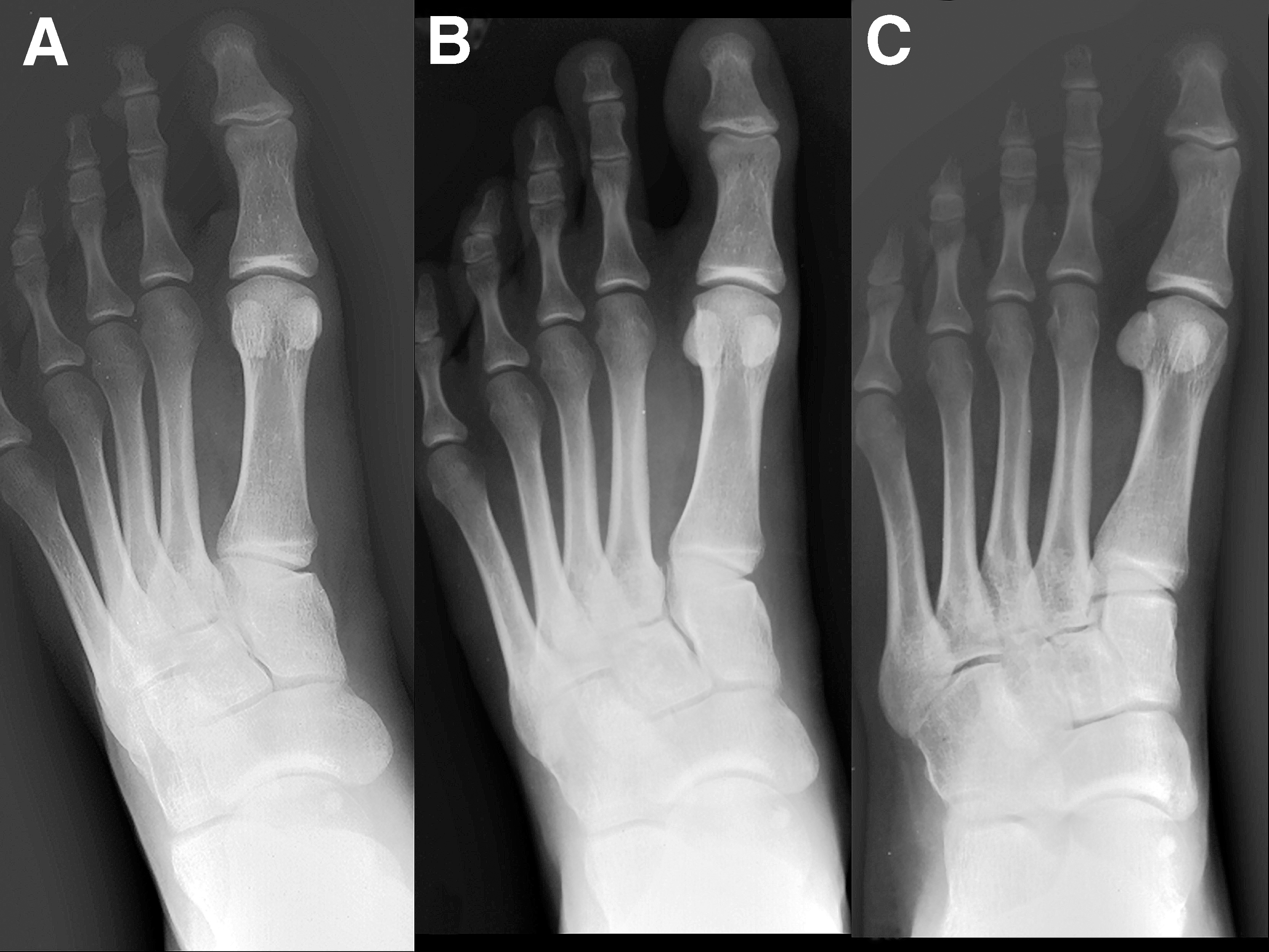
The images in this study serve as a general reference guide. The descriptions pertaining to the foot reflect the appearance of each bone in the “rectus” position. Slight variations in the appearance of each bone in any given patient may occur depending on the position of the extremity relative to the image receptor and x-ray beam. For example, a pronated or supinated foot will affect the overall appearance of the entire foot and each individual bone (Fig. 1). There also may be variations in radiographic appearance when weightbearing foot or ankle views are compared with nonweightbearing views. Therefore, it is important to concentrate on the correlation of gross anatomy to the radiograph and not on memorization of the appearance of each bone per se.
Figure 1.
Variances in the form and position of bones relative to one another as a result of foot position relative to the image receptor and x-ray beam: pronation (A), “rectus” (B), and supination (C) dorsoplantar foot views of the same individual.
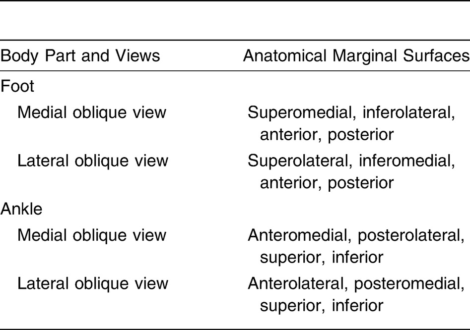
Confusion often arises in reference to interpretation of oblique views. In particular, correlation of radiographic bone margins to gross anatomy can be perplexing; however, the following system can be applied. The radiographic margin of each bone is predictable based on what I call the marginal or tangential surfaces concept. [23] All of the bones in each 2-D radiographic view, with standard positioning techniques, have four anatomical margins. A margin represents that aspect of the bone surface that is perpendicular or “tangent” to the x-ray beam. The anatomical surface or aspect of any bone can easily be determined for views performed in the three anatomical planes of the body. For example, in a dorsoplantar foot view (transverse plane), the medial, lateral, anterior, and posterior aspects of each bone are seen along their respective margins. This concept can also be applied to views that position the foot or ankle oblique to the three anatomical planes (Table 1). Pathologic abnormalities noted at the margin of a bone in the radiograph can then be directly correlated to the symptomatic area of the patient's extremity clinically or vice versa. However, be aware that the radiographic margin of a bone may not directly correlate to the description of a gross anatomical specimen in an anatomy textbook; the position of each bone in the patient's foot relative to the radiographic image receptor must then be considered. The cuboid, for example, is set in the foot such that its superior surface is oblique, almost 45° in the frontal plane, to the image receptor in a dorsoplantar view. In this extreme case, the inferomedial aspect of the cuboid bone specimen is viewed at the margin in the dorsoplantar view, not the medial aspect. However, throughout this article, the gross anatomical specimens pictured in the photographs are positioned as they would be found in the foot relative to the radiographic image receptor for all of the views; so, when the reader correlates a foot skeleton to the radiograph, the bones must be positioned in a similar manner. Generally speaking, the marginal/tangential surfaces concept offers a unique approach to understanding the seemingly perplexing radiographic anatomy of bones in the oblique views and for correlating to the patient and will be discussed further in a separate publication (work in progress).
Table 1.
Anatomical Marginal Surfaces Seen with Oblique Radiographic Views.
As noted earlier, the gross specimen is 3-D and the radiograph is 2-D. To avoid confusion and establish consistency when correlating radiographic anatomy to the bone specimen, the following format is used. Description of the gross specimen's osteological structure in the 3-D coordinate system includes use of the following standard terms: anterior, posterior, superior, inferior, medial, lateral, and any combinations thereof. These terms strictly apply to the gross anatomical specimen. However, terms used to describe radiographic anatomy in the 2-D coordinate system (ie, the radiograph) include mediad, laterad, anteriad, posteriad, superiad, and inferiad. The use of these latter terms strictly applies to the 2-D radiograph (Table 2). (The suffix -ad can be used in two contexts [24]: directional [in this sense, the term anteriad would mean in an anterior direction] and relational [here, the term anteriad would mean pertaining to the anterior margin of a bone surface in the radiograph].)
Table 2.
Terminology Used for Describing Image Marginal Surfaces in the Two-dimensional Radiograph.
The radiographic image is formed after x-rays passing through the body part are attenuated by structures of differing thickness, density, and atomic number. This is known as differential absorption. A surface, border, or landmark of the 3-D bone will be viewed in the 2-D radiograph as a margin or will be superimposed. The term margin refers to the projection of the bone–soft-tissue interface in a particular plane onto the image receptor and appears as the sharply defined edge of the bony structure. The radiographic margin corresponds to the surface of any structure that is perpendicular or tangent to the x-ray beam. Superimposition refers to the remainder of the osseous and soft-tissue structures that appear as shadows of differing radiodensities between the margins of a bone. A margin, however, is not always isolated; it may further be superimposed on another bone or even on the same bone if the latter's anatomical form is flat or irregular (concave, for example) and not spherical.
The radiographic anatomy of each bone is discussed for each standard view of the foot and ankle. Pertinent osteological features are discussed before the radiographic anatomy description of each bone. This format was chosen instead of separate sections on osteology and radiographic anatomy so that the reader can quickly refer to and correlate the two disciplines. When appropriate, variant presentations are described.
The image presentations, by including two perspectives (“front” and “back”) of every bone, help the reader to think in layers and, through deductive reasoning, anticipate the radiographic appearance of the bone. Thinking in layers will aid the reader's appreciation of 3-D and the application of spatial relationships to the 2-D radiograph. These images have not been labeled so that the reader can view them without distraction or clutter; a supplementary publication (work in progress) will present each image with labels as a pictorial atlas.
The findings from the original project, due to its broad scope, have been divided into five parts: the lower leg (the focus of this paper), the greater tarsus, the lesser tarsus, the metatarsals, and the phalanges.
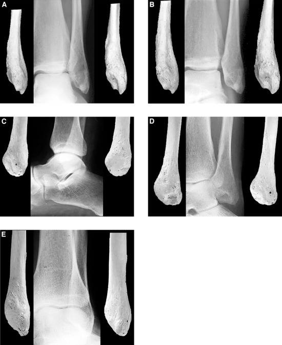
Distal Tibia (Fig. 2)
Figure 2.
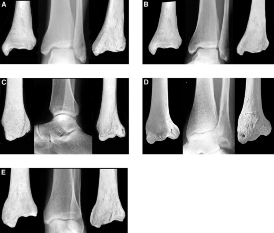
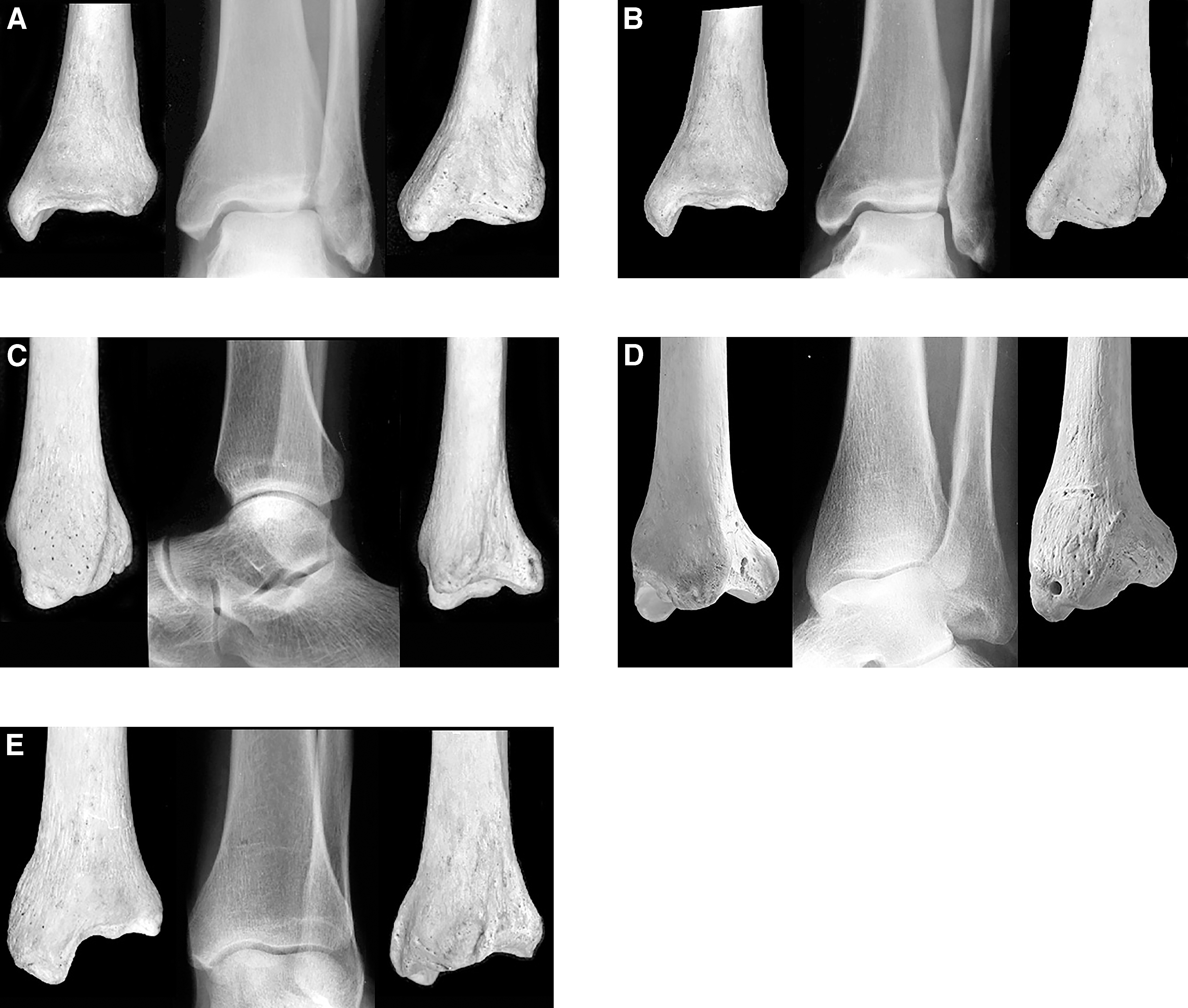
Distal tibia. A, Anteroposterior ankle view (anterior perspective, radiograph, posterior perspective); B, mortise ankle view (anterior perspective, radiograph, posterior perspective); C, lateral ankle view (medial perspective, radiograph, lateral perspective); D, medial oblique ankle view (anterolateral perspective, radiograph, posteromedial perspective); E, lateral oblique ankle view (anteromedial perspective, radiograph, posterolateral perspective).
Osteology
The distal tibial shaft has five surfaces: medial, lateral, anterior, posterior, and inferior. The medial malleolus is found at its inferomedial end.
Shaft
The medial surface is continuous with the medial surface of the medial malleolus. The upper arm of the flexor retinaculum and, occasionally, the oblique superomedial band of the inferior extensor retinaculum insert onto this surface.
The lateral surface of the distal tibia is triangular. Its base is inferior and its apex is superior. It is concave transversely and flat vertically. This surface, also known as the fibular notch, provides a small facet for the distal fibula. Two large tubercles form the anterior and posterior borders of the fibular notch. The interosseous tibiofibular ligament inserts onto the apex of the lateral surface.
The anterior surface is convex transversely and flat to mildly concave vertically. It is bordered inferiorly by the talar articular surface. A large crest or ridge runs transversely across the inferior aspect of the anterior surface, parallel to the inferior border. Extensions of the flexor and extensor retinacula may attach to the anterior surface. A large tubercle is found along the inferolateral aspect of this surface. It is known as the anterolateral tibial tubercle and borders the fibular notch anteriorly. It provides insertion for the anterior tibiofibular ligament.
The posterior surface is covered with rugosities along its inferior aspect. A well-defined, narrow ridge demarcates the posterior surface's medial border. The tibialis posterior tendon runs along the posterior surface adjacent to this ridge. The large posterolateral tibial tubercle is found at the inferolateral border of the posterior surface. It borders the fibular notch posteriorly and provides insertion for the posterior tibiofibular ligament.
The inferior surface is entirely articular with the talus. It is also known as the tibial plafond. This surface is convex transversely and concave from anterior to posterior. The posterior and anterior borders reflect the apex of the convexity and the edges of the concavity. The posterior border extends further inferiorly than does the anterior border. The lateral aspects of both borders are continuous with the posterolateral and anterolateral tibial tubercles. The inferior surface continues medially as the lateral surface of the medial malleolus.
Malleolus
The tibial malleolus projects in an inferomedial direction from the shaft. There are medial, anterior, posterior, and lateral surfaces and an inferior end.
The inferior end comprises two colliculi and an intercollicular groove. The anterior colliculus is the larger of the two colliculi. It also extends more inferiorly than does the posterior colliculus. The anterior colliculus provides insertion for numerous ligaments. The anterior superficial tibiotalar, tibionavicular, and tibioligamentous fascicles insert onto its anterior aspect. The tibiocalcaneal ligament originates from the medial aspect, and the superficial posterior tibiotalar ligament inserts onto the posterior aspect. The deep anterior tibiotalar ligament originates from the tip of the anterior colliculus and from the intercollicular groove. The deep posterior tibiotalar ligament originates from the intercollicular groove and the anterior aspect of the posterior colliculus.
Most of the medial surface is subcutaneous. It provides insertion for the medial and flexor aponeuroses. This surface is convex transversely and vertically. Part of the deltoid ligament inserts along its inferior aspect. The medial surface is the widest malleolar surface; it is rugous superiorly. A ridge runs obliquely across this surface from anterosuperior to posteroinferior. It is continuous with the inferior aspect of the posterior calcaneus.
The anterior surface is narrow and angular. Superiorly, it projects inferomedially and forms a 45° angle with the distal tibia. A segment of the superior extensor retinaculum inserts here. At its midsection, it angulates 45° inferolateral to the superior section and is vertical. It is continuous with the anterior colliculus inferiorly. The oblique superomedial band of the inferior extensor retinaculum inserts onto the anterior surface.
The posterior surface is very small. A smooth, triangular gliding facet for the tibialis posterior tendon occupies most of this surface. It also serves as the insertion site for the deep aponeurosis. A narrow, well-defined ridge marks the border between the posterior and medial surfaces. The lateral surface is entirely articular with the talus.
Radiographic Anatomy
Anteroposterior Ankle View (Weightbearing) (Fig. 2A)
The tibial malleolus projects in an inferomedial direction from the distal tibia. Its mediad margin corresponds to the medial surface; the laterad margin corresponds to the medial articular surface for the talus. The inferiad margin is the anterior colliculus; it is triangular, with the apex inferiorly. A thick, transverse sclerosis demarcates the base of this triangle; this represents the inferior surface of the posterior colliculus and intercollicular groove. The remainder of the tibial malleolus is radiolucent relative to the distal tibia; this is a normal finding. Numerous curvilinear striations of decreased density run parallel and adjacent to the medial margin; these correspond to the rugosities along the medial surface.
The distal tibia's inferiad margin represents the central aspect of the tibial plafond; it continues mediad as the laterad margin of the medial malleolus. Several shadows can be identified around the inferiad margin. The posterior border of the tibial plafond is superimposed on the joint space inferiad to the inferiad margin; its inferolateral extent is superimposed near the center of the talar dome. The posterior border then turns approximately 90° and runs superiad as a somewhat linear increased density. At this point it becomes the posterolateral tubercle. The anterior border of the tibial plafond appears as a vague, convex shadow along the center of the tibiotalar joint that is superimposed on the posterior border and the joint space. It is superimposed on the distal fibula laterad and continues superiad as the anterolateral tibial tubercle. The ill-defined, transverse sclerosis that runs parallel to the inferiad margin represents a remnant of the physis. The irregular sclerosis between this and the inferiad margin corresponds to the superimposed transverse ridges along the anterior and posterior distal tibial surfaces.
The laterad margin of the distal tibia corresponds to the border between the anterior and lateral surfaces. The lateral aspect of the anterolateral tibial tubercle is viewed along the inferiad aspect of this margin. The posterolateral tibial tubercle is superimposed on the distal tibia and adjacent to the mediad margin of the distal fibula. It is seen as a well-defined, vertical sclerosis that is continuous inferiad with the laterad extent of the plafond's posterior border. This increased density becomes ill defined as it merges with the laterad cortex superiad.
The mediad margin corresponds to the posterior aspect of the distal tibia's medial surface. It is continuous with the mediad margin of the tibial malleolus.
Mortise Ankle View (Weightbearing) (Fig. 2B)
The tibial malleolus is isolated. The anterior colliculus is found at the inferior tip of the malleolus. It is triangular in shape. A thick, transverse sclerosis demarcates the base of this triangle. This represents the inferior aspect of the posterior colliculus and intercollicular groove. The indistinct junction between the malleolus and the distal tibia is radiolucent relative to the remainder of the distal tibia. This is a normal finding. The mediad and laterad margins correspond to the medial surface and talar articular surface, respectively.
The inferiad margin corresponds to the center of the tibial plafond. The broad, ill-defined, transverse sclerosis located superiad and parallel to this landmark represents a remnant of the physis. Ill-defined increased densities are visible between these two outlines. These radiodensities correspond to the ridges along the inferior aspects of the anterior and posterior surfaces. The posterior border of the tibial plafond is superimposed on the talar dome. It is continuous mediad with the tibial malleolus posterior colliculus. The latter is superimposed on the joint space superomediad. The posterior border of the tibial plafond appears as a crescentic increased density that is superimposed on the talar dome. The posterior border ends laterad at the posterolateral tubercle. The anterior border of the tibial plafond is seen as a linear increased density running obliquely through the mediad half of the tibiotalar joint space. It continues collinear with the talar dome until it meets the fibula. The anterior border ends laterad at the anterolateral tubercle.
The laterad margin corresponds to the border between the lateral and anterior distal tibial surfaces. The lateral aspect of the anterolateral tubercle is found along the inferiad aspect of this margin. The anterolateral tubercle is superimposed on the fibula. The distance between the anterolateral and posterolateral tubercles is shorter than that seen in the anteroposterior ankle view. The vertical increased density that travels superiad from the posterolateral tubercle is the border between the posterior and lateral surfaces.
The mediad margin corresponds to the distal tibia's convex medial surface. It is continuous inferiad with the tibial malleolus.
Lateral Ankle View (Weightbearing) (Fig. 2C)
The anteriad margin of the distal leg corresponds to its anterior surface. It can be divided into two sections, superiad and inferiad, which are separated by the ridge along the inferior aspect of the anterior surface. The superiad section is larger and convex. The inferiad section is concave and intra-articular.
The posteriad margin corresponds to the lateral aspect of the posterior surface; it is smooth and convex. The convexity corresponds to the posterior aspect of the posterolateral tubercle.
The inferiad margin corresponds to the inferior articular surface or tibial plafond. An ill-defined, crescentic increased density parallels this surface superiad. This represents a remnant of the physis.
The tibial malleolus is superimposed on the talar dome. However, the inferiad margins of the anterior and posterior colliculi and the anteriad margin of the anterior colliculus are perceptible.
Medial Oblique Ankle View (Weightbearing) (Fig. 2D)
The anteriad margin of the distal tibia corresponds to the border between the anterior and medial surfaces. The tibial malleolus is only partly isolated. The anterior and posterior colliculi are superimposed on the talar dome. The anterior colliculus is inferomediad relative to the posterior colliculus.
The inferiad margin corresponds to the tibial plafond. The posterior border of the plafond is fully superimposed on the talar dome and is not visible.
The posteriad margin corresponds to the posterolateral tubercle and the lateral aspect of the posterior surface. The former is superimposed on the distal fibula. The anterolateral tubercle is superimposed on the distal tibia. It is identified as a curvilinear sclerosis that runs parallel to the anteromediad margin of the distal fibula. The anterolateral tubercle becomes ill defined as it merges with the cortex.
Lateral Oblique Ankle View (Weightbearing) (Fig. 2E)
The posteriad margin of the distal tibia corresponds to the border between the posterior and lateral surfaces. It continues inferiad as the posterior colliculus. The anterior colliculus can also be seen. It begins mediad as a linear increased density that is superimposed on the posterior colliculus. It then dips inferiad, superimposed on the talar dome.
The inferiad margin corresponds to the tibial plafond. The posterior border of the plafond is superimposed on the talar dome.
The anteriad margin corresponds to the lateral aspect of the anterior surface. The anterolateral tibial tubercle is viewed along this margin inferiad; it is superimposed on the distal fibula. The posterolateral tubercle projects inferiad and is superimposed on the talar dome and distal fibula.
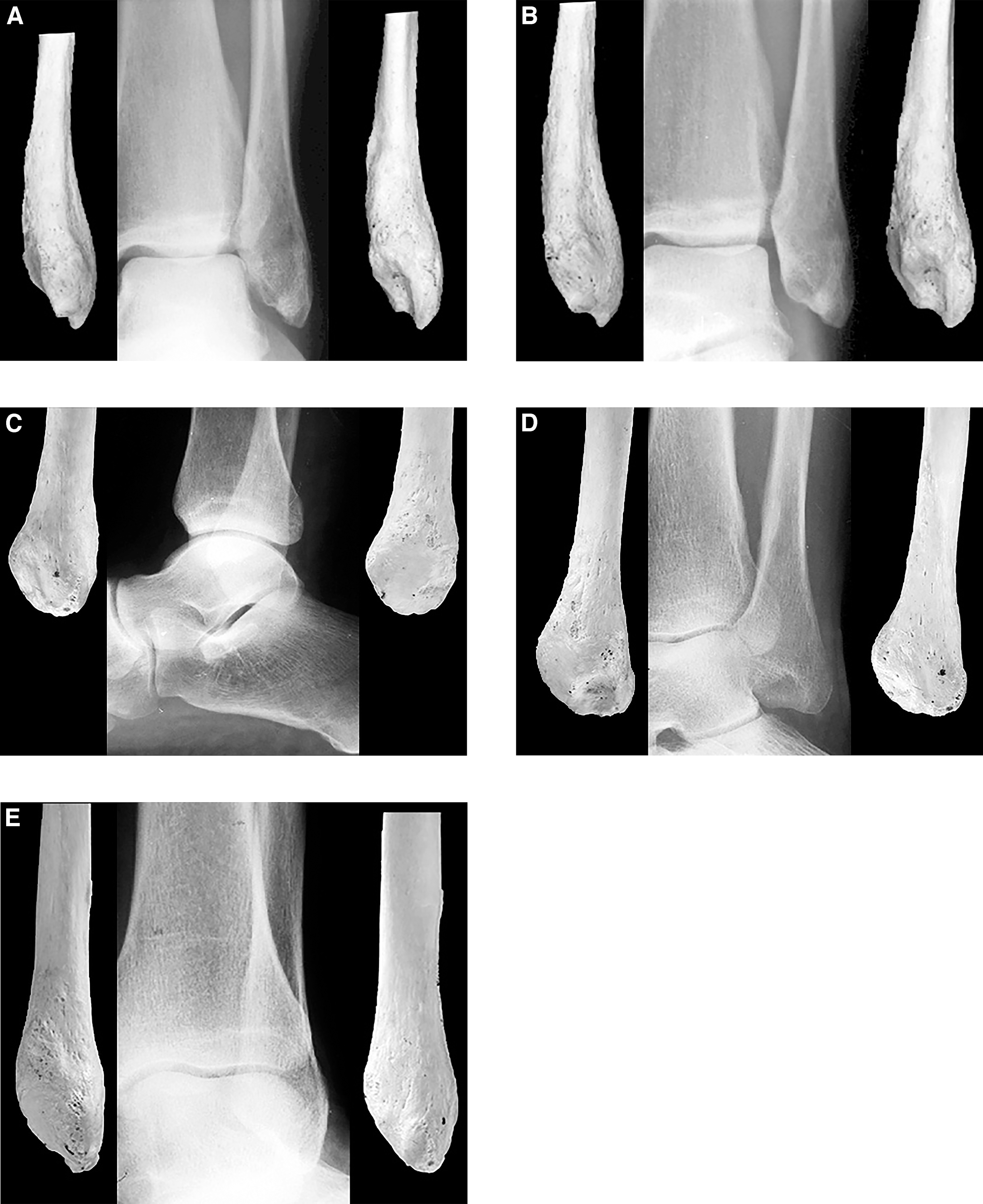
Distal Fibula (Fig. 3)
Figure 3.
Distal fibula. A, Anteroposterior ankle view (anterior perspective, radiograph, posterior perspective); B, mortise ankle view (anterior perspective, radiograph, posterior perspective); C, lateral ankle view (lateral perspective, radiograph, medial perspective); D, medial oblique ankle view (posteromedial perspective, radiograph, anterolateral perspective); E, lateral oblique ankle view (anteromedial perspective, radiograph, posterolateral perspective).
Osteology
The distal fibular shaft has four surfaces: anterolateral, posterolateral, medial, and anteromedial. The inferior end is the fibular malleolus.
Shaft
The distal fibula's anterolateral surface is triangular. Its apex is superior and its base is inferior. The superior apex is formed at the inferior subdivision of the middle fibular shaft's anterior border. The anterior and posterior branches of the anterior border form the anterior and posterior boundaries of the anterolateral surface, respectively. It primarily is subcutaneous.
The posterolateral surface is roughly rectangular. Its anterior boundary is the posterior branch of the middle fibula's anterior border. The posterior boundary is the well-defined posterior border of the distal fibula. The upper and lower boundaries are not defined and merge imperceptibly with the middle fibular shaft and malleolus, respectively. The peroneus brevis muscle originates from the posterolateral surface.
The interosseous crest forms the anterior boundary of the medial surface. The posterior border of the distal fibula serves as this surface's posterior boundary. The deep transverse fascia inserts onto the posterior border. The inferior boundary is marked by the surface for the syndesmotic recess anteriorly and the posterior tubercle posteriorly. The posterior tibiofibular ligament inserts onto this tubercle. Fibers of the flexor hallucis longus muscle originate from the upper aspect of this surface; the interosseous tibiofibular ligament inserts along the lower aspect.
The anteromedial surface is very narrow. Its upper boundary is not defined; it continues with the middle fibular shaft. The lower boundary is formed by the surface for the syndesmotic recess. The anterior branch of the fibula's anterior border represents the lateral boundary. This boundary ends inferiorly at the tubercle for insertion of the anterior tibiofibular ligament. The interosseous crest forms the medial boundary. The interosseous tibiofibular ligament inserts along the lower aspect of this surface. The peroneus tertius muscle inserts onto the upper aspect.
Malleolus
The fibular malleolus has anterolateral, posterior, medial, and inferomedial surfaces.
The anterolateral surface is continuous with the distal fibula's anterolateral surface and is subcutaneous. A tubercle is found at its superoanterior corner. The anterior tibiofibular ligament inserts here. Two small colliculi mark the anterior and posterior aspects of the inferior boundary. The posterior colliculus is slightly larger and extends more inferiorly. The anterior talofibular ligament originates from the inferoanterior boundary of the anterolateral surface, below the tubercle and onto the anterior colliculus.
The posterior surface is a continuation of the distal tibia's posterolateral surface and posterior aspect of the medial surface. It is wide superiorly and narrow inferiorly. The peroneus brevis tendon glides across a sulcus along the lateral aspect of the posterior surface. A tubercle or ridge is found at its superomedial aspect. The posterior tibiofibular ligament inserts onto this tubercle.
The medial surface is triangular. The base of this triangle is superior, formed by the surface for the syndesmotic recess. The apex is inferior. The medial surface is primarily articular with the talus.
The digital fossa is the primary landmark on the inferomedial surface. The posterior talofibular ligament inserts onto the lower aspect of this fossa. The superior boundary of the inferomedial surface is marked by the tubercle for the posterior tibiofibular ligament insertion. The inferior boundary is mildly concave between the two colliculi. The calcaneofibular ligament originates from the intercollicular surface. Sharply defined crests mark the superoanterior and posterior boundaries of the digital fossa.
Radiographic Anatomy
Anteroposterior Ankle View (Weightbearing) (Fig. 3A)
The shaft's laterad margin corresponds to the posterior branch of the anterior border. The mediad margin corresponds to its medial surface.
The fibular malleolus appears roughly triangular. The base of the triangle blends with the distal fibular shaft at the level of the tibial plafond. Its apex is inferiad and extends below the level of the tibial malleolus.
The inferiad margin corresponds to the inferior aspects of the anterior and posterior colliculi. The posterior colliculus corresponds to the apex of the malleolus and is isolated. The anterior colliculus appears as a diffuse, circular radiodensity just superomediad to the apex. This is a normal finding and should not be misinterpreted as pathology. The ill-defined, crescentic increased density that runs superolaterad from the anterior colliculus follows the path of the sulcus for the peroneal tendon. Another area of increased density is seen along the mediad aspect of the malleolus adjacent to the superolaterad corner of the talar dome. This corresponds to the superoanterior crest of the digital fossa. A well-defined radiolucency appears between this latter increased density, the superimposed shadow of the anterolateral tibial tubercle, and the crescentic sclerosis that follows the sulcus for the peroneal tendon. This is a normal finding and must not be misinterpreted as a geographic lytic lesion. A linear radiodensity connects the two increased densities corresponding to the superoanterior crest of the digital fossa and the anterior colliculus. This represents the posterior boundary of the digital fossa. An oval area of decreased density is seen inferomediad to this linear sclerosis. This represents the digital fossa viewed en face. The talar articular surface cannot be identified.
The mediad margin corresponds to the posterior aspect of the talar articular surface. The laterad margin corresponds to the posterior aspect of the anterolateral surface.
Mortise Ankle View (Weightbearing) (Fig. 3B)
The fibular malleolus appears trapezoidal in the mortise view. The mediad margin corresponds to the talar articular surface. The posterior colliculus is isolated at the inferior apex of the malleolus. The anterior colliculus is superimposed but is seen as a diffuse, circular increased density superomediad to the posterior colliculus. An oval radiolucency can be identified between the anterior colliculus and the mediad margin. This corresponds to the digital fossa. Superiad to the fossa, an increased density is seen that is formed by the superimposed shadows of the anterior and posterior tibiofibular ligament tubercles.
The laterad margin corresponds to the posterior border between the anterolateral and posterior malleolar surfaces. The shaft's laterad margin corresponds to the posterior branch of its anterior border.
The mediad margin of the distal fibula corresponds to the interosseous crest. The malleolus blends with the distal fibula at the level of the tibial plafond.
Lateral Ankle View (Weightbearing) (Fig. 3C)
The inferiad margin of the fibular malleolus lies at the level of the posterior subtalar joint. This corresponds to the apex of the posterior colliculus. An oval-shaped radiolucency is found just superiad to this margin. This corresponds to the fibular fossa. It could be misinterpreted as a geographic lytic lesion.
The posteriad margin corresponds to its posterior surface. The anteriad margin of the fibular malleolus is directed anterosuperiad. It corresponds to the border between the anterolateral and medial surfaces. Inferiad it is superimposed on the talar body; superiad, it is superimposed on the tibial malleolus anterior colliculus.
The posteriad margin of the distal fibular shaft corresponds to its posterior border. The shaft's anteriad margin corresponds to the anterior branch of the anterior border.
Medial Oblique Ankle View (Weightbearing) (Fig. 3D)
The anteriad margin of the malleolus corresponds to the border between the anterolateral and medial surfaces. It is superimposed on the tibia and not clearly seen. The talar articular facet is not visible.
The inferiad margin corresponds to the inferior aspects of the anterior and posterior colliculi and the intercollicular groove. The oval radiolucency seen superiad and between the two colliculi is the fossa. It is viewed en face.
The posteriad margin corresponds to the border between the posterior and anterolateral malleolar surfaces.
The posteriad and anteriad margins of the shaft correspond to its posterolateral surface and interosseous crest, respectively. The crest is partially superimposed on the distal tibia.
Lateral Oblique Ankle View (Weightbearing) (Fig. 3E)
The distal fibula is superimposed on the talus and tibia. The anteriad margin of the shaft corresponds to the anteroinferior segment of the distal fibular lateral surface. The shaft's posteriad margin corresponds to its posterior border. The posteriad margin of the malleolus corresponds to the superimposed superoanterior and posterior boundaries of the digital fossa. It is further superimposed on the talus and is barely perceptible. The posterior tubercle of the malleolar medial surface is also found along this margin superiad. The anteriad margin of the malleolus corresponds to its anterolateral surface. The inferiad margin corresponds to the posterior colliculus.
Soft Tissues
A few soft-tissue structures and landmarks are recognized in ankle radiographs. Most are identified in the lateral view. The Achilles tendon can usually be identified, although “spot” light enhancement may be necessary. If the ankle joint axis is not perpendicular to the image receptor, which frequently occurs in the weightbearing view of a cavus or metatarsus adductus foot, the Achilles tendon shadow cannot be sharply identified. In these cases, it is necessary to pull the heel away from the image receptor to align the ankle joint axis perpendicular to the image receptor.
A triangular-shaped radiolucency seen posterior to the distal leg in the lateral view is known as the posterior triangle; it also is referred to as Kager's triangle and Toygar's triangle. Its posterior boundary is the Achilles tendon, its inferior boundary is the calcaneus, and its anterior boundary is the flexor hallucis longus. [25] Rarely, an anomalous muscle, the accessory soleus, obliterates this radiolucent triangle. [26,27]
Funding
None reported.
Conflicts of Interest
None reported.
References
- MorganA, KimPS, ChristmanRA:Radiographic anatomy of the talus. JAPMA93: 449, 2003.
- ChristmanRA, LyP:Radiographic anatomy of the first metatarsal. JAPMA80: 177, 1990.
- ChristmanRA:Radiographic anatomy of the calcaneus: part I. Inferior surface. JAPMA77: 549, 1987.
- ChristmanRA:Radiographic anatomy of the calcaneus: part II. Posterior surface. JAPMA77: 581, 1987.
- ChristmanRA:Radiographic anatomy of the calcaneus: part III. Superior surface. JAPMA77: 633, 1987.
- ChristmanRA:Radiographic anatomy of the calcaneus: part IV. Lateral and medial surfaces. JAPMA78: 11, 1988.
- WeissmanSD: Radiology of the Foot, 2nd Ed, Williams & Wilkins, Baltimore, 1989.
- ForresterDM, KricunME, KerrR: Imaging of the Foot and Ankle, Aspen Publishers, Rockville, MD, 1988.
- BerquistTH: Imaging of the Foot and Ankle, 3rd Ed, Lippincott Williams & Wilkins, Philadelphia, 2011.
- CrimJR, CracchioloA, HallRL: Imaging of the Foot and Ankle, Lippincott-Raven, London, 1996.
- MeschanI: An Atlas of Anatomy Basic to Radiology, WB Saunders, Philadelphia, 1975.
- WickeL: Atlas of Radiologic Anatomy, 3rd Ed, Urban & Schwarzenberg, Baltimore, 1982.
- HamiltonWJ, SimonG, HamiltonI: Surface and Radiologic Anatomy,5th Ed, Williams & Wilkins, Baltimore, 1971.
- McMinnRMH, HutchingsRT, LoganBM: Color Atlas of Foot and Ankle Anatomy,Appleton-Century-Crofts, New York, 1982.
- BryanGJ: Johnson and Kennedy Radiographic Skeletal Anatomy, 2nd Ed, Churchill Livingstone, New York, 1982.
- BirknerR: Normal Radiologic Patterns and Variances of the Human Skeleton, Urban & Schwarzenberg, Baltimore, 1978.
- WolfeJN, KapdiCC, MurphyHS: Atlas of Xeroradiographic Anatomy of Normal Skeletal Systems,Charles C Thomas, Springfield, IL, 1978.
- MontagneJ, CherrotA, GalmicheJM: Atlas of Foot Radiography, Masson Publishing, New York, 1981.
- KayeJJ, BohneWH:A radiographic study of the ligamentous anatomy of the ankle. Radiology125: 659, 1977.
- GoergenTG, DanzigLA, ResnickD, et al: Roentgenographic evaluation of the tibiotalar joint. J Bone Joint Surg Am59: 874, 1977.
- ShereffMJ, JohnsonKA:Radiographic anatomy of the hindfoot. Clin Orthop Relat Res177: 16, 1983.
- RheaJT, DeLucaSA, SheehanJ:Radiographic anatomy of the tarsal bones. Med Radiogr Photogr59: 2, 1983.
- ChristmanRA: Foot and Ankle Radiology, Churchill Livingstone, St Louis, 2003.
- Funk and Wagnalls New Comprehensive International Dictionary of the English Language,Ferguson Publishers, New York, 1982.
- LieberGA, LemontH:The posterior triangle of the ankle: determination of its true anatomic boundary. JAPMA72: 363, 1982.
- NideckerAC, von HochstetterA, FredenhagenH:Accessory muscles of the lower calf. Radiology151: 47, 1984.
- PercyEC, TelepGN:Anomalous muscle in the leg: soleus accessorium. Am J Sports Med12: 447, 1984.


© 2014 American Podiatric Medical Association