


Abstract
Digital and interdigital corns are common painful foot conditions encountered by podiatrists during the course of their practice. These corns can often be treated with conservative techniques, although they tend to eventually recur. Currently, no single treatment exists that is efficient, long-lasting, minimally invasive, and easy to administer. This article describes two cases where hyaluronic acid gel injections were used to improve symptoms associated with digital and interdigital corns located in a nonweightbearing area. Both patients tolerated the intervention well and showed considerable improvement of their condition for a substantial period of time after the intervention without developing adverse reactions. Hyaluronic acid gel injections could very well represent an interesting therapeutic alternative for digital and interdigital corns located in nonweightbearing areas.
Corns are considered mechanical dermatologic lesions. They are caused by excessive pressure or friction being applied to the skin. Digital corns are usually located on the dorsal or distal plantar aspects of the toes and are made of compacted dry keratin. Conversely, interdigital corns are located in the deep web spaces or on the inner sides of the toes and are often composed of wet macerated skin. [1] When corns are the result of an identifiable cause, the treatment should be aimed, when possible, at modifying the source of the mechanical stress, whether it is inadequate footwear or flawed biomechanics. When this is not possible, symptomatic management can be achieved with padding, debridement, orthotics, or surgery. [2] Unfortunately, those therapeutic alternatives do not always sufficiently improve the patient's quality of life. Additionally, in the case of debridement, the patient requires regular follow-up appointments.
To replace the loss of fibrofatty tissue consequent to chronic corns and prevent foot ulceration, fluid silicone injections have been used with good success rates. However, in order for the implant to be secured into the foot's soft tissues, up to 10 injections of 0.1 mL of fluid must be made at 1-month intervals. [3] In addition, medical-grade fluid silicone is not widely available and the preparation that has beed used in the past was not convenient in a clinical setting. [4]
Newer dermal fillers have been developed primarily for the aesthetic medicine market. [5] Currently, the most widely used dermal filler is hyaluronic acid gel. Although it is not as permanent as silicone, hyaluronic acid gel is considered long lasting, with a durability of up to 24 months, and safe, since it is biosynthesized and therefore free of any animal products. [5,6] It also has an advantage over silicone; it is injectable in larger quantities, reducing the number of treatments needed in order to achieve satisfactory results. Finally, because of cross-linking, it stays viscous in the soft tissues while remaining easy to inject through a small gauge needle as a result of its dynamic viscosity. [6,7,8]
Although corns treated with dermal fillers do not always show significant improvement upon visual inspection, a clinically relevant reduction of plantar pain is usually seen following a single injection. The following two cases demonstrate the use of hyaluronic acid gel injections in the treatment of digital and interdigital corns.
Case 1
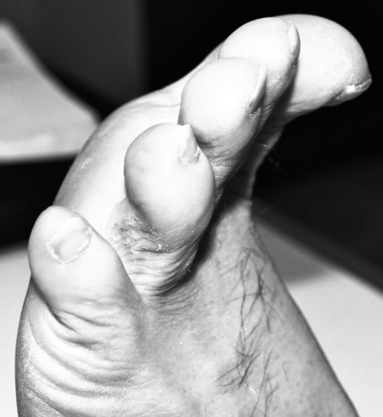
A 58-year-old man with an unremarkable medical and surgical history presented with three painful interdigital corns grouped on the lateral aspect of his left fourth toe (Fig. 1). The lesions had been present for more than 30 years. The patient, a construction worker, recalled that they appeared a few months after he sprained his fourth toe at work. He had the corns trimmed by podiatrists on several occasions but they always recurred weeks after treatment and became symptomatic again. Different padding devices were tried in the past, all of them being impractical or uncomfortable for the patient. The corns never ulcerated nor did they become infected. Upon examination, we noted that the slightly macerated 2 × 2-mm corns were located above the distal end of the proximal phalanx of the fourth toe. No lesion was seen on the fifth toe and no gross toe deformity could explain their occurrence. The tibialis posterior and dorsalis pedis pulses were easily palpable, and no sign of ischemia could be observed. Documentation of adequate digital arterial perfusion was made in order to prevent application of the projected technique in contraindicated circumstances.
Figure 1.
Preinjection view of the lateral side of the left fourth toe showing interdigital corns.
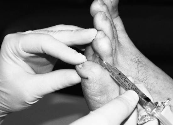
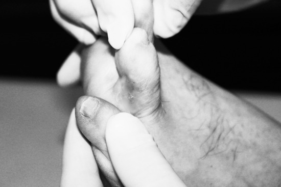
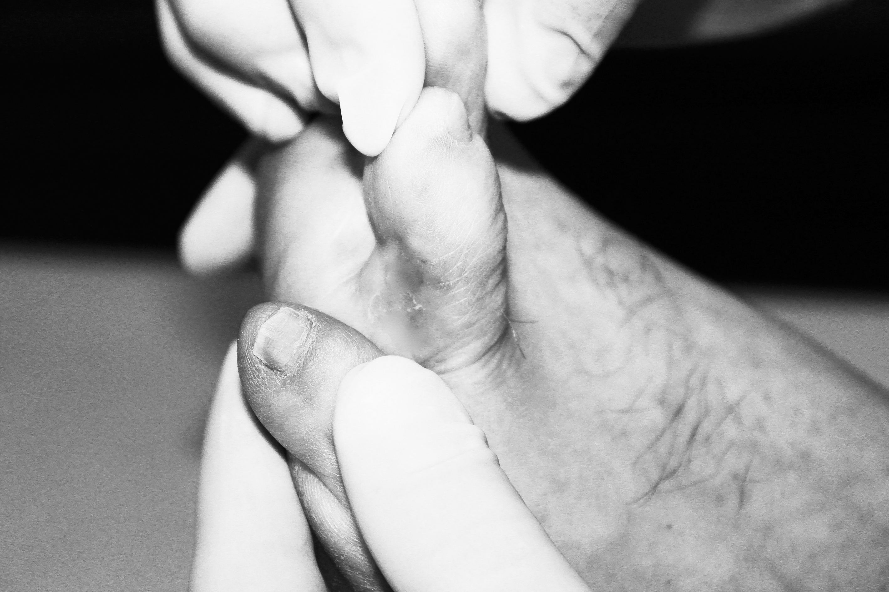
Since the patient was interested in a more permanent treatment alternative, he was offered a local injection of hyaluronic acid gel sold under the label Redexis Ultra (Prollenium Medical Technologies Inc, Toronto, Canada). After debridement of the corns in the usual aseptic manner, 2.0 mL plain lidocaine was used as a digital anesthesia. A 27-gauge needle was then inserted at the subcutaneous level precisely underneath the lesions (Fig. 2), and 0.5 mL of gel was injected in a fanning technique until the skin showed its first sign of discoloration. The needle was withdrawn and a simple dressing was placed over the toe. No particular care had to be taken afterward. The patient was seen 8 months later because he felt the skin between his toes began to cause discomfort again. No skin lesions were identifiable at that time. Nevertheless, a second similar injection of 0.5 mL hyaluronic acid gel was repeated with the same technique. At a follow-up visit, 36 months after the initial visit, the patient remained asymptomatic and interdigital corns did not reappear (Fig. 3).
Figure 2.
Injection of hyaluronic acid gel at the subcutaneous level using the fanning technique.
Figure 3.
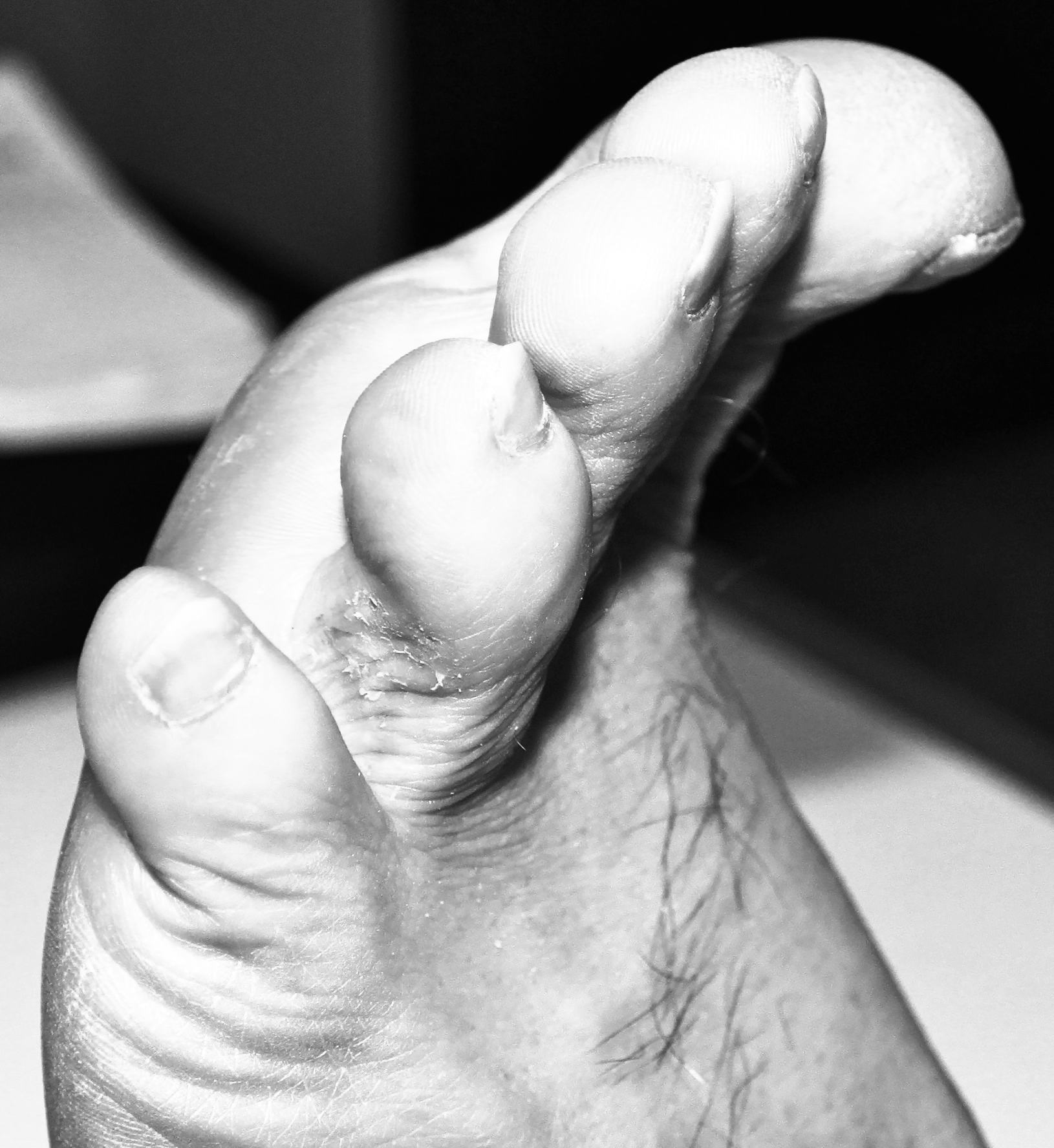
View of the lateral side of the left fourth toe 2.5 years after the first injection.
Case 2
A 48-year-old man with an unremarkable medical and surgical history presented with a painful digital corn on the distal aspect of his right second toe. The patient, an industrial worker, reported that it appeared following a work-related injury 20 years earlier. A large metal beam had crushed his foot, but from what he could recall, no bones had been broken at the time of the accident. Nevertheless, a progressive foot deformity developed in the years following the accident. The patient had his corn debrided every 4 weeks by a podiatrist but would remain symptom-free for only a few weeks after each treatment (Fig. 4). Different padding devices were tried in the past and the patient wore custom-made orthotics, which helped slightly. The corn was never ulcerated or infected. On examination, a semi-flexible hammertoe deformity was present from the second to the fourth toes and a thick corn occupied the whole distal surface of the second toe. The tibialis posterior and dorsalis pedis pulses were easily palpable, and no sign of ischemia could be observed. Once again, documentation of adequate digital arterial perfusion was made in order to prevent application of the projected technique in contraindicated circumstances.
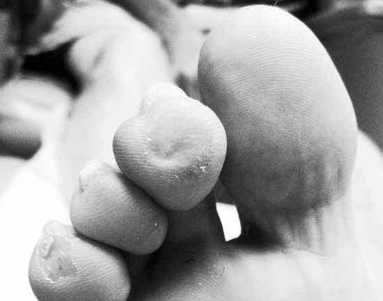
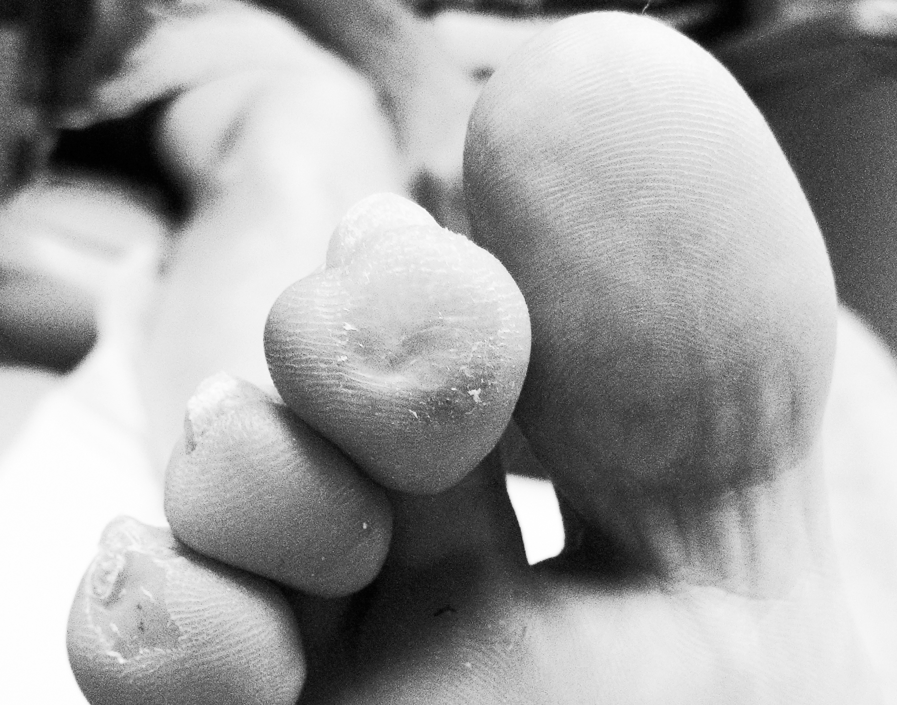
Figure 4.
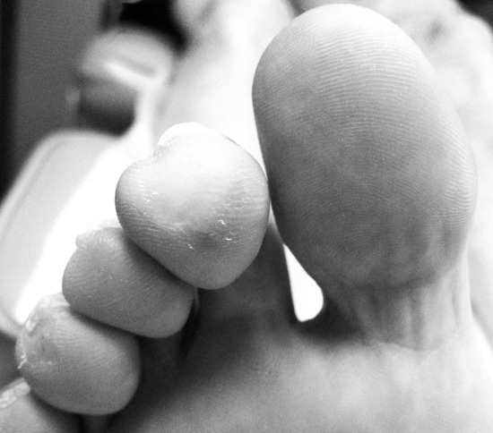
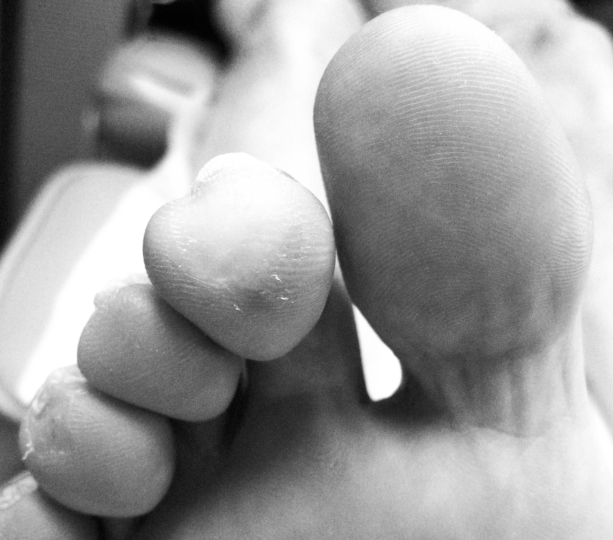
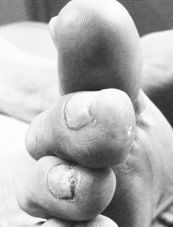
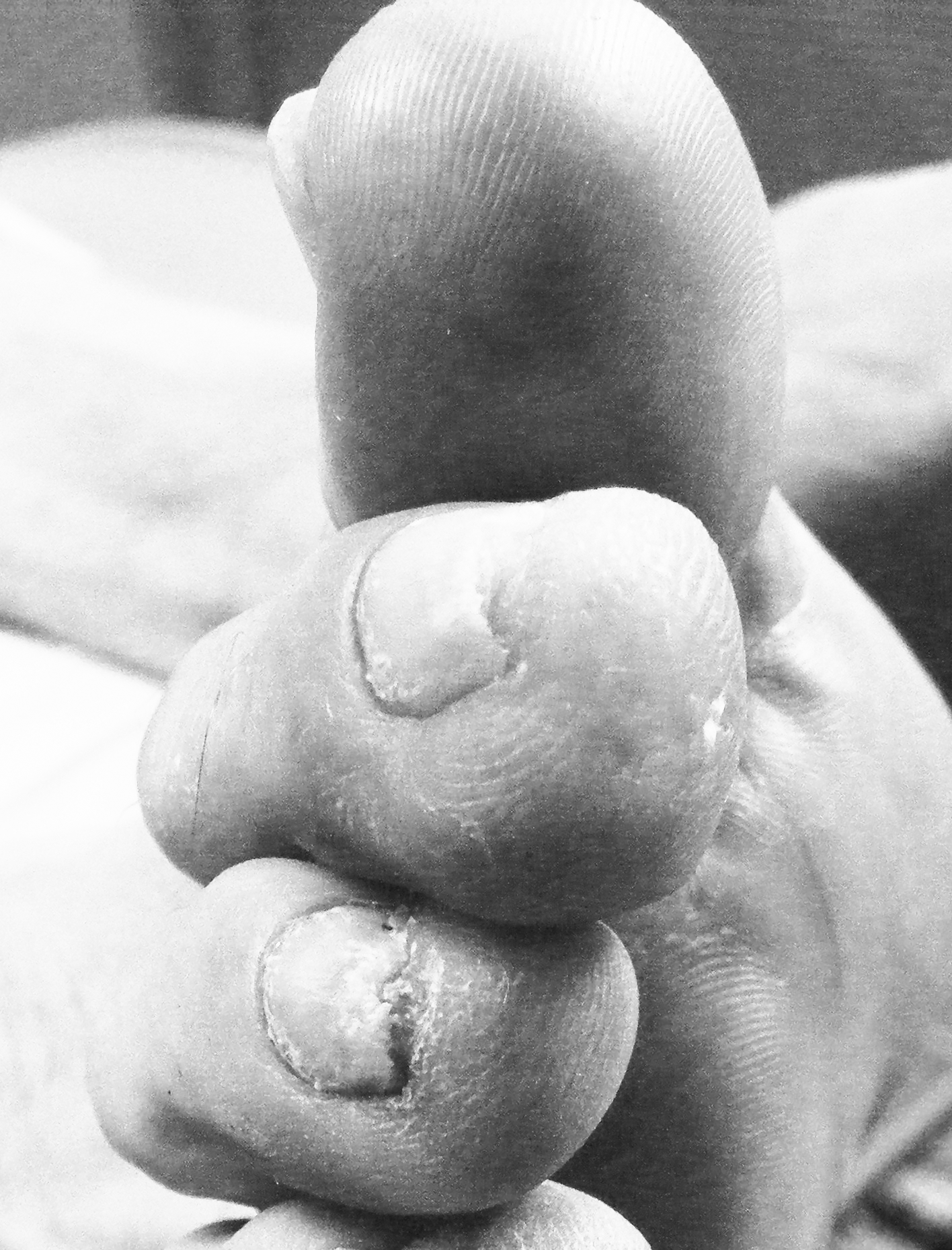
Pre-injection view of the right second toe after debridement of a painful distal plantar corn.
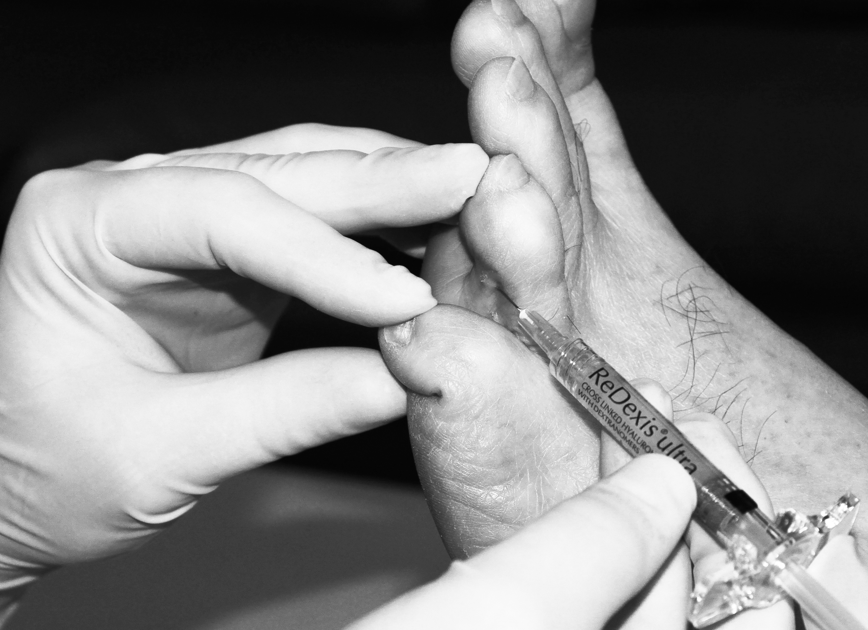
Since the patient did not want surgery, a local injection of Redexis Ultra hyaluronic acid gel was offered. After debridement of the corns in the usual aseptic manner, 2.0 mL plain lidocaine was used as a digital anesthesia. A 27-gauge needle was then inserted at the subcutaneous level, and 0.6 mL of gel was injected in a fanning technique until the skin showed local discoloration (Fig. 5). The needle was withdrawn and a simple dressing was placed over the toe. The patient was asked to wear a silicone toecap for the week following the injection. At the 1-month follow-up, the patient noted that his symptoms were significantly less than what they were prior to the procedure. Although the hammertoe deformities remained, only mild distal digital discomfort was reported by the patient. He agreed to a second injection of 0.3 mL of hyaluronic acid gel following the same technique. At the 2-month follow-up, the patient no longer complained of digital corns and did not report any pain despite having recently changed positions at work and now had to walk significantly more during the day (Fig. 6).
Figure 5.
Post-injection view of the right second toe.
Figure 6.
View of the right second toe 2 months after the first injection.
Discussion
Patients presenting to podiatric clinics for painful foot lesions should go through a careful history and physical examination to rule out other dermatologic conditions that could have a similar presentation to corns. Differential diagnosis includes, but is not limited to, warts, foreign bodies, skin tumors, and eccrine poroma. [1,2] Once the diagnosis is made, an attempt at conservative measures such as shoe modifications, padding, or debridement should be made. Although scalpel debridement offers immediate relief, this is almost always short-lived and symptoms usually recur in weeks, [9,10] significantly impairing the patient's quality of life. [11] Since the surgical option is not a panacea, [12] newer, less invasive treatment alternatives should be explored. If the patient is healthy and is interested in a more durable treatment, dermal fillers can be proposed as an alternative.
Dermal fillers have the advantage of being injectable in quantities sufficient to alleviate the patient's symptoms in only one to several sessions. [10] This was not the case with silicones. Indeed, the patients required a series of monthly injections of 0.1 mL in order to obtain a fibroblastic reaction within the skin and prevent fluid migration. [3] Also, hyaluronic acid offers more versatility since it is not permanent like silicones, and will be reabsorbed over an 18- to 24-month period. [6] Finally, the commercialized hyaluronic acid products are much more convenient in the clinical setting because they are widely available and usually come in pre-filled sterile syringes.
Conclusions
Digital and interdigital corns are common conditions that may benefit from local subcutaneous injection of hyaluronic acid gel. Although most of these lesions can be treated conservatively, many appear to recur. This minimally invasive procedure is an interesting therapeutic alternative that appears to be comfortable for the patient and safe and effective in the long term. Nonetheless, to this day, no randomized, controlled trial has evaluated the potential benefits that could result from this approach, nor has it been compared with more traditional treatment options.
Funding
None reported.
Conflicts of Interest
None reported.
References
- DockeryGL, CrawfordME:“Mechanical Injuries,”inCutaneous Disorders of the Lower Extremity, p235, WB Saunders, Philadelphia, 1997.
- LuckeT, MunroC, RobertsD, et al: “Dermatological Conditions of the Foot and Leg”inNeale's Disorders of the Foot, 7th Ed, edited byDLorimer, GFrench, MO'Donnell, et al, p23, Churchill Livingstone Elsevier, Philadelphia, 2006.
- Balkin SW: ‘‘Injectable Silicone and the Foot''inNeale's Disorders of the Foot, 6th Ed, edited byDLorimer, GFrench, MO'Donnell, et al, p471, Churchill Livingstone, Edinburgh, 2002.
- BalkinSW:Injectable silicone and the foot: a 41-year clinical and histologic history. Dermatol Surg31: 1555, 2005.
- HottaT:Dermal fillers: the next generation. Plast Surg Nurs24: 14, 2004.
- PriceRD, BerryMG, NavsariaHA:Hyaluronic acid: the scientific and clinical evidence. J Plast Reconstr Aesthet Surg60: 1110, 2007.
- NarinsRS, CohenJL, BeerK:“Injectable Skin Fillers,”inRoenigk's Dermatologic Surgery, 3rd Ed, edited byRKRoenigk, JLRatz, HHRoenigk, p705, Informa Healthcare, New York, 2007.
- VedamurthyM:Soft tissue augmentation: use of hyaluronic acid as dermal filler. Indian J Dermatol Venereol Leprol70: 383, 2004.
- RedmondA, AllenN, VernonW:Effect of scalpel debridement on the pain associated with plantar keratosis. JAPMA89: 515, 1999.
- DavysHJ, TurnerDE, HelliwellPS, et al: Debridement of plantar callosities in rheumatoid arthritis: a randomized controlled trial. Rheumatology44: 207, 2005.
- SpinkMJ, MenzHB, LordSR:Distribution and correlates of plantar hyperkeratotic lesions in older people. J Foot Ankle Res2: 8, 2009.
- MenzHB:“Keratotic disorders”inFoot Problems in Older People: Assessment and Management, HB Menz, p 61, Churchill Livingstone Elsevier, Philadelphia, 2008.
© 2014 American Podiatric Medical Association