


Abstract
Background: Plantar pressure measurements are commonly used to evaluate foot function in chronic musculoskeletal conditions. However, manually identifying anatomical landmarks is a source of measurement error and can produce unreliable data. The aim of this study was to evaluate intratester reliability associated with manual masking of plantar pressure measurements in patients with gout. Methods: Twenty-five patients with chronic gout (mean disease duration, 22 years) were recruited from rheumatology outpatient clinics. Patients were excluded if they were experiencing an acute gout flare at the time of assessment, had lower-limb amputation, or had diabetes mellitus. Manual masking of peak plantar pressures and pressure-time integrals under ten regions of the foot were undertaken on two occasions on the same day using an in-shoe pressure measurement system. Test-retest reliability was assessed by using intraclass correlation coefficients, SEM, 95% limits of agreement, and minimal detectable change. Results: Mean peak pressure intraclass correlation coefficients ranged from 0.92 to 0.97, with SEM of 8% to 14%. The 95% limits of agreement ranged from−150.3 to 133.5 kPa, and the minimal detectable change ranged from 30.8 to 80.6 kPa. For pressure-time integrals, intraclass correlation coefficients were 0.86 to 0.94, and SEM were 5% to 29%, with the greater errors observed under the toes. The 95% limits of agreement ranged from −48.5 to 48.8 kPa/sec, and the minimal detectable change ranged from 6.8 to 21.0 kPa/sec. Conclusions: These findings provide clinicians with information confirming the errors associated with manual masking of plantar pressure measurements in patients with gout. (J Am Podiatr Med Assoc 101(5): 424–429, 2011)
Gout is an inflammatory arthritis that typically presents as an acute monoarthritis of rapid onset. In 90% of cases, the foot is affected. [1] Patients with gout often experience recurrent acute flares, and in the presence of persistent hyperuricemia, chronic tophaceous disease may develop, with subsequent bone erosion and disability. [2, 3] The resultant inflammation, erosion, and deformity of gout may impair gait, causing plantar pressure measurements to fall outside the reference range. [4]
Plantar pressure measurements have become an important component in the evaluation of foot function, particularly in the assessment of musculoskeletal conditions, such as rheumatoid arthritis, but they have not been assessed in chronic gout. [5–9] The ability of pressure equipment to consistently and reliably measure the loading characteristics is crucial for clinical decision making. [4, 10] Deschamps et al [11] reported that clinicians are challenged by two sources of measurement error relating to plantar pressure measurements: the technical characteristics of the equipment used and the accurate identification of anatomical structures on pressure-based footprints (masking). Although masking can be undertaken automatically, [4, 5, 7] with each footprint divided automatically into regions by a software program that uses a geometric algorithm, [12] manual masking is often required and depends on the clinician identifying anatomical regions of the plantar surface of the foot. [11, 13] In the latter technique, the accuracy of masking is a potential source of measurement error. Only two studies [11, 14] have reported on the reliability of using manual masks in plantar pressure analysis. De Cock [14] reported good reliability of manual masking of ten regions in a large cohort of barefoot runners.
Deschamps [11] reported on the intrarater and interrater reliability in one anatomical region of the foot in ten individuals with a history of hallux valgus. In these groups of participants, the degree of impairment was relatively low. Despite the widespread use of plantar pressure technology, only two studies [11, 14] have directly addressed the issue of manual masking. The clinical rationale for having sound knowledge of the repeatability of such measures relates to the prescription of foot orthoses and footwear. In many instances, the clinician may want to assess the immediate effects of such treatment on plantar pressure to ensure that they are having the intended effect. It is important that the clinician is aware of the error associated with taking such measures when evaluating the effects of short-term or immediate interventions. The aim of this study was to evaluate the intratester reliability of manual masking of plantar pressure measurements in patients with chronic gout.
Methods
Twenty-five adults with gout were recruited from rheumatology outpatient clinics in Auckland, New Zealand. The Northern X Regional ethics committee, New Zealand approved the study, and patients provided written informed consent. All of the patients had a history of chronic gout according to the American College of Rheumatology diagnostic criteria [15]; we excluded those experiencing an acute gout flare at the time of assessment, those with a lower-limb amputation, and those with rheumatologic conditions such as rheumatoid arthritis and diabetes mellitus. Patients with diabetes were excluded from the study to ensure that the foot characteristics of gout could be specifically assessed. Information was recorded relating to age, sex, ethnicity, body mass index, disease duration, and clinical characteristics of gout.
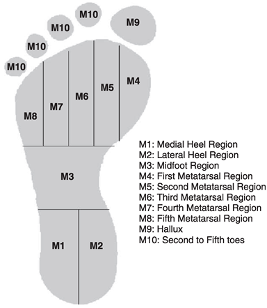
Each patient’s plantar pressure measurements were determined twice on the same day by the same rater (D.S.). The mean (SD) interval between each occasion was 3.0 (1.5) hours. Plantar pressure readings were obtained with the F-Scan mobile system (Tekscan Inc, South Boston, Massachusetts), which incorporates insoles with 960 different pressure-sensing locations (sensels) and a spatial resolution of 4 sensels/cm2. The plantar foot was masked into ten regions, and the mean peak pressure and pressure-time integrals were calculated. In accordance with the method of Cavanagh and Ulbrecht, [16] the plantar surface of the foot was masked into ten regions and included the regional division of the foot into the lateral heel, medial heel, midfoot, first metatarsal region, second metatarsal region, third metatarsal region, fourth metatarsal region, fifth metatarsal region, hallux, and second to fifth toes. Manual masking was undertaken based on the protocols used by automated masking software (Research Foot, version 5.24, software). The heel comprised the first 0% to 30% of foot length, the midfoot the next 30% to 60%, the forefoot the following 60% to 85%, and the hallux/toe the remaining 85% to 100%. The heel region width was divided into two equal parts and the midfoot into one part. The forefoot region width was divided into equal fifths, creating five forefoot regions. The hallux/toe region width was also divided into two parts, with the hallux region occupying the medial 40% and the second to fifth toe region occupying the lateral 60% (Fig. 1). At each condition, we used only the entire foot contact period to analyze the peak pressures and pressure-time integrals.
Before the tests, the pressure insoles were calibrated according to the manufacturer’s instructions, and before data acquisition, the zero-setting procedure was performed as recommended by Tekscan Inc. After a step calibration routine, the participant undertook a seven-stride protocol. The first and last strides were excluded, and the remaining five strides were averaged from each patient walking trial. Before data acquisition, each patient was instructed to walk freely in the laboratory to reproduce their typical gait and to feel comfortable in the laboratory. [17, 18] Three trials of the five-stride protocol were recorded. After each occasion, the examiner removed the pressure insole and used the same pressure insole for the next occasion. Data analysis was conducted with the F-Scan, version 5.24, software package (Tekscan Inc).
Figure 1.
Masking regions of the foot.
Intratester reliability was assessed by calculation of the intraclass correlation coefficient (ICC) with the 95% confidence interval. Shrout and Fleiss [19] suggested that ICC values greater than 0.75 represent excellent reliability; 0.40 to 0.74, fair to good reliability; and less than 0.40, poor reliability. Bland and Altman [20] suggested that for most measures, the within-subjects SD, as an index of measurement error, is to be preferred to the ICC because it has a more direct interpretation that can be applied to individual measurements. Therefore, 95% limits of agreement were used to evaluate within-subject variation, which represents the 95% likely range for the difference between a subject’s scores over the two occasions. SEM calculations were also undertaken. Furthermore, based on the standard error of measurement, the minimal detectable change score (MDC) was calculated with the following formula: MDC = z scorelevel of confidence×SDbaseline×√[2×( 1-r )], where the z score is 1.645 (90% level of confidence), SD is the standard deviation at baseline, and r is the ICC for test-retest reliability. [21]
Results
Clinical Characteristics
Of the 25 patients in the study, 19 (76%) were male, 17 (68%) were non-Polynesian, and 8 (32%) were of Maori or Pacific ethnicity. The mean (SD) age was 61.2 (11.7) years, and the mean (SD) body mass index (calculated as weight in kilograms divided by the square of the height in meters) was 32.1 (5.6). The mean (SD) disease duration was 22 (13) years, with a flare frequency of 2.92 per year.
Peak Plantar Pressure Measurements
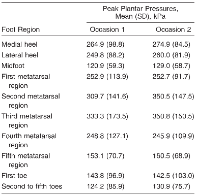
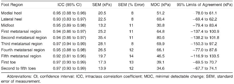
The peak plantar pressure values for each region are presented in Table 1. The test-retest ICC for peak plantar pressures across the ten regions was found to be excellent (range, 0.92–0.97) (Table 2). The measurement error between the two testing occasions was low (range, 13.2–35.4 kPa). Reporting SEM as a percentage error, the results ranged from 8% to 14% across all of the foot regions. The minimal detectable change and the 95% limits of agreement for all of the peak plantar pressure measurements are reported in Table 2.
Table 1.
Peak Plantar Pressure Descriptive Information
Pressure-Time Integral Measurements
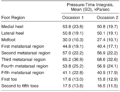
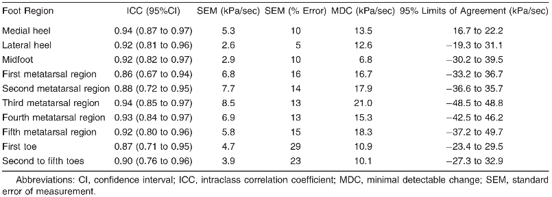
Table 3 presents the descriptive information for pressure-time integrals on repeated testing occasions. The ICCs for the pressure-time integrals ranged from 0.86 to 0.94. The SEM for pressure-time integrals was relatively small for all of the regions under the foot (Table 4); however, it was notably higher under the hallux and the toes, with a 29% error under the hallux and a 23% error under the second to fifth toes (Table 4). The minimal detectable change and the 95% limits of agreement for all pressure-time integral measurements are reported in Table 4.
Table 2.
Reliability Indices for Mean Peak Pressure
Discussion
Any measuring system needs to be of proven reliability and validity before being applied in a clinical setting so that clinicians may be ensured of reproducible and meaningful results. [11, 22, 23] The findings of the present study provide clinicians with the level of error associated with same-day measures of plantar pressures. Such information would be valuable in treatment settings where a clinician may want to evaluate whether change is due to a treatment intervention or to measurement error.
Table 3.
Pressure-Time Integral Descriptive Information
Clinical interpretation of gait data involves identifying the problem, proposing the possible causes, and selecting solution strategies. [10] The results of the present study demonstrate excellent intratester reliability of masking of mean peak pressures in patients with gout. This observation was particularly true in regions of the foot where relatively high loading typically occurs during gait, such as the rearfoot. Generally, lower reliability was found in the typically less-loaded areas, such as the toes.
The intratester reliability measurements for the pressure-time integrals were also excellent, with ICCs ranging from 0.86 to 0.94. However, the lower ICCs and the high SEM values under all of the toes suggest an increase in measurement errors in this specific region of the foot. Possible explanations for the observed errors are the masking of the forefoot region. A recent study [11] reported that small-sized masking to define pressure characteristics in the forefoot can cause unreliable data. The present study used absolute plantar pressure values and did not consider the size of the region.
This study has limitations. The number of participants was relatively small but was comparable with that of other studies of gait and foot pressure. [4, 6] The cohort may not be representative of all patients with chronic gout, and future studies should evaluate the reliability of manual masking across a range of patients with gout that includes those with acute attacks and those in remission.
Table 4.
Reliability Indices Relating to Pressure-Time Integrals
Conclusions
This study has shown good to excellent intratester reliability for manual masking of plantar pressure measurements in patients with chronic gout. Good to excellent ICCs with low measurement errors under the foot indicated that these measurements are sufficiently reliable to be undertaken by a clinician or researcher. We did not evaluate interrater reliability, but the present study provides baseline measurements for intrarater measurement error using a standardized protocol. This protocol can be built on latterly to generate interrater reliability. Subsequent to this, the method may be applied to explore population variability. These results will allow clinicians and researchers to more confidently use plantar pressure parameters in evaluating patients with gout and to enhance the delivery of treatment in short-term clinical scenarios. These results might also prove useful in the planning of appropriate intervention strategies.
Acknowledgment
We thank all of the participants and staff at the Rheumatology Department, Greenlane Hospital, Auckland, New Zealand, for their participation and assistance, and Alex Sanders for assistance with data collection.
Financial Disclosure: This study was funded by the Faculty of Rehabilitation and Occupation Studies, AUT University, and by JF Findlay Trust. | |
Conflict of Interest: None reported. |
References
- Underwood, M. :Diagnosis and management of gout. .BMJ332::1315. ,2006. .
- Dalbeth, N, BClark, KGregory, et al. :Mechanisms of bone erosion in gout: a quantitative analysis using plain radiography and computed tomography. .Ann Rheum Dis68: 1290, 2009.
- Dalbeth, N, JCollis, KGregory, et al. :Tophaceous joint disease strongly predicts hand function in patients with gout. .Rheumatology46.:1804, 2007.
- Gurney, JK, UGKersting, and DRosenbaum. :Between-day reliability of repeated plantar pressure distribution measurements in a normal population. .Gait Posture27: 706, 2008.
- Turner, DE, PSHelliwell, KLSiegel, et al. :Biomechanics of the foot in rheumatoid arthritis: identifying abnormal function and the factors associated with localised disease “impact.”.Clin Biomech23: 93, 2009.
- Woodburn, J and PSHelliwell. :Relation between heel position and the distribution of forefoot plantar pressures and skin callosities in rheumatoid arthritis. .Ann Rheum Dis55: 806, 1996.
- Vidmar, G and PNovak. :Reliability of in-shoe plantar pressure measurements in rheumatoid arthritis patients. .Int J Rehabil Res32: 36, 2009.
- van der Leeden, M, MSteultjens, JHDekker, et al. :Forefoot joint damage, pain and disability in rheumatoid arthritis patients with foot complaints: the role of plantar pressure and gait characteristics. .Rheumatology45: 465, 2006.
- Firth, J, DTurner, WSmith, et al. :The validity and reliability of PressureStat for measuring plantar foot pressures in patients with rheumatoid arthritis. .Clin Biomech22: 603, 2007.
- Schwartz, MH, JPTrost, and RAWervey. :Measurement and management of errors in quantitative gait data. .Gait Posture20: 196, 2004.
- Deschamps, K, IBirch, JMcInnes, et al. :Inter- and intra-observer reliability of masking in plantar pressure measurement analysis. .Gait Posture30: 379, 2007.
- Cavanagh, PR, MMRodgers, and AIiboshi. :Pressure distribution under symptom-free feet during barefoot standing. .Foot Ankle Int7: 262, 1987.
- Zammit, GV, HBMenz, SEMunteanu, et al. :Plantar pressure distribution in older people with osteoarthritis of the first metatarsophangeal joint (hallux limitus/ rigidus). .J Orthop Res26: 1665, 2008.
- DeCock, De, DClercq, TWillems, et al. :Temporal characteristics of foot roll-over during barefoot jogging: reference data for young adults. .Gait Posture21: 432, 2005.
- Wallace, SL, HRobinson, ATMasi, et al. :Preliminary criteria for the classification of the acute arthritis of primary gout. .Arthritis Rheum20: 895, 1977.
- Cavanagh, PR and JSUlbrecht. :Clinical plantar pressure measurement in diabetes: rationale and methodology. .Foot4: 123, 1992.
- Hennig, EM and DRosenbaum. :Pressure distribution patterns under the feet of children in comparison with adults. .Foot Ankle Int11: 306, 1991.
- Bacarin, TA, ICSacco, and EMHennig. :Plantar pressure distribution patterns during gait in diabetic neuropathy patients with a history of foot ulcers. .Clinics (São Paulo)64: 113, 2009.
- Shrout, PE and JLFleiss. :Measurement reliability and agreement in psychiatry. .Statist Methods Med Res7: 301, 1998.
- Bland, J and DAltman. :Applying the right statistics: analyses of measurement studies. .Ultrasound Obstet Gynecol22: 85, 2003.
- McPoil, T, BVicenzino, MCornwall, et al. :Reliability and normative values for the foot mobility magnitude: a composite measure of vertical and medial-lateral mobility of the midfoot. .J Foot Ankle Res2: 6, 2009.
- Ota, S, SRWard, YJChen, et al. :Concurrent criterion-related validity and reliability of a clinical device used to assess lateral patellar displacement. .J Orthop Sports Phys Ther36: 645, 2006.
- Rome, K and NHanchard. :Within-day reliability of gait parameters associated with rheumatoid arthritis. .Musculoskeletal Care3: 17, 2005.
© 2011 American Podiatric Medical Association