


Abstract
Background: The intention of this investigation was to longitudinally describe and compare the plantar pressure distribution in orthostatic posture and gait throughout pregnancy. Methods: A prospective longitudinal observational study was conducted with six pregnant women (mean ± SD age, 32 ± 3 years) with a mean ± SD weight gain of 10.0 ± 1.4 kg. Peak pressure, contact time, contact area, and maximum force in five plantar areas were evaluated using capacitive insoles during gait and orthostatic posture. For 1 year, the plantar pressures of pregnant women were evaluated the last month of each trimester. Comparisons among plantar areas and trimesters were made by analysis of variance. Results: For orthostatic posture, no differences in contact time, contact area, peak pressure, and maximum force throughout the trimesters were found. During gait, peak pressure and maximum force of the medial rearfoot were reduced from the first to third and second to third trimesters. Maximum force increased at the medial forefoot from the first to second trimester. Contact area increased at the lateral rearfoot from the second to third trimester and at the midfoot from the first to third trimester. Contact time increased at the midfoot and medial and lateral forefoot from the first to third trimester. Conclusions: Pregnant women do not alter plantar pressure during orthostatic posture, but, during gait, the plantar loads were redistributed from the rearfoot (decrease) to the midfoot and forefoot (increase) throughout pregnancy. These adjustments help maintain the dynamic stability of the pregnant woman during locomotion. (J Am Podiatr Med Assoc 101(5): 415–423, 2011)
During pregnancy, the musculoskeletal system undergoes a series of ligament, joint, and postural modifications [1, 2] that can result in painful discomfort in the spine, [3–5] hips, [6, 7] knees, and feet. [8] Most of these changes are mainly due to high hormone levels, such as progesterone, estrogen, and relaxin. [9] The orthostatic posture also undergoes a series of modifications, such as pelvic anteversion, lumbar lordosis increase, [2, 3] head retification, [4] and longitudinal plantar arch decrease. These changes in the plantar arch are mainly due to the increased volume, [10, 11] length, and width of the feet of pregnant women. [11] All of these postural modifications will potentially lead to compensatory mechanisms that may overload some body segments and provoke pain and discomfort. [4–8] In addition, the following uterine growth increases the load at the anterior part of the trunk and provokes perturbations in the center of gravity. [2–7] These perturbations result in greater oscillation of the center of pressure, which leads to an anteroposterior disequilibrium. [12, 13]
In orthostatic posture, the pregnant woman uses different support strategies to maintain stability, such as foot repositioning to increase her support base, [14] which culminates in overloading the heel. [14, 15] This effort to maintain the equilibrium in orthostatic posture can also be manifested during locomotor skills. [16]
So far, the main kinematic findings of pregnant women’s gait are an increase in hip extension and abduction and in ankle extension at the end of the stance phase. [17] During late pregnancy, a woman’s steps are shorter, her support base is wider, and her stride length is greater. [14, 16] However, Taves et al [18] and Golomer et al [19] did not observe any changes in the speed, length, and width of steps during late pregnancy. Regarding kinetic changes, Lymbery and Gilleard [16] observed a greater lateral component of the ground reaction force in late pregnancy compared with in the postpartum period. These authors [16] also observed a shift in the center of pressure more medially and anteriorly at 100% of the stance phase in late pregnancy. Block et al [20] observed a more pronated pattern of the rearfoot and midfoot during gait. Nyska et al [15] and Goldberg et al [21] observed increased contact time and peak pressure in the rearfoot during gait [15, 21] and orthostatic posture [15] in the late trimester of gestation. Ribas and Guirro, [12] however, did not observe any differences in peak pressure and contact area during orthostatic posture among the three trimesters evaluated in a transversal study design.
To date, it still remains controversial [12, 15, 21] whether, during gait, loads over the plantar surface change progressively while body mass is increasing, center of gravity is dislocating forward, and orthostatic posture is changing throughout gestational ages. A better understanding of these biomechanical adjustments would result in more appropriate prophylactic treatment for foot pain symptoms in pregnant women. [8, 15]
The aim of this study was to longitudinally describe and compare the plantar pressure distribution in orthostatic posture and gait throughout the gestation trimesters. The premise was that plantar pressure in orthostatic posture and gait will increase progressively throughout pregnancy in the rearfoot to compensate for the anterior displacement of the pregnant woman’s center of gravity.
Methods
Study Participants
From January 1, 2008, to November 30, 2008, healthy pregnant women were recruited at the low-risk prenatal care unit at University Hospital -HU, São Paulo, SP, Brazil. Six women volunteered to undergo a longitudinal evaluation of the plantar pressure distribution throughout gestation (mean ± SD age of 32 ± 3 years and mean ± SD weight gain of 10.0 ± 1.4 kg). Before participating in this study, all of the women signed an informed consent form approved by the Ethics Committee for Analysis of Research Projects of the Clinics Hospital, Faculty of Medicine, University of São Paulo, Brazil.
The inclusion criteria were normal pregnancy (absence of maternal disease), pregnant carrying only one fetus (nonmultiple pregnancy), previous pregnancy, body mass index (calculated as weight in kilograms divided by the square of height in meters) less than 30, [22] weight gain smaller than 12 kg throughout gestation, and gestational age younger than 12 weeks (dating established by date of last menstruation and by sonographic measurement of the crown-to-rump length in the first trimester). The exclusion criteria were absence of claudication (leg cramping); previous musculoskeletal abnormalities (arthritis or osteoporosis); injury (fractures) in the previous 6 months [16]; foot deformities; neurologic, vascular, or cognitive problems [15,21]; gestational diabetes; and preeclampsia.
Plantar Pressure Measurements: Orthostatic Posture and Gait
The plantar pressure distribution (contact time, contact area, peak pressure, and maximum force) was evaluated statically (orthostatic posture) and dynamically (gait). The plantar pressure distribution was always evaluated in the last month of each trimester (first, second, and third trimesters) in an effort to standardize the procedure and to obtain more reliable data. The first trimester was evaluated between 10 and 12 (median of 11) weeks, the second trimester between 22 and 23 (median of 23) weeks, and the third trimester between 32 and 35 (median of 33) weeks.
Initially, the pregnant women were positioned in the orthostatic posture with both feet on level ground, a 7.5-cm block of ethylene vinyl acetate was placed between the feet, and three trials of 1 min each were performed. [15] The mean value of all of the trials was used for statistical analysis.
Capacitive insoles from the pedar-X system (Novel, Munich, Germany) were used to measure plantar pressure at a sampling rate of 100 Hz. The insoles were 2.5 mm thick and contained a matrix of 99 capacitive pressure sensors with a spatial resolution of 1.6 to 2.2 cm2. An antiskid sock was used to attach the insole sensors to the feet of the volunteers and was connected to a conditioner placed inside a backpack juxtaposed on the back of the volunteers evaluated. The pedar-X system has the advantage of acquiring multiple steps without requiring the study participant to alter his or her gait, [23] and pressure data averaged from multiple trials are far more reliable than are those from single-trial acquisition. [24, 25]
After that, the women walked on a 10-m walkway at a self-selected cadence and controlled speed between trials and trimester. A digital metronome was used to limit gait cadence to a range of 96 to 112 steps per minute to avoid great differences among participants. Gait cadence was determined from the metronome during initial testing, and the same cadence was used for all subsequent testing. Before gait evaluation, the women were instructed to walk freely to reproduce their daily gait pattern and to adapt to the laboratory environment. Three trials were acquired during gait, and a mean of 30 steps (excluding the first and last steps per study participant) were used for statistical analysis purposes.
Although the pedar-X system is most appropriate for evaluating shod gait, we used the system to evaluate barefoot gait to avoid the influence of the volunteer’s shoes on plantar pressure. In addition, the main goal was to study the complex behavior of the foot-floor interaction [26] without any other interference.
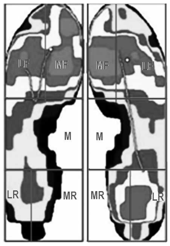
Pressure was evaluated in five areas for better understanding of the plantar pressure distribution on the medial and lateral sides of the rearfoot and forefoot during gait (Fig. 1). The plantar surface was first divided into three larger areas: rearfoot (30% of foot length), midfoot (30% of foot length), and forefoot and toes (40% of foot length), following the scheme established by Cavanagh and Ulbrecht. [27] The rearfoot and forefoot were subdivided, respectively, into medial rearfoot (50% of rearfoot width), lateral rear-foot (50% of rearfoot width), medial forefoot (55% of forefoot width), and lateral forefoot (45% of forefoot width). These areas were defined in the Creation of Any Mask software (Novel), and they are proportional to the length and width of the insoles.
Statistical Analysis
Contact time, contact area, peak pressure, and maximum force followed a normal distribution (Shapiro-Wilk test, P > .05) and a homogeneous variance distribution (Levene test, P > .05). For gait comparisons, the mean for each plantar pressure variable was calculated across steps, excluding the first and last steps per pregnancy. For statistical analysis purposes, pressure data from only one foot of each pregnant woman (randomly selected) were analyzed, and the mean pressures of 30 steps per pregnancy were compared between the trimesters of gestation. The variables were compared among trimesters in five plantar areas during gait and posture using general linear models for repeated-measures analysis of variance (3 × 5), followed by Scheffé post hoc testing (α = .05).
Figure 1.
Regions of the plantar surface studied during gait and orthostatic posture: medial rearfoot (MR), lateral rearfoot (LR), midfoot (M), medial forefoot (MF), and lateral forefoot (LF).
Results
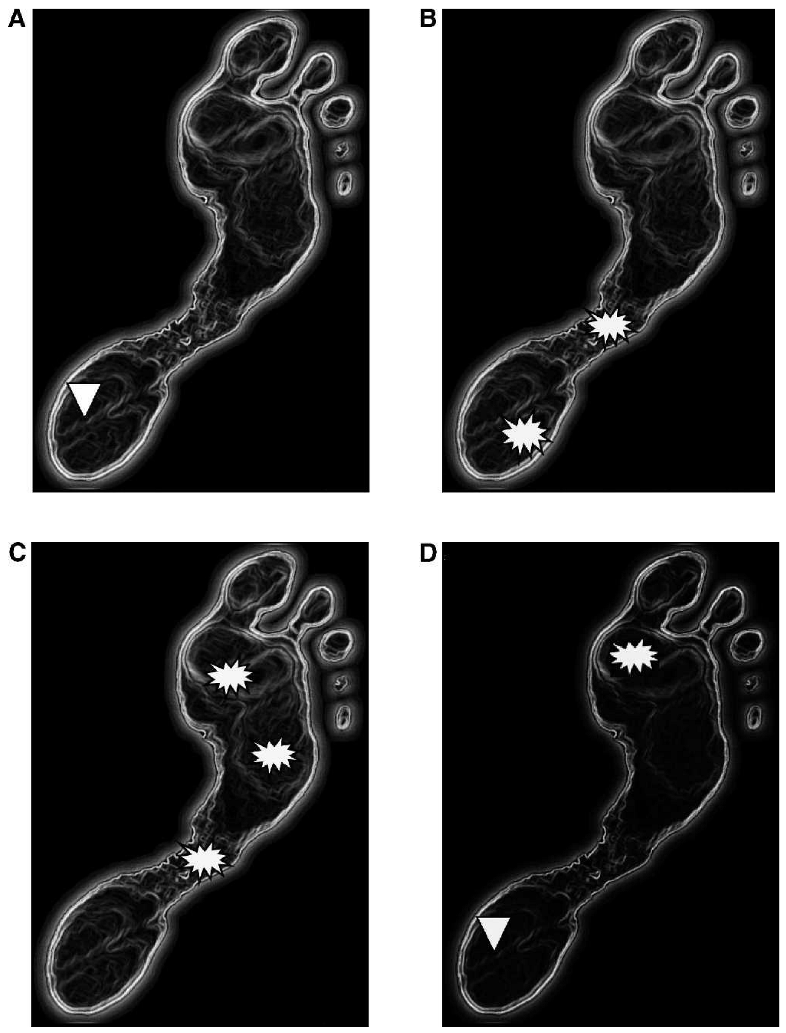
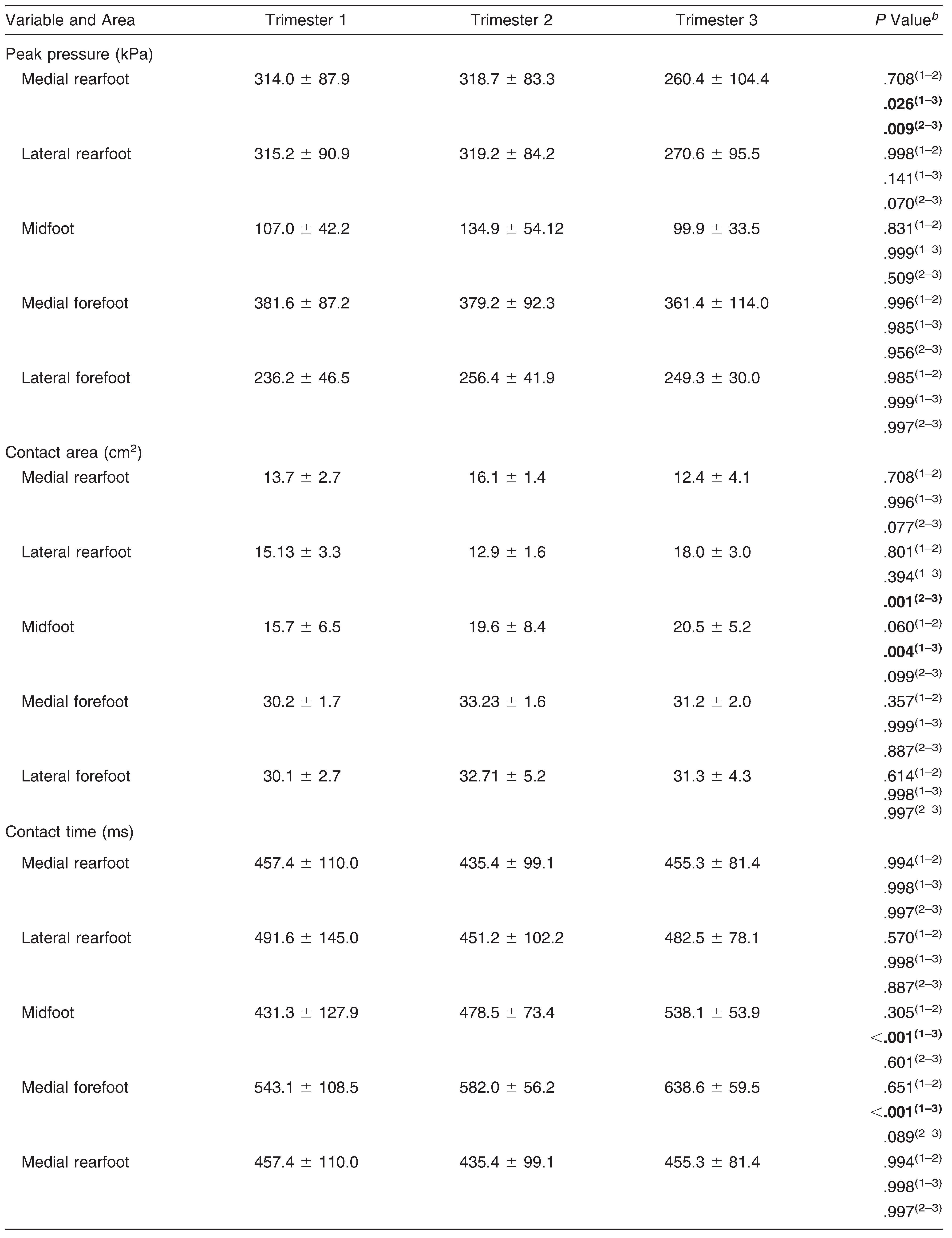
There were no significant differences in any pressure variables during orthostatic posture for all of the plantar areas throughout gestation (Table 1). During gait, throughout gestation, peak pressure at the medial rearfoot significantly reduced from the first to third and second to third trimesters (Table 2). A significant increase in contact area was observed at the lateral rearfoot from the second to third trimester and at the midfoot from the first to third trimester. Contact time increased significantly at the midfoot and the medial and lateral forefoot from the first to third trimester (Fig. 2). Maximum force decreased in 39% from the first to third trimester and in 47% from the second to third trimester in the medial rearfoot and increased in 16% from the first to second trimester in the medial forefoot.
Table 1.
Plantar Pressure Distribution of Various Foot Areas During Orthostatic Posture in Each Trimester of Pregnancy a
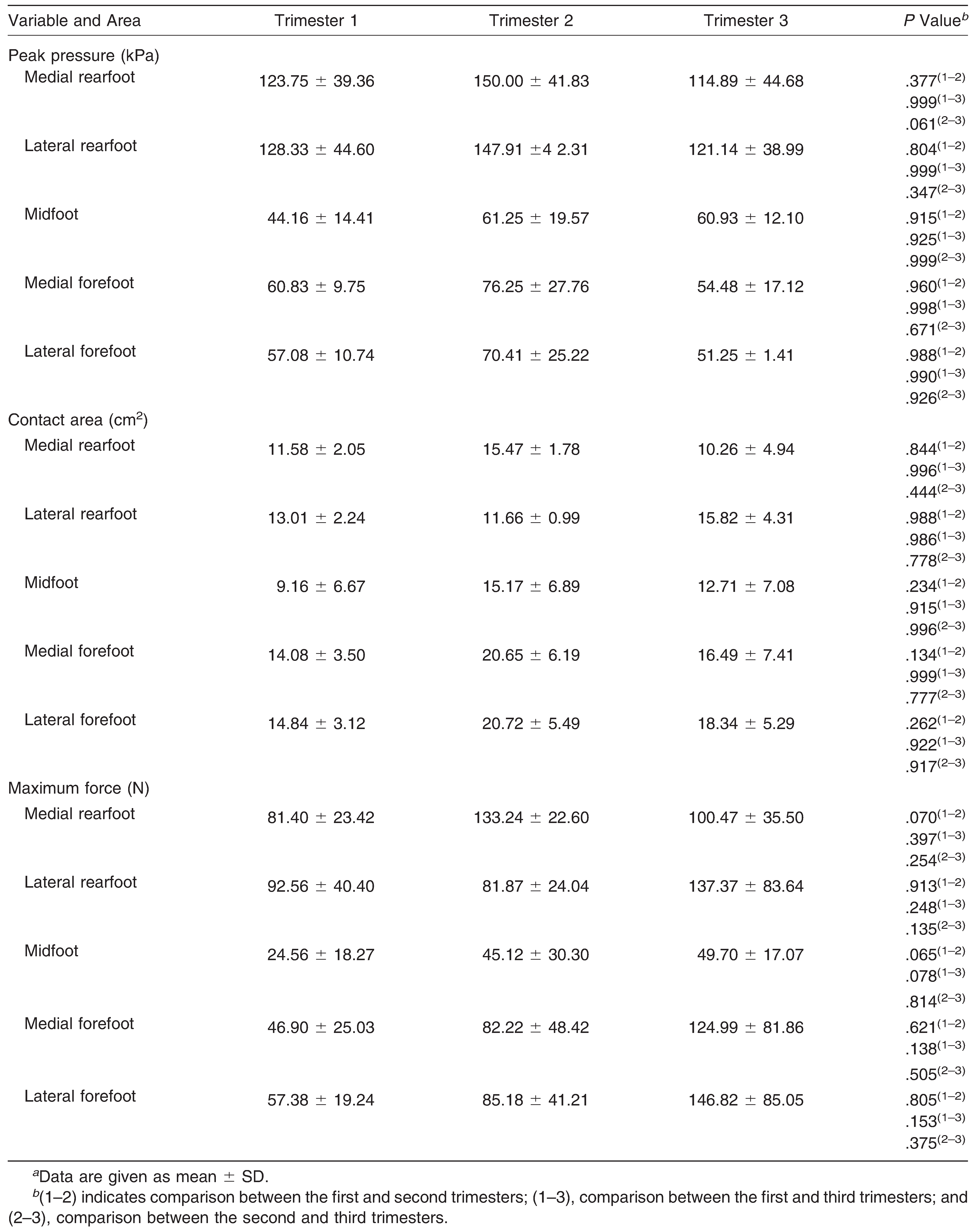
Figure 2.
Representation of changes in peak pressure, contact time, contact area, and maximum force throughout the gestation trimesters during gait. A, Peak pressure on the medial rearfoot from the first to third and second to third trimesters. B, Contact area on the lateral rearfoot from the second to third trimester and on the midfoot from the first to third trimester. C, Contact time on the midfoot and on the medial and lateral forefoot from the first to third trimester. D, Maximum force on the medial rearfoot from the first to third and second to third trimesters and on the medial forefoot from the first to second trimester. Decrease  Increase

Increase
Discussion
The aim of this study was to describe longitudinally the plantar pressure distribution during orthostatic posture and gait in pregnant women. The main results did not support the initial premise because the plantar pressures during orthostatic posture remained unaltered throughout the gestational trimesters. However, during gait, there was an increase in contact time at the anterior parts of the foot (forefoot and midfoot), an increase in contact area at the midfoot, and a decrease in peak pressure and maximum force at the medial rearfoot throughout the gestational trimesters.
Table 2.
Plantar Pressure Distribution of Various Foot Areas During Gait in Each Trimester of Pregnancya
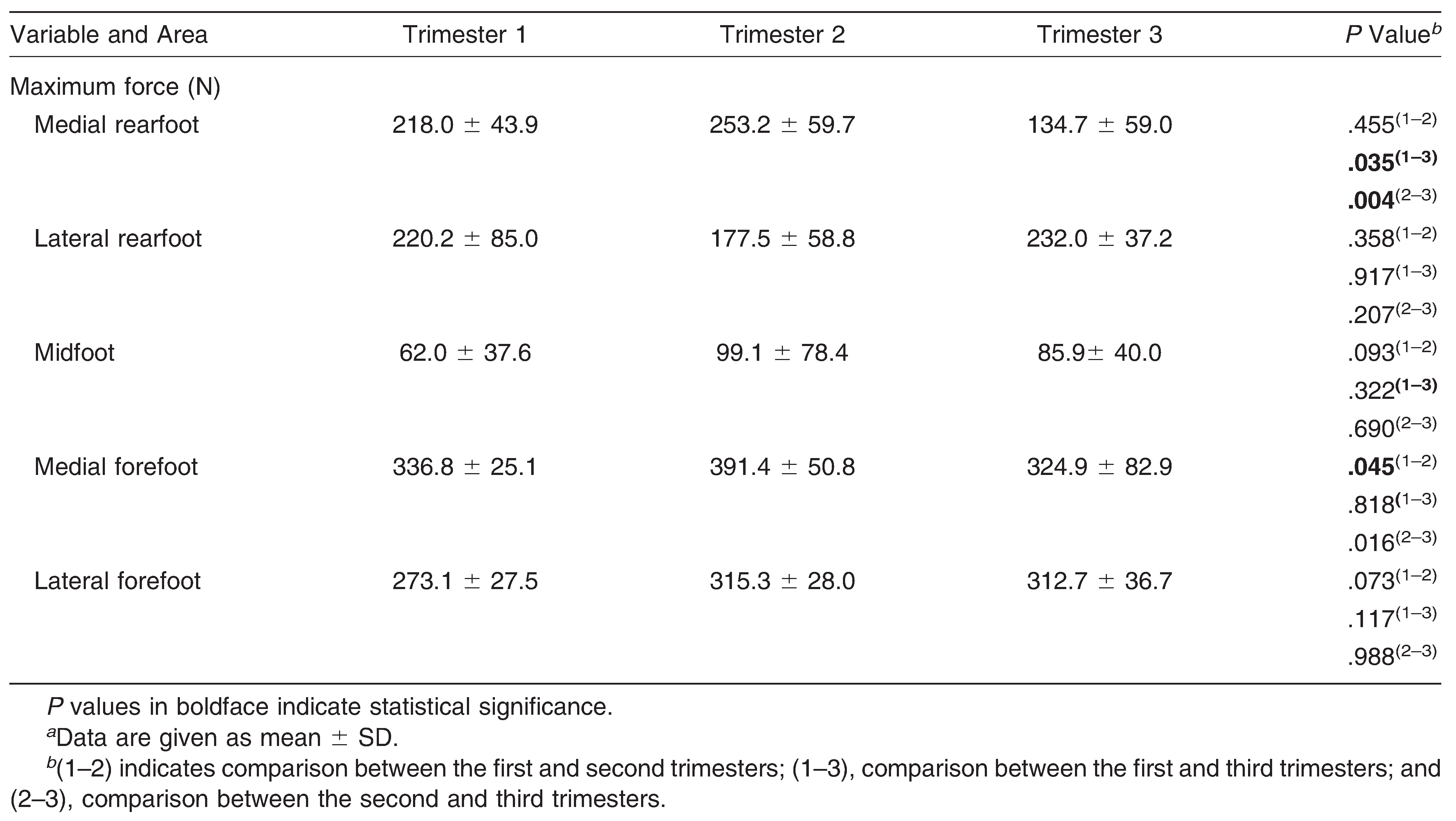
The orthostatic posture results corroborated the findings of Ribas and Guirro, [12] who transversally evaluated peak pressure and contact area in groups of pregnant women at their respective gestation trimesters. Nyska et al, [15] on the other hand, found contact area and peak pressure at the rearfoot to be 12% greater in the last week of pregnancy during orthostatic posture than in a group of nonpregnant women. However, the authors did not consider the mechanical and postural adaptations of each pregnant woman throughout gestation. The present longitudinal study observed no significant differences in plantar pressure variables during orthostatic posture for pregnant women. The explanation for the absence of differences in plantar pressure during orthostatic posture throughout the trimesters might be a possible progressive increase in postural stability that enabled the pregnant women to maintain this pressure distribution regardless of the overloads generated on the plantar surface.
During gait, the most notable changes throughout the gestational trimesters were the increases in contact area and contact time at the midfoot and forefoot and the peak pressure and maximum force reductions at the rearfoot at the end of the third trimester. The peak pressure results differ from the observations of Nyska et al, [15] who observed higher peaks in pregnant women but only at the last week of the third trimester. These authors did not follow gestational changes throughout the pregnancy, and this can explain the differences observed.
The results of loads, contact time, and contact area indicate a potential alteration in the foot rollover process throughout the pregnancy in a way that the midfoot and the forefoot played a different role during the stance phase of gait. This rollover process (the weight acceptance, midstance, and push-off phases during gait) should be performed in such a way to contact the plantar regions in a progressive temporal order from the heel to the metatarsal heads and hallux. [28] This probable alteration in the foot rollover process led to the observed changes in the pressure, force, and contact time distribution. There was an evident contribution of the anterior parts of the foot in the rollover, moving the plantar loads from the posterior parts of the foot (rearfoot) to more anterior parts (forefoot), increasing contact area at the midfoot and contact time at the midfoot and forefoot.
Another suitable explanation for the increase in contact area at the midfoot during gait can be based on the studies by Alvarez et al, [10] Wetz et al, [11] and Block et al [20] that pointed out anthropometric and anatomical alterations in the plantar surface. According to these authors, these changes can be caused by ligament structure modifications, edema accumulation, and increased foot volume and size. This can lead to lowering the longitudinal plantar arch, increasing the contact area over the midfoot. However, the present study cannot confirm this premise because we did not measure anthropometric characteristics of feet during pregnancy.
The reduction in the loads (peak pressure and maximum force) at the rearfoot and the increase in force at the forefoot and contact area at the midfoot may also be explained by the pregnant woman’s attempt to maintain equilibrium, especially during the third trimester, despite the weight gain. [22, 29] This weight change contributes greatly to increase the dislocation of the center of gravity and, consequently, to a greater dynamic postural instability. Thus, the changes in the loads and plantar support distribution that pregnant women undergo to initiate gait is understandable given that a greater risk of falling is prevalent in 25% of this population. [13] This demonstrates a pregnant woman’s attempt at controlling the plantar loads during the foot rollover process to perform gait throughout the gestation trimesters. Lymbery and Gilleard [16] studied women during late pregnancy and also suggest that pregnant women may adapt their gait to maximize stability in the stance phase of gait and to control mediolateral motion of the foot.
Despite the small sample, which was a limitation in the present study, this work adds to the existing evidence regarding the effects of the pregnancy on walking. This work also contributes to the literature by longitudinally investigating gestation throughout the trimesters. These results reveal an important effect of gestational trimester on the foot rollover process, increasing the force, area, and contact time of the anterior parts of the foot. Further kinematic evaluations focused on the lower limbs, particularly on the foot, may better explain the foot and ankle adjustments throughout pregnancy and would provide a better understanding of the plantar pressure distribution findings of the present study.
Conclusions
Pregnant women did not alter plantar pressure during orthostatic posture throughout gestation. However, during gait, there was a redistribution of the plantar loads from the rearfoot to the forefoot due to decreasing peak pressure and force at the rearfoot and increasing force, contact area, and contact time at the midfoot and forefoot throughout gestation. These adjustments in the plantar loads help maintain the dynamic stability of pregnant women during locomotion.
Financial Disclosure: None reported. | |
Conflict of Interest: None reported. |
References
- Borg-Stein, J, SADugan, and JGruber. :Musculoskeletal aspects of pregnancy. Am J Phys Med Rehabil 84: 180, 2005.
- Gilleard, WL, JCrosbie, and RSmith. :Static trunk posture in sitting and standing during pregnancy and early postpartum. Arch Phys Med Rehabil 83: 1739, 2002.
- Bullock, J, JGwendolen, and MBullock. :The relationship of low back pain to postural changes during pregnancy. Aust J Physiother 33: 11, 1987.
- Franklin, ME and TConner-Kerr. :An analysis of posture and back pain in the first and third trimesters of pregnancy. J Orthop Sports Phys Ther 28: 133, 1998.
- Dumas, GA, JGReid, LAWolfe, et al. :Exercise, posture, and back pain during pregnancy. Clin Biomech (Bristol, Avon) 10: 104, 1995.
- Paul, JA, HSalle, and MHFrings-Dresen. :Effect of posture on hip joint moment during pregnancy, while performing a standing task. Clin Biomech (Bristol, Avon) 11: 111, 1996.
- Moore, K, GDumas, and JReid. :Postural changes associated with pregnancy and their relationship with low back pain. Clin Biomech 5: 169, 1990.
- Vullo, VJ, JKRichardson, and EAHurvitz. :Hip, knee, and foot pain during pregnancy and the postpartum period. J Fam Pract 43: 63, 1996.
- Calguneri, M, HABird, and VWright. :Changes in joint laxity occurring during pregnancy. Ann Rheum Dis 41: 126, 1982.
- Alvarez, R, IAStokes, DEAsprinio, et al. :Dimensional changes of the feet in pregnancy. J Bone Joint Surg Am 70: 271, 1988.
- Wetz, H, JHentschel, BDrerup, et al. :Form-und Größenveränderungen des Fußes während der Schwangerschaft. Orthopäde 35: 1124, 2006.
- Ribas, S and EGuirro. :Analysis of plantar pressure and postural balance during different phases of pregnancy. Brazilian J Phys Ther 11: 391, 2007.
- Butler, EE, IColon, MLDruzin, et al. :Postural equilibrium during pregnancy: decreased stability with an increased reliance on visual cues. Am J Obstet Gynecol 195: 1104, 2006.
- Bird, AR, HBMenz, and CCHyde. :The effect of pregnancy on footprint parameters: a prospective investigation. JAPMA 89: 405, 1999.
- Nyska, M, DSofer, APorat, et al. :Planter foot pressures in pregnant women. Isr J Med Sci 33: 139, 1997.
- Lymbery, J and WGilleard. :The stance phase of walking during late pregnancy: temporospatial and ground reaction force variables. JAPMA 95: 247, 2005.
- Foti, T, JRDavids, and ABagley. :A biomechanical analysis of gait during pregnancy. J Bone Joint Surg Am 82: 625, 2000.
- Taves, C, JCharteris, and JWall. :The kinematics of treadmill walking during pregnancy. Physiother Can 34: 321, 1982.
- Golomer, E, DDucher, GSArfi, et al. :Simple locomotion and during load carrying in pregnant women [in French]. J Gynecol Obstet Biol Reprod (Paris) 20: 406, 1991.
- Block, RA, LAHess, EVTimpano, et al. :Physiologic changes in the foot during pregnancy. JAPMA 75: 297, 1985.
- Goldberg, J, MPBesser, and LSilverstein-Selby. :Changes in foot function throughout pregnancy. Obstet Gynecol 97: 395, 2001.
- Jensen, RK, SDoucet, and TTreitz. :Changes in segment mass and mass distribution during pregnancy. J Biomech 29: 251, 1996.
- Shaw, JE, CHvan Schie, ALCarrington, et al. :An analysis of dynamic forces transmitted through the foot in diabetic neuropathy. Diabetes Care 21.:1955, 1998.
- Bus, SA and Ade Lange. :A comparison of the 1-step, 2-step and 3-step protocols for obtaining barefoot plantar pressure data in the diabetic neuropathic foot. Clin Biomech 20: 892, 2005.
- Gurney, JK, UGKersting, and DRosenbaum. :Between-day reliability of repeated plantar pressure distribution measurements in a normal population. Gait Posture 27: 706, 2008.
- Macellari, V, CGiacomozzi, and RSaggini. :Spatial-temporal parameters of gait: reference data and a statistical method for normality assessment. Gait Posture 10: 171, 1999.
- Cavanagh, PR and JSUlbrecht. :“Biomechanical Aspects of Foot Problems in Diabetes,”. inThe Foot in Diabetes, ,2nd Ed, ed byBoulton, AJM, HConnor, and PRCavanagh, p25, John Wiley & Sons Ltd, Chichester, UK, 1994.
- Sacco, ICN, ANHamamoto, AAGomes, et al. :Role of ankle mobility in foot rollover during gait in individuals with diabetics neuropathy. Clin Biomech 24: 687, 2009.
- Cogswell, ME, MKSerdula, DWHungerford, et al. :Gestational weight gain among average-weight and overweight women: what is excessive?Am J Obstet Gynecol 172 ( pt 1:):705, 1995.
© 2011 American Podiatric Medical Association