


Simple Summary
A common treatment for liver cancer involves guiding a thin tube into an artery that supplies the tumor and delivering tiny radioactive beads through the tube. In some patients, the tumor also draws blood from vessels outside the liver, such as the artery beneath the diaphragm or the artery supplying the gallbladder. The usual approach is to block those vessels to redirect blood flow through the liver’s arteries before delivering the beads. We reviewed published reports in which doctors instead delivered the beads directly through vessels outside the liver. Across eleven studies, doctors delivered the beads successfully through vessels outside the liver in every patient treated this way. Serious side effects were uncommon, and no study reported radiation damage to the liver. Many tumors responded to treatment. Because liver tumors often draw blood from several vessels, almost all patients also received beads through a liver artery. The contribution of delivery through vessels outside the liver therefore remains unclear.
Abstract
Hepatocellular carcinoma (HCC) often recruits an arterial blood supply from parasitized extra-hepatic vessels. Yttrium-90 (Y-90) radioembolization can be delivered directly through these vessels as an alternative to embolizing them to redistribute flow. This comprehensive review describes the feasibility, safety, and reported tumor response of that approach. PubMed, Embase, and Scopus were searched to June 2026, and outcomes were summarized descriptively. We included five retrospective cohorts (109 reported participants) and six case reports. All five cohorts were at serious risk of bias. The studies reported delivery through the inferior phrenic, cystic, adrenal, internal mammary, and superior mesenteric arteries. Delivery through the extra-hepatic artery was completed in every treated patient. In the four cohorts reporting tumor response, the objective response ranged from 68% to 100% and complete response from 21.1% to 76.5%, with wide confidence intervals, but nearly all patients also received hepatic-artery Y-90, so response cannot be attributed to the extra-hepatic route alone. Grade ≥ 3 toxicity occurred in 5.0% and 7.0% of patients in the two cohorts reporting it, and no radioembolization-induced liver disease was reported. No study compared direct delivery with flow redistribution, so the evidence supports feasibility and preliminary safety in selected patients rather than efficacy, and comparative studies with standardized dosimetry and reporting may be helpful.
1. Introduction
While hepatic resection and transplantation are the primary curative treatments for hepatocellular carcinoma (HCC), their application is frequently limited by the severe shortage of donor organs, specific tumor characteristics, and the severity of the patient’s underlying liver dysfunction [1,2,3,4].
Radiation segmentectomy, the ablative form of yttrium-90 (Y-90) radioembolization, is used as a bridge to liver transplantation. In selected patients with unresectable early-stage HCC, it is considered a curative intent therapy, supported by high rates of complete radiologic response and complete pathologic necrosis at explant [5,6,7,8,9,10,11]. For patients with intermediate and advanced stage HCC, transarterial radioembolization (TARE) has similarly taken a more prominent role with recent studies showing excellent outcomes compared to transarterial chemoembolization (TACE) and with contemporary dosimetry [12,13,14,15].
Accurate evaluation of hepatic arterial anatomy is therefore essential, as variant hepatic artery anatomy occurs in 45% to 75% of individuals. Meticulous pre-procedural mapping is needed to delineate the tumor’s arterial supply, enable superselective therapeutic delivery, and avoid non-target embolization [16,17,18]. Particularly in large, peripheral, or previously treated tumors, tumor perfusion may shift from native hepatic arterial branches to extra-hepatic collateral arteries [19,20].
Extra-hepatic collateral pathways are frequently recruited in HCC and can constitute the dominant tumor supply [19]. In a 2006 prospective study of 479 consecutive patients with newly diagnosed HCC, Chung et al. found extra-hepatic collateral arterial supply in 17% of patients at initial presentation. Collateral recruitment increased as these patients underwent repeated chemoembolization [21]. The right inferior phrenic artery (IPA) accounted for approximately half of all extra-hepatic collateral vessels identified [21]. Other studies have demonstrated similar findings as well with prevalence of 43.3% for tumors larger than 5 cm at initial presentation, and escalating from 47% at the second TACE session to 98% by the third session [21,22]. Failure to identify and treat these vessels is a well-recognized cause of incomplete response and local recurrence following locoregional therapy [19,23].
The IPA is the most common parasitized extra-hepatic artery [24,25,26]. It most frequently supplies tumors that are touching the diaphragm or are located in the bare area of the liver [20,27]. The IPA has a complex anatomy with ascending and descending branches, superior suprarenal branches, and potential systemic-to-pulmonary shunts through the azygoesophageal branch, all of which must be characterized before safe microsphere delivery [20,28]. The cystic artery, which conventionally arises from the main trunk of the right hepatic artery in approximately half of patients, is clinically relevant for lesions adjacent to the gallbladder fossa, where its deep branches may function as true tumor-feeding vessels rather than merely a source of non-target embolization risk [20,29]. Radiation-induced cholecystitis is a recognized complication of radioembolization, caused by inadvertent deposition of microspheres in the gallbladder wall. After lobar hepatic-artery radioembolization, where the gallbladder is irradiated as an off-target structure, symptomatic cholecystitis requiring cholecystectomy has been reported in 0.6% to 5.4% of patients [29,30,31].
Despite the proven efficacy of Y-90 TARE for HCC treated via intrahepatic arterial supply, its application via extra-hepatic arterial routes has been limited by concerns about non-target radiation to the diaphragm, adrenal glands, lungs, and gallbladder, and by the technical demands of superselective catheterization of small-caliber collateral vessels [28,32]. The conventional approach to extra-hepatic tumor supply has been embolization of parasitized collateral arteries to redirect arterial inflow back through intrahepatic branches before Y-90 delivery. In 2011, Abdelmaksoud et al. demonstrated that this redistribution strategy was technically successful in 94–96% of territories but acknowledged that flow redistribution may be incomplete or unpredictable, and that other collateral arteries may be recruited to replace the embolized vessel [33]. More recently, several groups have demonstrated that direct Y-90 delivery through extra-hepatic arteries (principally the IPA and cystic artery) is technically feasible with acceptable safety and encouraging tumor response [28,32,34,35,36,37]. However, these studies are constrained by small sample sizes, diverse patient populations, and inconsistent reporting of adverse events and efficacy endpoints.
Given the clinical importance of extra-hepatic arterial supply in HCC and the expanding technical capability to deliver Y-90 directly through collateral vessels, this comprehensive review describes the feasibility and safety of TARE delivered through extra-hepatic arteries in patients with HCC and summarizes the reported tumor responses. It also uses the cohort and case-report evidence to set out the anatomical and technical factors that bear on choosing between direct extra-hepatic delivery and hepatic delivery with flow redistribution, and to identify the evidence still needed to guide that choice.
2. Materials and Methods
2.1. Study Design
This comprehensive review was conducted to describe the feasibility, safety, and reported tumor response of TARE delivered through extra-hepatic arteries in patients with HCC. We chose a comprehensive rather than a systematic review format because the aims go beyond the effect of a single intervention. The review also sets out the anatomy and technique of each route and the factors that bear on choosing between direct extra-hepatic delivery and hepatic delivery with flow redistribution, and it identifies priorities for future studies. Also, no included study had a comparison group, and nearly all cohort patients also received Y-90 through the hepatic artery, so the effect of the extra-hepatic route could not be separated from that of the hepatic treatment. The literature search and study selection were still carried out systematically, so that the clinical evidence was captured as completely as the three databases allowed and the selection can be reproduced. The selection process is shown in a flow diagram based on the Preferred Reporting Items for Systematic Reviews and Meta-Analyses (PRISMA 2020) template [38]. Because few studies were eligible and they were clinically heterogeneous, the evidence was synthesized as a structured narrative in line with the Synthesis Without Meta-analysis (SWiM) guidance rather than by statistical pooling [39].
2.2. Search Strategy
A comprehensive electronic search was conducted in PubMed, Embase, and Scopus from database inception to 24 June 2026. Search terms combined Medical Subject Headings (MeSH) and free-text words: “Hepatocellular Carcinoma,” “HCC,” “Yttrium-90,” “Y-90 radioembolization,” “extra-hepatic,” “inferior phrenic artery,” “cystic artery,” “adrenal artery,” and “internal mammary artery.” Boolean operators (“AND” “OR”) were applied to refine search sensitivity. The detailed search strategy for each database is provided in Supplementary File S1.
2.3. Inclusion Criteria
Studies were eligible if they reported outcomes of TARE delivered through an extra-hepatic artery, such as the inferior phrenic, cystic, adrenal, or internal mammary artery, in adults aged 18 years or older with HCC. For this review, an extra-hepatic artery was any tumor-feeding vessel other than the hepatic artery supplying that tumor’s territory. One eligible group comprised systemic collaterals recruited from outside the hepatic circulation, such as the inferior phrenic, adrenal, and internal mammary arteries. The other comprised the cystic artery, which arises from the hepatic arterial tree but supplies the gallbladder and may be recruited by an adjacent tumor. Both were eligible, and results are reported separately by route. No comparator was required, as controlled studies were not anticipated. Each study had to report at least one response outcome (tumor response, complete response (CR), partial response (PR), stable disease (SD), progressive disease (PD), or objective response rate (ORR)) or one safety outcome (procedure-related adverse events, hepatobiliary or hematologic toxicity, or pulmonary complications). Retrospective and prospective cohort studies, case series, and single-patient case reports were all eligible. Descriptive response and safety estimates were based on cohorts and case series alone. Case reports were summarized separately to describe the range of clinical and technical findings. Only full-text, peer-reviewed publications in English were included.
2.4. Exclusion Criteria
Studies were excluded if they (i) did not involve Y-90 delivery through an extra-hepatic artery, (ii) were reviews, editorials, or conference abstracts without full peer-reviewed data, or (iii) were non-English publications.
2.5. Study Selection
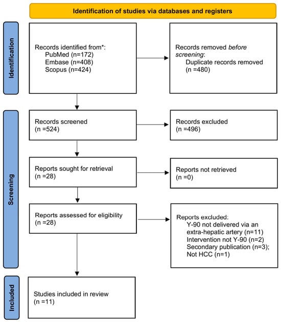
Two reviewers (A.Si. and V.P.K.) independently screened titles and abstracts against the eligibility criteria in Section 2.3 and Section 2.4, and full texts of potentially eligible studies were retrieved for detailed assessment. Discrepancies were resolved by consensus. The study selection process is shown in Figure 1.
Figure 1.
Flow diagram of study identification, screening, and inclusion, based on the PRISMA 2020 template. Of 1004 records identified across PubMed, Embase, and Scopus, 524 were screened after duplicate removal, 28 underwent full-text assessment, and 11 studies were included (five retrospective cohorts and six single-patient case reports). * Number of records identified in each database searched.
2.6. Outcomes and Data Extraction
Two reviewers (A.Si. and V.P.K.) independently extracted data using a predefined, standardized data extraction form. Extracted study-level data included patient demographics (mean or median age, sex distribution, liver function status, Barcelona Clinic Liver Cancer (BCLC) stage, tumor morphology, and presence of portal vein invasion), study characteristics (study design and sample size), and bibliographic information (first author, year of publication, and country of origin). Intervention-related data included details of extra-hepatic Y-90 radioembolization, such as arterial access route (e.g., inferior phrenic, cystic, adrenal, or internal mammary artery), type of Y-90 microspheres used, administered activity, absorbed dose and the dosimetry model used where reported, number of treatment sessions, and technical success of extra-hepatic delivery. For the single-patient case reports, the arterial route, microsphere type, administered activity and/or absorbed dose, tumor size, tumor response, and reported complications were extracted separately.
The extraction of outcome data followed established safety and response endpoints. Tumor response (CR, PR, SD, PD, and ORR) was extracted as each study defined it, along with the response criteria used, such as the modified Response Evaluation Criteria in Solid Tumors (mRECIST), and the tumor that was assessed. Technical success, overall survival, and tumor progression or recurrence were also extracted where reported. Overall toxicity of any grade, grade ≥ 3 adverse events, and specific clinical toxicities such as pain, fever, fatigue, nausea and vomiting, and ascites were all included in the safety outcomes. Several laboratory abnormalities were identified, including decreases in serum albumin levels and hematological toxicities and increases in aspartate aminotransferase, alanine aminotransferase, alkaline phosphatase, and total bilirubin. All extracted data were cross-checked for accuracy, and discrepancies between reviewers were resolved through discussion and consensus.
2.7. Risk of Bias Assessment
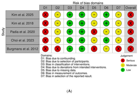
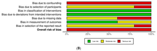
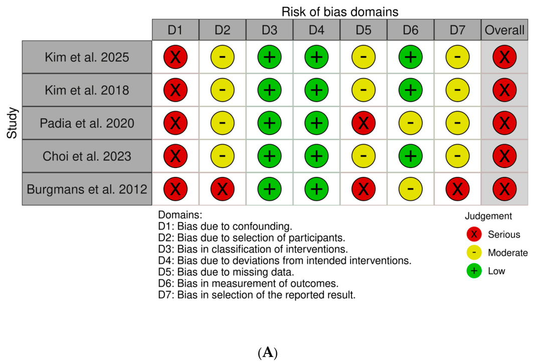
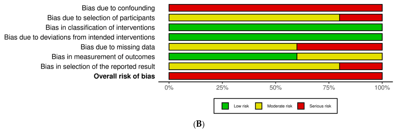
The Risk Of Bias In Non-randomized Studies of Interventions (ROBINS-I) tool was used to assess the risk of bias in included cohort studies. The seven domains assessed were confounding, selection of participants, classification of interventions, deviations from intended interventions, missing data, outcome measurement, and selection of reported results. Each domain was rated as low, moderate, or serious risk, and the overall risk of bias for each study was determined based on the highest domain-level risk. Results were visualized using summary and traffic light plots (Figure 2A,B) made with the robvis web app (https://mcguinlu.shinyapps.io/robvis/, acccessed on 10 November 2026) [40]. The six case reports were appraised separately with the tool described by Murad et al. in 2018, which assesses selection, ascertainment of exposure and outcome, causal attribution, and reporting [41].
Figure 2.
(A) ROBINS-I traffic-light plot showing the individual risk-of-bias judgment for each cohort study [28,32,34,35,36] across the seven ROBINS-I domains and the overall rating. (B) ROBINS-I risk-of-bias summary plot for the five cohort studies, showing the proportion of studies judged at low, moderate, or serious risk of bias across the seven ROBINS-I domains and the overall assessment.
2.8. Data Synthesis
Outcomes were synthesized using a structured narrative approach (SWiM) [39]. For each outcome, study-level event counts and crude proportions were tabulated and summarized as a range across studies, organized by arterial route and treatment intent. Exact (Clopper–Pearson) 95% confidence intervals were calculated in R (version 4.6.0) for each study’s response proportions. The cohort studies contributed descriptive response and safety estimates, while the case reports were summarized separately to describe the clinical and technical spectrum without contributing to rate estimates. Statistical pooling was not undertaken because of the small number of studies, the clinical heterogeneity in tumor characteristics, dosimetry, and treatment intent, and the non-uniform outcome definitions across studies. Per-study data were presented in summary tables.
3. Results
3.1. Study Selection Process
The database search identified 1004 records (PubMed, n = 172; Embase, n = 408; Scopus, n = 424). After 480 duplicates were removed, 524 unique records underwent title and abstract screening, of which 496 were excluded. The remaining 28 reports were assessed in full text. The 17 excluded reports included 11 in which Y-90 was not delivered through an extra-hepatic artery (it was administered via the hepatic artery, with extra-hepatic vessels embolized only for flow redistribution or non-target protection), 2 that involved a non–Y-90 intervention (one holmium-166 radioembolization and one chemoembolization of the extra-hepatic supply), 3 because they were not primary reports (a review article, an invited commentary, and a symposium paper), and 1 that involved a non-HCC population. Eleven studies met the inclusion criteria (five retrospective cohort studies and six single-patient case reports) and were included in the synthesis [28,32,34,35,36,37,42,43,44,45,46]. The study selection process is illustrated in Figure 1.
3.2. Study Characteristics and Patient Demographics
Eleven studies were included, comprising five retrospective cohort studies (published 2012–2025; with 109 reported participants) and six single-patient case reports (published 2019–2026). Three cohorts delivered Y-90 through the inferior phrenic artery and two through the cystic artery. Table 1 shows the baseline characteristics, arterial route, and treatment details for each cohort, and Table 2 gives the number of patients in each category. Of the 109 reported participants treated through an extra-hepatic artery, 103 were evaluable for response and 104 also received hepatic-artery Y-90 in the same session. The remaining 5, all reported by Padia et al. in 2020 [35], were treated through the cystic artery alone. In 2012, Burgmans et al. [32] reported safety outcomes only.
Table 1.
Baseline characteristics, extra-hepatic arterial route, and treatment delivery of the five included cohort studies.
Table 2.
Patient numbers by category in the five cohort studies.
Three case reports described treatment through parasitized adrenal arteries [37,42,43], two through the internal mammary artery, and one through the right inferior phrenic artery together with a superior mesenteric artery branch in fibrolamellar HCC [44,45,46]. The adrenal and superior mesenteric routes appear only in the case reports. The internal mammary artery was also used in one patient in the 2018 study by Kim et al. [28]. Treated tumors ranged from 2.5 to 14 cm and four of six were treated with ablative intent (Table 3).
Table 3.
Delivery artery, tumor characteristics, activity, response, and adverse events in the six single-patient case reports.
Two cohorts, both from Seoul, reported the cause of liver disease. Hepatitis B predominated in both cohorts, affecting 29 of 43 patients (67%) in the 2025 study by Kim et al. [36] and 18 of 24 (75%) in the 2023 study by Choi et al. [34]. Hepatitis C, alone or with hepatitis B, was reported in 5 and 2 patients, respectively. Alcohol was identified as the cause in one patient in the Kim et al. 2025 cohort [36]. No cause was identified in 8 patients in the Kim et al. 2025 cohort and 4 in Choi et al.’s cohort. The other three cohorts did not report etiology. Padia et al. 2020 [35] stated that all 17 patients had cirrhosis, whereas the other cohorts gave Child-Pugh class without stating whether every patient was cirrhotic (Table 1). Among the case reports, the underlying disease was metabolic dysfunction-associated steatotic liver disease in two patients (reported as non-alcoholic fatty liver disease or steatohepatitis), hepatitis C in one, and alpha-1-antitrypsin deficiency with alcohol-related liver disease in one. The underlying liver disease was not stated for a patient with HIV, and the fibrolamellar tumor arose without underlying liver disease.
3.3. Risk of Bias
All five cohort studies were rated as having a serious overall risk of bias using ROBINS-I. Confounding drove these ratings because none had a control group or could adjust for baseline liver function, tumor burden, or prior treatment. Domain-level judgments for each study are shown in Figure 2A,B. This uniformly serious rating limits confidence in the cohort findings and is why they were summarized descriptively rather than pooled.
Exposure ascertainment, follow-up, and reporting were adequate in all six case reports assessed with the tool described by Murad et al. in 2018 [41], and selection was unclear in each, as expected for single-patient reports [41]. Outcome ascertainment was objective throughout, with imaging used to assess response in every case and pathologic necrosis assessed at explant in two, although two reports did not name formal response criteria. The principal limitation was causal attribution: in two reports (Cortizo Vidal et al. 2019 [43]; Casadaban and Padia 2023 [45]) Y-90 was also delivered through the hepatic artery in the same session, so the response could not be attributed to the extra-hepatic route alone, whereas the remaining four delivered Y-90 exclusively through extra-hepatic arteries (Supplementary Table S1).
3.4. Outcomes
Four cohorts reported tumor response, all using mRECIST (Table 4). The objective response rate ranged from 68.4% (95% confidence interval [CI] 43.4–87.4%) in the 2018 study by Kim et al. [28] to 100% (95% CI 80.5–100%) in the 2020 study by Padia et al. [35], and the complete response rate from 21.1% to 76.5% in the same two cohorts. With 17 to 43 evaluable patients per cohort, the confidence intervals for complete response spanned 31 to 43 percentage points. Progressive disease was reported only in the 2018 study by Kim et al. [28] (4 of 19 patients), and stable disease in no more than two patients in any cohort.
Table 4.
Tumor response by study in the four cohorts that reported it.
Most of these patients also received hepatic-artery Y-90 in the same session, including all patients in Kim et al. 2025 [36], Kim et al. 2018 [28], and Choi et al. 2023 [34], and 12 of the 17 in Padia et al. 2020 [35], whose five cystic-artery-only patients were not reported separately (Table 2). All four cohorts measured response in the tumor supplied by the extra-hepatic artery, and because that tumor also received hepatic-artery Y-90 in most patients, the cohort data cannot show how much of the response is due to the extra-hepatic route. Treatment through extra-hepatic arteries alone appears only in four case reports, each of which described some tumor response, including complete pathologic necrosis at explant in one (Table 3).
Within each arterial route, CR was more frequent in the cohorts with smaller tumors and ablative treatment intent. Among the cystic-artery cohorts, the complete response rate was 76.5% in Padia et al. 2020 [35], which had the smallest tumors in the review, compared with 29.2% in Choi et al. 2023 [34], where median tumor diameter was more than twice as large. The IPA cohorts followed the same pattern: 44.2% in Kim et al. 2025 [36], restricted to BCLC stage A or B disease, and 21.1% in Kim et al. 2018 [28], the most advanced of the four cohorts reporting response (Table 1 and Table 4).
Across routes, CR rates aligned more closely with tumor size and treatment intent than with the artery used, although with four studies that differed in design, patient selection, and dosimetry it is not possible to say which factor accounts for the differences. Complete response rates in the two cystic-artery cohorts alone ranged from 29.2% to 76.5%, and one IPA cohort (Kim et al. 2025 [36], 44.2%) fell between those values, while the rate in the other (Kim et al. 2018 [28]) was lower at 21%.
Delivery through the extra-hepatic artery was completed in every treated patient in all five cohorts, although only two cohorts defined technical success. Burgmans et al. 2012 [32] defined it as no significant activity outside the liver on post-treatment Y-90 imaging, and Padia et al. 2020 [35] defined it as mapping and infusion completed without technical complications by Society of Interventional Radiology criteria. In Kim et al. 2025 [36], one of the 44 patients with inferior phrenic supply received bland embolization instead, because the tumor blush was mixed with a systemic-to-pulmonary shunt. Of the two cohorts that reported survival, Kim et al. 2025 [36] followed patients for a median of 31.7 months and reported overall survival of 83.9% at 2 years and 70.9% at 5 years, local tumor progression-free survival of 81.7% at 1 year and 52.9% at 3 years, and a median progression-free survival of 14.8 months. Complete response in that cohort was associated with longer local tumor progression-free survival (p = 0.018) and progression-free survival (p = 0.032), but not with overall survival (p = 0.74). Padia et al. 2020 [35] reported overall survival of 94% at 6 months and 68% at 12 months, with no target tumor progression during a median follow-up of 9 months, although 60% of patients had progression of non-target tumors at 1 year. The other three cohorts did not report survival or progression over time, and their mean or median follow-up ranged from 4.5 to 9 months (Table 1).
3.5. Safety Outcomes
All five cohorts graded adverse events with the Common Terminology Criteria for Adverse Events (CTCAE), but in different versions: 3.0 in Burgmans et al. 2012 [32], 4.03 in Kim et al. 2018 [28], and 5.0 in the other three, with Kim et al. 2025 [36] also using the Society of Interventional Radiology (SIR) classification. Two cohorts reported an overall rate of severe (grade ≥ 3) toxicity. The rates were 1 of 20 patients (5.0%) in Kim et al. 2018 [28] and 3 of 43 (7.0%) in Kim et al. 2025 [36]. The single grade ≥ 3 event in Kim et al. 2018 [28] was a femoral artery pseudoaneurysm related to vascular access rather than a direct treatment toxicity (Table 5). The three grade ≥ 3 events in Kim et al. 2025 [36] were lymphocyte depletion in two patients and severe postembolization syndrome in one. Any-grade toxicity was reported by three cohorts but is not comparable across them: rates of 0%, 55%, and 93% (Burgmans et al. 2012 [32], Kim et al. 2018 [28], Kim et al. 2025 [36]) reflect differing definitions: Burgmans recorded only events attributable to extra-hepatic delivery (none observed), whereas the Kim cohorts captured a broad range of clinical and laboratory events. No radioembolization-induced liver disease (REILD) was reported in any included study, and radiation-induced lung injury was limited to one patient in Kim et al. 2025 [36] with symptomatic radiation pneumonitis (Table 5).
Table 5.
Adverse events and laboratory abnormalities by study, with outcome-specific denominators.
Clinical toxicities were predominantly mild. Pain was the most common, with per-study rates of 15.0% to 50.0%. The lowest rate (Kim et al. 2018 [28]) reflected its recording of intraprocedural pain only, whereas the other cohorts captured both intraprocedural and post-procedural pain. Other clinical toxicities were infrequent across the reporting cohorts and included fever (per-study range 12.5–14.0%), fatigue (7.0–23.5%), nausea/vomiting (4.2–16.3%), and ascites (2.3–17.6%) (Table 5).
Pulmonary imaging findings and hepatobiliary laboratory abnormalities are reported per study in Table 5. Because 104 of the 109 cohort patients also received hepatic-artery Y-90 in the same session, most adverse events in the cohorts, whether clinical, laboratory, or imaging, cannot be attributed to the extra-hepatic route alone. Reporting also differed, with Padia et al. 2020 [35] capturing only grade 3 or higher laboratory events while the others reported all grades.
Safety in the case reports was consistent with the cohort experience, with two route-specific observations. Delivery through the internal mammary artery produced self-limiting, non-target skin ulceration over the epigastric region in one patient, due to the artery supplying the chest/abdominal wall [45]. Conversely, treatment of parasitized adrenal arteries was not complicated by adrenal crisis in any of the three adrenal cases, though one team gave prophylactic medication in anticipation of that risk (Table 3).
4. Discussion
This comprehensive review synthesizes the evidence for Y-90 radioembolization delivered through extra-hepatic arteries in HCC, across the inferior phrenic, cystic, adrenal, and internal mammary routes. Delivery through these routes was completed in every treated patient, and severe toxicity was uncommon, with no REILD reported. The objective response rate ranged from 68% to 100% in the four cohorts that reported it, but nearly all of those patients also received hepatic-artery Y-90, so the response cannot be credited to the extra-hepatic route alone.
Complete response varied more across cohorts than across arterial routes. Tumor size and treatment intent may explain this, but the cohorts also differed in patient selection and dosimetry, so four studies cannot separate these factors. Padia et al. treated the smallest tumors (median 3.8 cm) at a median of 340 Gy to perfused tissue, whereas the other cohorts treated substantially larger tumors. That gradient may reflect the more complex perfusion of larger tumors rather than any limitation of the extra-hepatic route. A tumor that has recruited extra-hepatic supply has a more complex blood supply than one fed entirely from the hepatic artery, and response depends on delivering an adequate dose to viable tumor regardless of the route. Lower response in the larger-tumor cohorts may therefore reflect incomplete identification and treatment of every feeding vessel rather than the route of administration. The case reports are consistent with this pattern. Of the four patients treated with ablative intent, three achieved complete responses and one had more than 95% necrosis at explant, although two of the four also received hepatic-artery Y-90 [37,42,43,45]. Of the two patients treated for downstaging, one had a reduction in tumor size from 3.8 to 2.4 cm, and the other had a 20% reduction in the volume of a 14-cm fibrolamellar tumor. Neither report stated the response criteria [44,46]. Extra-hepatic Y-90 radioembolization therefore covers a range of clinical scenarios rather than a single, uniform intervention.
All four progressive disease events occurred in Kim et al. 2018 [28], which had the most advanced cohort (60% BCLC-C, 35% infiltrative tumors, 55% portal vein invasion), while the other three cohorts reported none. Three of the four occurred in infiltrative tumors that could not be measured, and the fourth in the cohort’s second-largest measurable tumor at 13.6 cm. Progression clustered in these large and infiltrative tumors, in which identifying and treating every feeding vessel is most difficult. Given dose heterogeneity, outcomes for large tumors are known to be worse [47]. The conventional alternative to direct extra-hepatic Y-90 delivery (embolization of parasitized arteries to redistribute flow) carries its own limitations, including incomplete redistribution and recruitment of new collateral pathways [33,48]. Kim et al. 2025 [36] noted a preference for direct IPA treatment over redistribution because proximal IPA occlusion may redirect flow through intercostal or internal mammary arteries rather than the hepatic artery, potentially leaving tumor-feeding territories undertreated [36,49].
Reported doses are not directly comparable across cohorts because they refer to different tissue volumes (Table 1). At the combined-route level, Kim et al. 2025 [36] reported a post-treatment tumor dose of 629 Gy and Kim et al. 2018 [28] reported 152 Gy across all perfused tissue. Because nearly all patients also received hepatic-artery Y-90, these response rates cannot show that the extra-hepatic route alone delivered an effective dose. The more direct evidence comes from the two adrenal-artery case reports without hepatic-artery treatment, in which a tumor dose of 2037 Gy and an administered activity of 1.82 GBq were followed by complete necrosis and more than 95% necrosis, respectively, at explant (Table 3) [37,42].
In terms of safety, the rates of grade ≥3 toxicity in the two cohorts that reported it (5.0% and 7.0%) are consistent with established Y-90 safety profiles, although the cohorts used different CTCAE versions and recording periods. One severe event was attributed to the extra-hepatic route itself. A patient in Kim et al. 2025 [36] who received a virtual tumor absorbed dose of 1000 Gy from resin microspheres through the IPA developed severe postembolization syndrome with reduced diaphragmatic movement and atelectasis and required 13 days in hospital. Postembolization syndrome is the most common complication of hepatic Y-90, reported in 20 to 70% of patients, and is usually mild enough that admission is rarely needed [30]. The frequency of pain (per-study range 15–50%) overlaps the 13–39% reported after standard hepatic Y-90 radioembolization, although the cohorts recorded pain differently [50]. For IPA-delivered therapy, pain reflects diaphragmatic irritation or postembolization syndrome involving the treated tumor bed with possible hepatic capsular irritation. For cystic artery delivery, right upper quadrant pain may represent subclinical cholecystitis; however, no patient in either the Padia et al. 2020 [35] or Choi et al. 2023 [34] cohort required invasive intervention for symptomatic radiation cholecystitis. This may also be the result of low microsphere density treatments.
REILD has been reported in 1–5.4% of patients after hepatic radioembolization, with higher rates in those with impaired baseline liver function or prior liver-directed therapy [50,51,52]. No patient in this review developed REILD. Extra-hepatic delivery deposits microspheres into the tumor’s parasitized bed rather than through the native hepatic arterial tree, which may limit hepatic parenchymal exposure. In Kim et al. 2025 [36] the IPA supplied a mean of 10.4% of total tumor volume [36]. That is not the entire explanation, however, as every cohort patient but five also received hepatic-artery Y-90 in the same session. Most were Child-Pugh A, and the Kim et al. 2025 protocol preserved at least 30% of functional liver volume from irradiation [36]. The more direct evidence comes from the four case reports of patients treated exclusively through extra-hepatic arteries with no concurrent hepatic infusion, none of which reported REILD [37,42,44,46].
Symptomatic radiation pneumonitis is also rare after Y-90 radioembolization, reported in 1.8% of 732 patients in a Korean multicenter study and an estimated 0.1% in a systematic review [53,54]. One patient across this review developed it, a self-resolving event graded 1 on the SIR scale in Kim et al. 2025 [36] (Table 5). Extra-hepatic territories can communicate with the pulmonary circulation, so lung shunt assessment matters as much here as in hepatic delivery [55]. The low rate probably reflects meticulous mapping: systemic-to-pulmonary shunt identification on cone-beam CT and prophylactic embolization of azygoesophageal branches when found [28,36]. Even so, route-specific risks remain clinically relevant: diaphragmatic irritation or injury after inferior phrenic delivery, which in one Kim et al. 2025 [36] patient presented as severe postembolization syndrome with reduced diaphragmatic movement; non-target skin injury after internal mammary delivery [45]; radiation pneumonitis through systemic-to-pulmonary shunts, which were present in 18 of 43 patients in Kim et al. 2025 [36]; and symptomatic radiation cholecystitis after cystic artery delivery. With so few treated patients, events of each kind cannot be excluded.
4.1. Clinical Considerations
No included study compared direct extra-hepatic delivery with embolization and flow redistribution, so the evidence supports the technical feasibility of direct delivery but not its independent efficacy or any advantage over redistribution. The cohorts do show how operators made the choice, and Table 6 summarizes the factors they reported. Direct delivery was chosen when a tumor-only branch could be catheterized and non-target branches could be embolized or bypassed, and when embolizing the feeding vessel for flow redistribution was expected either to fail, because other collaterals would take over [28,36], or to injure the organ it supplies, as with the gallbladder after cystic artery occlusion [34,35]. It was avoided when the feeder could not be catheterized or when a significant shunt or non-target gastric or esophageal enhancement could not be managed. In one series, it was also avoided when the feeder supplied less than 10% of the tumor. These are reported practices rather than validated criteria: Burgmans et al. 2012 left such small feeders untreated [32], whereas Kim et al. 2018 treated the inferior phrenic artery regardless of its share [28]. Proximal delivery that includes non-target branches may be reasonable once significant shunts and enteric supply have been excluded, as in Kim et al. 2025, where microspheres were infused into the main trunk of the right IPA in 23 of 43 patients [36]. As discussed for cystic artery delivery, a low microsphere density may be advisable.
Table 6.
Factors reported by the included cohorts when choosing between direct extra-hepatic delivery and embolization of the feeder.
The current literature cannot answer several questions that follow from these observations. The first is whether the share of the tumor supplied by the feeder should guide treatment, since one group used a 10% cut-off and another treated regardless of share, and no study compared the outcomes. Criteria that define when direct treatment is unsafe also need to be established, beyond the shunts and non-target enhancement that led individual groups to stop. No absorbed-dose thresholds exist for the organs at risk in this setting, such as the gallbladder, diaphragm, adrenal glands, and skin, and it is not known which protective steps, such as embolizing non-target branches, premedicating before adrenal treatment, or applying ice packs to the skin, reduce organ-specific toxicity. Dosimetry of the extra-hepatic territory was planned and reported differently in every cohort and needs a common methodology. Finally, both glass and resin microspheres were used, and resin was used in the one patient with severe postembolization syndrome after inferior phrenic delivery. Given the inherent differences in microsphere activity and microsphere density, a direct comparison of the two platforms is difficult.
Future studies would be more useful if they reported the same core items: the share of tumor perfusion supplied by each extra-hepatic feeder and how it was measured, the absorbed dose to the treated territory and to adjacent organs at risk using a multicompartment dosimetry model, response by mRECIST for the territory fed by the extra-hepatic artery as well as for the whole tumor, and adverse events graded with a stated CTCAE version and attributed to the route where possible. Prospective registries that record both direct delivery and embolization with flow redistribution would allow the two approaches to be compared, ideally with adjustment for tumor burden and liver function, which no study in this review could do.
4.2. Limitations
This review has several limitations. The evidence base is small and uncontrolled: five retrospective cohorts at serious risk of bias, and six single-patient reports subject to selection and publication bias. Patients were treated through the extra-hepatic artery when operators judged the route technically feasible and clinically appropriate, which exposes the cohorts to confounding by indication. Case reports also tend to describe technically successful treatments and may under-report complications. Outcomes were therefore synthesized descriptively rather than pooled, and the per-study figures should be read as a range. The clinical evidence came from a systematic search, but the anatomical and technical literature used to interpret it was selected for relevance and may not be complete. All cohorts used mRECIST. The case reports used mRECIST or the Liver Imaging Reporting and Data System (LI-RADS), or did not specify their response criteria. Toxicity reporting also differed. Padia et al. 2020 [35] reported laboratory events only at grade ≥ 3, and Kim et al. 2018 [28] reported only intraprocedural pain in its assessment of pain. Extra-hepatic dosimetry varied most of all (Table 1). Only Burgmans et al. 2012 [32] measured the phrenic lung shunt fraction directly; the Kim cohorts estimated it from the hepatic artery and used single-compartment models, and Padia et al. 2020 [35] and Choi et al. 2023 [34] named no model at all. Both Kim cohorts also equated perfused tissue volume with tumor volume, which Kim et al. 2018 [28] note overestimates the delivered dose because the thin diaphragm is difficult to contour. Treatment intent and delivered dose are not the same thing, and this review can report the first far more reliably than the second, because the definition of an ablative dose has evolved over time and the metrics that define it are not interchangeable. Administered activity, absorbed dose to the perfused volume, absorbed dose to tumor, and microsphere number all appear in this literature, and reporting was incomplete for every one of them: Burgmans et al. 2012 [32] and Choi et al. 2023 [34] gave no absorbed dose at all, Padia et al. 2020 [35] reported a perfused-angiosome dose against an intended threshold of 200 Gy, and the two Kim cohorts reported doses referring to different volumes; Kim et al. 2025 [36] prescribed 500 to 1000 Gy for glass microspheres and 400 to 700 Gy for resin. The two Seoul National University inferior phrenic artery cohorts overlapped in early 2017 and neither reports whether patients were shared, so their counts were not combined, and the total is reported participants, not confirmed unique patients. Survival was reported by only two cohorts, and only Kim et al. 2025 [36] followed patients beyond 1 year. The central limitation is attribution. All but five cohort patients, as well as two of the six patients described in case reports, received hepatic-artery Y-90 in the same session. The five cystic-artery-only patients in Padia et al. 2020 [35] were not reported separately. Only four case reports described extra-hepatic delivery alone.
5. Conclusions
Y-90 radioembolization through parasitized extra-hepatic arteries was technically feasible in every treated patient, and severe toxicity was uncommon in the cohorts that reported it. Objective response was observed in most patients in the four cohorts that reported it, but nearly all of them also received hepatic-artery Y-90, and no study compared direct delivery with embolization and flow redistribution. The evidence therefore supports feasibility and preliminary safety rather than independent efficacy or superiority. The decision to treat through an extra-hepatic artery depends less on which vessel is involved than on whether the dose can be confined to tumor, which in practice requires reaching tumor-only branches and controlling shunts and non-target branches. Prospective studies that report the share of tumor supplied by each feeder, territory and organ-at-risk dosimetry, and standardized response and toxicity outcomes and compare direct delivery with redistribution may help define the role of extra-hepatic Y-90 delivery.
Supplementary Materials
The following supporting information can be downloaded at: https://www.mdpi.com/article/10.3390/curroncol33110706/s1, Supplementary File S1: Search strategies for PubMed, Embase, and Scopus; Table S1: Methodological quality of the six case reports (Murad et al. 2018 [41] tool).
Author Contributions
Conceptualization, V.P.K.; methodology, A.S. (Areeb Siddiqui), M.L.J.S., M.S.M. and V.P.K.; validation, A.S. (Areeb Siddiqui), J.D.K., S.I., A.S. (Abin Sajan), K.G.-R., J.R. and V.P.K.; formal analysis, A.S. (Areeb Siddiqui), A.S. (Abin Sajan), R.M.H. and V.P.K.; investigation, A.S. (Areeb Siddiqui), K.N.-T., T.S.M. and V.P.K.; data curation, A.S. (Areeb Siddiqui) and V.P.K.; writing—original draft preparation, A.S. (Areeb Siddiqui); writing—review and editing, J.D.K., S.I., K.N.-T., T.S.M., A.S. (Abin Sajan), R.M.H., K.G.-R., J.R., M.L.J.S., M.S.M. and V.P.K.; visualization, A.S. (Areeb Siddiqui); supervision, V.P.K.; project administration, V.P.K. All authors have read and agreed to the published version of the manuscript.
Funding
This research received no external funding.
Institutional Review Board Statement
Not applicable. This review analyzed previously published studies and did not involve new research on human or animal subjects.
Informed Consent Statement
Not applicable.
Data Availability Statement
All data supporting this review were extracted from previously published studies, which are cited in the reference list. The extracted dataset is available from the corresponding author on reasonable request.
Conflicts of Interest
The authors declare no conflicts of interest.
Abbreviations
The following abbreviations are used in this manuscript:
| ALBI | albumin-bilirubin |
| ALP | alkaline phosphatase |
| ALT | alanine aminotransferase |
| AST | aspartate aminotransferase |
| BCLC | Barcelona Clinic Liver Cancer |
| CI | confidence interval |
| CR | complete response |
| CT | computed tomography |
| CTCAE | Common Terminology Criteria for Adverse Events |
| ECOG PS | Eastern Cooperative Oncology Group performance status |
| GBq | gigabecquerel |
| Gy | gray |
| HCC | hepatocellular carcinoma |
| IPA | inferior phrenic artery |
| LI-RADS | Liver Imaging Reporting and Data System |
| MAA | macroaggregated albumin |
| MIRD | medical internal radiation dose |
| mRECIST | modified Response Evaluation Criteria in Solid Tumors |
| MRI | magnetic resonance imaging |
| NR | not reported |
| ORR | objective response rate |
| PD | progressive disease |
| PR | partial response |
| PRISMA | Preferred Reporting Items for Systematic Reviews and Meta-Analyses |
| REILD | radioembolization-induced liver disease |
| RIPA | right inferior phrenic artery |
| ROBINS-I | Risk Of Bias In Non-randomized Studies of Interventions |
| SD | stable disease |
| SIR | Society of Interventional Radiology |
| SMA | superior mesenteric artery |
| SPECT | single-photon emission computed tomography |
| SWiM | Synthesis Without Meta-analysis |
| TACE | transarterial chemoembolization |
| TARE | transarterial radioembolization |
| Y-90 | yttrium-90 |
References
- Adam, R.; Azoulay, D.; Castaing, D.; Eshkenazy, R.; Pascal, G.; Hashizume, K.; Samuel, D.; Bismuth, H. Liver Resection as a Bridge to Transplantation for Hepatocellular Carcinoma on Cirrhosis: A Reasonable Strategy? Ann. Surg. 2003, 238, 508. [Google Scholar] [CrossRef] [Scilit] [PubMed]
- Martinino, A.; Bucaro, A.; Cardella, F.; Wazir, I.; Frongillo, F.; Ardito, F.; Giovinazzo, F. Liver Transplantation vs Liver Resection in HCC: Promoting Extensive Collaborative Research through a Survival Meta-Analysis of Meta-Analyses. Front. Oncol. 2024, 14, 1366607. [Google Scholar] [CrossRef] [Scilit] [PubMed]
- Hao, K.; Paik, A.J.; Han, L.H.; Makary, M.S. Yttrium-90 Radioembolization Treatment Strategies for Management of Hepatocellular Carcinoma. World J. Radiol. 2024, 16, 512–527. [Google Scholar] [CrossRef] [Scilit] [PubMed]
- Fite, E.L.; Makary, M.S. Advances and Emerging Techniques in Y-90 Radioembolization for Hepatocellular Carcinoma. Cancers 2025, 17, 1494. [Google Scholar] [CrossRef] [Scilit] [PubMed]
- Salem, R.; Padia, S.A.; Toskich, B.B.; Callahan, J.D.; Fowers, K.D.; Geller, B.S.; Johnson, G.E.; Kulik, L.; Patel, T.C.; Lewandowski, R.J.; et al. Radiation Segmentectomy for Early Hepatocellular Carcinoma Is Curative. J. Hepatol. 2025, 82, 1125–1132. [Google Scholar] [CrossRef] [Scilit] [PubMed]
- Gabr, A.; Kulik, L.; Mouli, S.; Riaz, A.; Ali, R.; Desai, K.; Mora, R.A.; Ganger, D.; Maddur, H.; Flamm, S.; et al. Liver Transplantation Following Yttrium-90 Radioembolization: 15-Year Experience in 207-Patient Cohort. Hepatology 2021, 73, 998–1010. [Google Scholar] [CrossRef] [Scilit] [PubMed]
- Singal, A.G.; Salem, R.; Pinato, D.J.; Pillai, A. Advances in Locoregional and Systemic Treatments for Hepatocellular Carcinoma. Gastroenterology 2025, 169, 585–599. [Google Scholar] [CrossRef] [Scilit] [PubMed]
- Gabr, A.; Riaz, A.; Johnson, G.E.; Kim, E.; Padia, S.; Lewandowski, R.J.; Salem, R. Correlation of Y90-Absorbed Radiation Dose to Pathological Necrosis in Hepatocellular Carcinoma: Confirmatory Multicenter Analysis in 45 Explants. Eur. J. Nucl. Med. Mol. Imaging 2021, 48, 580–583. [Google Scholar] [CrossRef] [Scilit] [PubMed]
- Salem, R.; Johnson, G.E.; Kim, E.; Riaz, A.; Bishay, V.; Boucher, E.; Fowers, K.; Lewandowski, R.; Padia, S.A. Yttrium-90 Radioembolization for the Treatment of Solitary, Unresectable HCC: The LEGACY Study. Hepatology 2021, 74, 2342–2352. [Google Scholar] [CrossRef] [Scilit] [PubMed]
- Berman, Z.T.; De la Garza-Ramos, C.; Montazeri, S.A.; Malone, C.D.; Sandow, T.; Gimenez, J.; Young, S.; Fidelman, N.; Pianka, K.T.; Sher, A.; et al. Consortium on Bridging Radiation Segmentectomy (COBRAS): A Multicenter Study of Complete Pathologic Necrosis in Hepatocellular Carcinoma. Radiology 2026, 319, e251923. [Google Scholar] [CrossRef] [Scilit] [PubMed]
- Kim, E.; Sher, A.; Abboud, G.; Schwartz, M.; Facciuto, M.; Tabrizian, P.; Knešaurek, K.; Fischman, A.; Patel, R.; Nowakowski, S.; et al. Radiation Segmentectomy for Curative Intent of Unresectable Very Early to Early Stage Hepatocellular Carcinoma (RASER): A Single-Centre, Single-Arm Study. Lancet Gastroenterol. Hepatol. 2022, 7, 843–850. [Google Scholar] [CrossRef] [Scilit] [PubMed]
- Dhondt, E.; Lambert, B.; Hermie, L.; Huyck, L.; Vanlangenhove, P.; Geerts, A.; Verhelst, X.; Aerts, M.; Vanlander, A.; Berrevoet, F.; et al. 90Y Radioembolization versus Drug-Eluting Bead Chemoembolization for Unresectable Hepatocellular Carcinoma: Results from the TRACE Phase II Randomized Controlled Trial. Radiology 2022, 303, 699–710. [Google Scholar] [CrossRef] [Scilit] [PubMed]
- Salem, R.; Gordon, A.C.; Mouli, S.; Hickey, R.; Kallini, J.; Gabr, A.; Mulcahy, M.F.; Baker, T.; Abecassis, M.; Miller, F.H.; et al. Y90 Radioembolization Significantly Prolongs Time to Progression Compared With Chemoembolization in Patients With Hepatocellular Carcinoma. Gastroenterology 2016, 151, 1155–1163.e2. [Google Scholar] [CrossRef] [Scilit] [PubMed]
- Guiu, B.; Bailly, C.; Vibert, E.; Tlili, G.; Mariano-Goulart, D.; Edeline, J.; Touchefeu, Y.; Durand, E.; Blanc, J.F.; Chalaye, J.; et al. Effectiveness and Safety of Selective Internal Radiation Therapy Using Yttrium-90 Glass Microspheres for Hepatocellular Carcinoma: Real-World Results from the Multi-Center Prospective PROACTIF Cohort of 989 Patients. EClinicalMedicine 2026, 95, 103884. [Google Scholar] [CrossRef] [Scilit] [PubMed]
- Garin, E.; Tselikas, L.; Guiu, B.; Chalaye, J.; Edeline, J.; De Baere, T.; Assenat, E.; Tacher, V.; Robert, C.; Terroir-Cassou-Mounat, M.; et al. Personalised versus Standard Dosimetry Approach of Selective Internal Radiation Therapy in Patients with Locally Advanced Hepatocellular Carcinoma (DOSISPHERE-01): A Randomised, Multicentre, Open-Label Phase 2 Trial. Lancet Gastroenterol. Hepatol. 2021, 6, 17–29. [Google Scholar] [CrossRef] [Scilit] [PubMed]
- Ishigami, K.; Zhang, Y.; Rayhill, S.; Katz, D.; Stolpen, A. Does Variant Hepatic Artery Anatomy in a Liver Transplant Recipient Increase the Risk of Hepatic Artery Complications After Transplantation? Am. J. Roentgenol. 2004, 183, 1577–1584. [Google Scholar] [CrossRef] [Scilit] [PubMed]
- Malviya, K.K.; Verma, A. Importance of Anatomical Variation of the Hepatic Artery for Complicated Liver and Pancreatic Surgeries: A Review Emphasizing Origin and Branching. Diagnostics 2023, 13, 1233. [Google Scholar] [CrossRef] [Scilit] [PubMed]
- Samuolyte, A.; Luksaite-Lukste, R.; Kvietkauskas, M. Anatomical Variations of Hepatic Arteries: Implications for Clinical Practice. Front. Surg. 2025, 12, 1593800. [Google Scholar] [CrossRef] [Scilit] [PubMed]
- Su, J.; Kapoor, S.; Brown, D.B. Intra- and Extrahepatic Collaterals: Pitfalls and Pathways for Locoregional Therapy. Semin. Interv. Radiol. 2018, 35, 069–080. [Google Scholar] [CrossRef] [Scilit] [PubMed]
- Kim, H.-C.; Miyayama, S.; Choi, J.W.; Kim, G.M.; Chung, J.W. Hepatocellular Carcinoma Supplied by the Inferior Phrenic Artery or Cystic Artery: Anatomic and Technical Considerations. RadioGraphics 2023, 43, e220076. [Google Scholar] [CrossRef] [Scilit] [PubMed]
- Chung, J.W.; Kim, H.-C.; Yoon, J.-H.; Lee, H.-S.; Jae, H.J.; Lee, W.; Park, J.H. Transcatheter Arterial Chemoembolization of Hepatocellular Carcinoma: Prevalence and Causative Factors of Extrahepatic Collateral Arteries in 479 Patients. Korean J. Radiol. 2006, 7, 257–266. [Google Scholar] [CrossRef] [Scilit] [PubMed]
- Zhao, Y.; Fang, Z.; Luo, J.; Liu, Q.; Xu, G.; Pan, H.; Wei, W.; Yan, Z. Evaluation of Extrahepatic Collateral Arteries in Hepatocellular Carcinoma in Three Independent Groups in a Single Center. Exp. Ther. Med. 2015, 10, 2366–2374. [Google Scholar] [CrossRef] [Scilit] [PubMed]
- Kim, H.-C.; Chung, J.W.; Lee, W.; Jae, H.J.; Park, J.H. Recognizing Extrahepatic Collateral Vessels That Supply Hepatocellular Carcinoma to Avoid Complications of Transcatheter Arterial Chemoembolization. RadioGraphics 2005, 25, S25–S39. [Google Scholar] [CrossRef] [Scilit] [PubMed]
- Li, Q.; Ao, G.-K.; Duan, F.; Wang, Z.-J.; Yan, J.-Y.; Wang, M.-Q. Incidence and Therapeutic Frequency of Extrahepatic Collateral Arteries in Transcatheter Arterial Chemoembolization of Hepatocellular Carcinoma: Experience from 182 Patients with Survival Time More than 3 Years. Eur. J. Radiol. 2015, 84, 2555–2563. [Google Scholar] [CrossRef] [Scilit] [PubMed]
- Chen, G.; Song, B.; Li, Z.; Yuan, Y. Ectopic Blood Supply of Hepatocellular Carcinoma as Depicted by Angiography with Computed Tomography: Associations with Morphological Features and Therapeutic History. PLoS ONE 2013, 8, e71942. [Google Scholar] [CrossRef] [Scilit] [PubMed]
- Tanabe, N.; Iwasaki, T.; Chida, N.; Suzuki, S.; Akahane, T.; Kobayashi, N.; Ishii, M.; Toyota, T. Hepatocellular Carcinomas Supplied by Inferior Phrenic Arteries. Acta Radiol. 1998, 39, 443–446. [Google Scholar] [CrossRef] [Scilit] [PubMed]
- Gwon, D.I.; Ko, G.-Y.; Yoon, H.-K.; Sung, K.-B.; Lee, J.M.; Ryu, S.J.; Seo, M.H.; Shim, J.-C.; Lee, G.J.; Kim, H.K. Inferior Phrenic Artery: Anatomy, Variations, Pathologic Conditions, and Interventional Management. RadioGraphics 2007, 27, 687–705. [Google Scholar] [CrossRef] [Scilit] [PubMed]
- Kim, H.-C.; Kim, Y.J.; Paeng, J.C.; Chung, J.W. Yttrium-90 Radioembolization of the Right Inferior Phrenic Artery in 20 Patients with Hepatocellular Carcinoma. J. Vasc. Interv. Radiol. 2018, 29, 556–563. [Google Scholar] [CrossRef] [Scilit] [PubMed]
- McWilliams, J.P.; Kee, S.T.; Loh, C.T.; Lee, E.W.; Liu, D.M. Prophylactic Embolization of the Cystic Artery Before Radioembolization: Feasibility, Safety, and Outcomes. Cardiovasc. Interv. Radiol. 2011, 34, 786–792. [Google Scholar] [CrossRef] [Scilit] [PubMed]
- Riaz, A.; Awais, R.; Salem, R. Side Effects of Yttrium-90 Radioembolization. Front. Oncol. 2014, 4, 198. [Google Scholar] [CrossRef] [Scilit] [PubMed]
- Parakh, S.; Gananadha, S.; Allen, R.; Yip, D. Cholecystitis after Yttrium-90 Resin Microsphere Radioembolization Treatment: Clinical and Pathologic Findings. Asian J. Surg. 2016, 39, 144–148. [Google Scholar] [CrossRef] [Scilit] [PubMed]
- Burgmans, M.C.; Kao, Y.H.; Irani, F.G.; Dames, E.L.; Teo, T.K.B.; Goh, A.S.W.; Chow, P.K.H.; Tay, K.H.; Lo, R.H.G. Radioembolization with Infusion of Yttrium-90 Microspheres into a Right Inferior Phrenic Artery with Hepatic Tumor Supply Is Feasible and Safe. J. Vasc. Interv. Radiol. 2012, 23, 1294–1301. [Google Scholar] [CrossRef] [Scilit] [PubMed]
- Abdelmaksoud, M.H.K.; Louie, J.D.; Kothary, N.; Hwang, G.L.; Kuo, W.T.; Hofmann, L.V.; Hovsepian, D.M.; Sze, D.Y. Embolization of Parasitized Extrahepatic Arteries to Reestablish Intrahepatic Arterial Supply to Tumors before Yttrium-90 Radioembolization. J. Vasc. Interv. Radiol. 2011, 22, 1355–1362. [Google Scholar] [CrossRef] [Scilit] [PubMed]
- Choi, R.; Lee, M.; Choi, J.W.; Kim, H.-C. Safety of Radioembolization via the Cystic Artery in Patients with Hepatocellular Carcinoma and Parasitized Arterial Supply. J. Vasc. Interv. Radiol. 2023, 34, 1802–1808. [Google Scholar] [CrossRef] [Scilit] [PubMed]
- Padia, S.A.; Johnson, G.E.; Lewandowski, R.J.; Gabr, A.; Toskich, B.B. Transarterial Yttrium-90 Radioembolization of Hepatocellular Carcinoma Perfused by the Cystic Artery: Multi-Institutional Feasibility Study. J. Vasc. Interv. Radiol. 2020, 31, 2022–2027. [Google Scholar] [CrossRef] [Scilit] [PubMed]
- Kim, H.-C.; Kim, D.H.; Suh, M.; Paeng, J.C.; Choi, J.W. High-Dose Radioembolization Limited by Lung Shunt for Hepatocellular Carcinoma Supplied by the Inferior Phrenic Artery. J. Vasc. Interv. Radiol. 2025, 36, 1427–1435.e3. [Google Scholar] [CrossRef] [Scilit] [PubMed]
- Okinedo, I.; Frageau, J.; Krishnarao, A.; Figueira, T.; Resnick, N.; Li, N. (Peter) Yttrium-90 Transarterial Radioembolization of a Hepatocellular Carcinoma via Parasitized Adrenal and Renal Capsular Arteries. J. Vasc. Interv. Radiol. 2026, 37, 108739. [Google Scholar] [CrossRef] [Scilit] [PubMed]
- Page, M.J.; McKenzie, J.E.; Bossuyt, P.M.; Boutron, I.; Hoffmann, T.C.; Mulrow, C.D.; Shamseer, L.; Tetzlaff, J.M.; Akl, E.A.; Brennan, S.E.; et al. The PRISMA 2020 Statement: An Updated Guideline for Reporting Systematic Reviews. BMJ 2021, 372, n71. [Google Scholar] [CrossRef] [Scilit] [PubMed]
- Campbell, M.; McKenzie, J.E.; Sowden, A.; Katikireddi, S.V.; Brennan, S.E.; Ellis, S.; Hartmann-Boyce, J.; Ryan, R.; Shepperd, S.; Thomas, J.; et al. Synthesis without Meta-Analysis (SWiM) in Systematic Reviews: Reporting Guideline. BMJ 2020, 368, l6890. [Google Scholar] [CrossRef] [Scilit] [PubMed]
- Sterne, J.A.; Hernán, M.A.; Reeves, B.C.; Savović, J.; Berkman, N.D.; Viswanathan, M.; Henry, D.; Altman, D.G.; Ansari, M.T.; Boutron, I.; et al. ROBINS-I: A Tool for Assessing Risk of Bias in Non-Randomised Studies of Interventions. BMJ 2016, 355, i4919. [Google Scholar] [CrossRef] [Scilit] [PubMed]
- Murad, M.H.; Sultan, S.; Haffar, S.; Bazerbachi, F. Methodological Quality and Synthesis of Case Series and Case Reports. BMJ Evid. Based Med. 2018, 23, 60–63. [Google Scholar] [CrossRef] [Scilit] [PubMed]
- Arotzarena, G.; Toskich, B.B.; Lewis, A.R.; Paz-Fumagalli, R. Ablative Radioembolization of Hepatocellular Carcinoma with Total Arterial Supply Originating from the Superior Adrenal Artery Achieved Complete Pathologic Necrosis. Radiol. Case Rep. 2023, 18, 196–199. [Google Scholar] [CrossRef] [Scilit] [PubMed]
- Vidal, L.L.C.; Frey, G.T.; Ritchie, C.; Lewis, A.; Paz-Fumagalli, R.; McKinney, M.; Toskich, B.B. Ablative Transarterial Radioembolization of a Parasitized Adrenal Artery for the Treatment of Hepatocellular Carcinoma. J. Vasc. Interv. Radiol. 2019, 30, 473–476. [Google Scholar] [CrossRef] [Scilit] [PubMed]
- Alhussaini, A.; Qazi, S.A.; AlZahrani, Y.A. Successful Transcatheter Arterial Radioembolization of Hepatocellular Carcinoma Via Left Internal Mammary Artery: A Case Report. Cureus 2022, 14, e22954. [Google Scholar] [CrossRef] [Scilit] [PubMed]
- Casadaban, L.; Padia, S.A. Yttrium-90 Radiation Segmentectomy from the Internal Mammary Arteries. In Extreme IR: Extraordinary Cases in Interventional Radiology and Endovascular Therapies; Haskal, Z.J., Ed.; Springer International Publishing: Cham, Switzerland, 2023; pp. 248–250. ISBN 978-3-031-24251-9. [Google Scholar]
- Ljuboja, D.; Weinstein, J.L.; Ahmed, M.; Sarwar, A. Extrahepatic Transarterial Radioembolization to Treat Fibrolamellar Hepatocellular Carcinoma: A Case Report. Radiol. Case Rep. 2020, 15, 2613–2616. [Google Scholar] [CrossRef] [Scilit] [PubMed]
- Pianka, K.T.; Collins, B.M.; Placek, S.; Robinson, S.; Gimenez, J.; Minocha, J.; Sandow, T.; Young, S.J.; Berman, Z.T. Ablative Radioembolization Dosing Thresholds for Glass Microspheres Predictive of Complete Imaging Response and Survival in Patients with Hepatocellular Carcinomas Larger than 4 cm: A Multi-Institution Retrospective Analysis. J. Vasc. Interv. Radiol. 2026, 37, 108961. [Google Scholar] [CrossRef] [Scilit] [PubMed]
- Alsultan, A.A.; van Roekel, C.; Barentsz, M.W.; Braat, A.J.A.T.; van Doormaal, P.J.; Lam, M.G.E.H.; Smits, M.L.J. The Efficacy of Coil Embolization to Obtain Intrahepatic Redistribution in Radioembolization: Qualitative and Quantitative Analyses. Cardiovasc. Interv. Radiol. 2020, 43, 391–401. [Google Scholar] [CrossRef] [Scilit] [PubMed]
- Kim, G.H.; Kim, J.; Ko, E.; Oh, M.; Sung, C.; Kim, J.H.; Gwon, D.I. Effectiveness and Safety of Transarterial Radioembolization Following Inferior Phrenic Artery Glue Embolization for Flow Redistribution in Hepatocellular Carcinoma. J. Vasc. Interv. Radiol. 2026, 37, 108784. [Google Scholar] [CrossRef] [Scilit] [PubMed]
- Laidlaw, G.L.; Johnson, G.E. Recognizing and Managing Adverse Events in Y-90 Radioembolization. Semin. Interv. Radiol. 2021, 38, 453–459. [Google Scholar] [CrossRef] [Scilit] [PubMed]
- Braat, M.N.G.J.A.; van Erpecum, K.J.; Zonnenberg, B.A.; van den Bosch, M.A.J.; Lam, M.G.E.H. Radioembolization-Induced Liver Disease: A Systematic Review. Eur. J. Gastroenterol. Hepatol. 2017, 29, 144. [Google Scholar] [CrossRef] [Scilit] [PubMed]
- Sangro, B.; Martínez-Urbistondo, D.; Bester, L.; Bilbao, J.I.; Coldwell, D.M.; Flamen, P.; Kennedy, A.; Ricke, J.; Sharma, R.A. Prevention and Treatment of Complications of Selective Internal Radiation Therapy: Expert Guidance and Systematic Review. Hepatology 2017, 66, 969. [Google Scholar] [CrossRef] [Scilit] [PubMed]
- Kim, H.-C.; Kim, G.M. Radiation Pneumonitis Following Yttrium-90 Radioembolization: A Korean Multicenter Study. Front. Oncol. 2023, 13, 977160. [Google Scholar] [CrossRef] [Scilit] [PubMed]
- Kis, B.; Gyano, M. Radiation Pneumonitis after Yttrium-90 Radioembolization: A Systematic Review. J. Vasc. Interv. Radiol. 2025, 36, 207–218. [Google Scholar] [CrossRef] [Scilit] [PubMed]
- Kappadath, S.C.; Lopez, B.P.; Salem, R.; Lam, M.G.E.H. Reassessment of the Lung Dose Limits for Radioembolization. Nucl. Med. Commun. 2021, 42, 1064. [Google Scholar] [CrossRef] [Scilit] [PubMed]

Disclaimer/Publisher’s Note: The statements, opinions and data contained in all publications are solely those of the individual author(s) and contributor(s) and not of MDPI and/or the editor(s). MDPI and/or the editor(s) disclaim responsibility for any injury to people or property resulting from any ideas, methods, instructions or products referred to in the content. |
© 2026 by the authors. Licensee MDPI, Basel, Switzerland. This article is an open access article distributed under the terms and conditions of the Creative Commons Attribution (CC BY) license.