


Simple Summary
Soft tissue sarcoma is a rare cancer that is sometimes removed without prior suspicion of malignancy, a situation called unplanned excision. Guidelines recommend prompt additional wide excision after such procedures, but some patients cannot undergo immediate re-excision because of their own wishes or delayed referral, and are instead followed with observation. Little is known about how these patients fare. We studied 32 such patients in whom immediate re-excision could not be performed. Local recurrence was found in 22 patients (68.8%), and most recurrences occurred within the first year. Recurrence was frequent even in subtypes considered to have relatively favorable local control, and even when imaging appeared clear. In contrast, overall survival remained favorable because recurrences could be treated with additional wide excision. These findings indicate that imaging alone cannot reliably exclude residual disease, and support recommending additional wide excision after unplanned excision whenever feasible.
Abstract
Unplanned excision (UE) of soft tissue sarcoma (STS) carries a high risk of local recurrence; additional wide excision is generally recommended. Some patients cannot undergo early re-excision because of patient preference, delayed referral, or an undefined extent of resection, and are observed instead. We retrospectively reviewed 32 patients with STS who had undergone UE at previous hospitals, who first visited our institution between 1997 and 2024, and in whom immediate additional wide excision could not be performed. Recurrence-free survival (RFS) was estimated using the Kaplan–Meier method. Local recurrence was detected in 22 of 32 patients (68.8%) over a median follow-up of 65.3 months, with 63.6% of recurrences occurring within 12 months. The 1-, 3-, and 5-year RFS rates were 56.2%, 32.5%, and 32.5% (median, 1.26 years). Recurrence also developed in 3 of 6 grade 1 tumors. In sensitivity analyses excluding patients referred only after recurrence (n = 20) and restricted to referral within 90 days (n = 22), recurrence still developed in 50.0% and 59.1%. The 5-year overall survival rate was 96.0%. UE of STS carries a high risk of early local recurrence, even in low-grade tumors; additional wide excision should therefore be recommended whenever feasible.
1. Introduction
Unplanned excision (UE) is an inevitable problem in the management of soft tissue sarcoma (STS). Because the initial procedure is usually performed by a surgeon without experience in sarcoma management, an intralesional or marginal procedure is often carried out without consideration of malignancy: preoperative imaging may not have been performed, the direction of the skin incision may not have been planned, and no attempt is made to achieve tumor-free surgical margins [1,2,3]. When a patient is referred to a sarcoma center after UE, additional wide excision of the tumor bed with generous margins around the previous excision cavity is the mainstay of additional treatment to reduce the risk of local recurrence, and it is generally recommended that such re-excision be performed at the earliest possible stage [4,5].
The oncological benefits of additional wide excision after UE have been demonstrated in several studies. Qureshi et al. reported that the local recurrence rate after planned excision of STS of the extremities and trunk was 11%, whereas the rate was 23.8% after UE despite subsequent oncological treatment [6]. Lewis et al. reported 5-year disease-free survival rates of 70% in patients treated without additional therapy and 88% in those who underwent subsequent re-resection [7]. In a report from the Bone and Soft Tissue Tumor Registry in Japan, Nakamura et al. described the characteristics of patients who underwent additional excision after UE and identified tumor size and resection margin status as factors influencing recurrence [8,9]. More recently, the reported rates of local recurrence-free survival (RFS) after re-excision for UE have ranged from 73.5% to 92.8% at 5 years [10].
However, additional wide excision cannot always be performed promptly in clinical practice. Some patients decline an additional operation because the tumor has already been removed and the wound has healed, some patients are referred long after the initial excision, and in some cases, the original tumor cannot be localized because of a massive postoperative hematoma or no residual tumor can be identified on imaging. In such situations, careful observation without immediate additional surgery may be selected. The temporal distribution of local recurrence after UE, including the proportion of recurrences occurring within the first postoperative year and the frequency of late recurrence beyond 5 years, has not been fully characterized. In addition, whether the risk of recurrence differs among histological subtypes remains unclear because soft tissue sarcomas comprise heterogeneous entities with distinct biological behaviors. Undifferentiated pleomorphic sarcoma (UPS) and myxofibrosarcoma (MFS) typically affect elderly patients and are known for their infiltrative growth and high local recurrence rates, whereas myxoid liposarcoma (MLS) occurs in younger patients and is characterized by a translocation-driven biology with a different recurrence pattern [11,12].
Therefore, the purpose of the present study was to investigate the clinical outcomes of patients with STS who underwent UE at previous hospitals and were subsequently referred to our institution, in whom immediate additional wide excision could not be performed because of patient preference, delayed referral, or an inability to define the extent of resection. We focused on (1) the incidence and temporal distribution of local recurrence, (2) the comparison of patient backgrounds between patients with and without local recurrence, and (3) the exploratory evaluation of recurrence patterns stratified by histological subtype (UPS/MFS, MLS, and other subtypes).
2. Materials and Methods
2.1. Patients
We retrospectively reviewed patients with STS who had undergone UE at previous hospitals and were subsequently referred to our institution between 1997 and 2024. Patients were identified by searching the institutional database of the Department of Orthopaedics, Kyoto Prefectural University of Medicine, in which all patients with bone and soft tissue tumors referred to our department are recorded. UE was defined as excision performed by a physician without experience or specialized knowledge in sarcoma management, or as excision performed without preoperative imaging. Procedures carried out with the intention of an excisional biopsy were excluded, on the basis of the referral letter and of information obtained from the referring physician and the patient. Fifty-two patients who met the definition of UE were referred to our institution during the study period. In principle, additional wide excision was performed at our institution when imaging revealed a residual tumor. Twenty of these patients were excluded because additional wide excision, chemotherapy, or radiotherapy was performed immediately after referral, without a period of observation. The remaining 32 patients were those in whom immediate additional wide excision, although indicated, could not be performed and who were therefore followed with careful clinical and radiological observation instead. Local recurrence was not detected at the first visit to our institution in 20 of these patients, whereas it was already present at the first visit in the remaining 12 patients, who had been managed with observation alone at the referring institution. None of the patients had received radiotherapy or chemotherapy between the initial UE and the detection of local recurrence. Additional wide excision was performed only when local recurrence was detected during follow-up. A total of 32 patients were included in the final analysis. Patients who developed local recurrence were classified into Group A (n = 22), and those who remained recurrence-free during follow-up were classified into Group B (n = 10). For subgroup analyses, histology was classified into three categories (UPS/MFS, MLS, and other histologies), and UPS and MFS were analyzed together because both are high-grade sarcomas that predominantly affect elderly patients and share an infiltrative growth pattern associated with high local recurrence rates [11].
This study was approved by the institutional review board of the University Hospital, Kyoto Prefectural University of Medicine (IRB No. ERB-C-2837), and the requirement for individual informed consent was waived because of the retrospective design.
2.2. Data Collection and Definitions
The following clinicopathological variables were collected: age at initial surgery, sex, histological subtype, maximum tumor diameter, surgical margin status of the initial UE (positive, negative, or unknown), tumor depth, histological grade, tumor location (extremity or trunk), anesthesia method at the initial surgery (local or general), the reason why immediate additional wide excision was not performed, the interval between the initial UE and the first visit to our institution, and final oncological status (no evidence of disease [NED], alive with disease [AWD], or died of disease [DOD]). Tumor size before UE was evaluated using magnetic resonance imaging (MRI), computed tomography (CT), ultrasound images, pathology specimens, and surgical records provided by the previous physician. Cases in which the tumor size could not be determined were excluded from the size-related analyses. The follow-up period was defined as the interval from the initial UE to the final clinical evaluation. The time to local recurrence was defined as the interval from the initial UE to the detection of local recurrence, at which point an additional wide excision was performed. Tumor depth was recorded relative to the muscular fascia as superficial (located above the fascia without contacting or invading it), deep (located beneath the fascia, superficial to the fascia with invasion through it, or involving both compartments), or unknown. Histological grade was assigned at our institution according to the FNCLCC system using the specimen obtained at the initial UE; grade was recorded as unknown when the specimen was unavailable for review and no grade had been reported. In 23 of the 32 patients (71.9%), the histological diagnosis and the assessment of the surgical margin were made at our institution by pathologists with expertise in soft tissue sarcoma. The reason why immediate additional wide excision was not performed was recorded as patient preference, delayed referral (the patient was referred only after local recurrence had developed), inability to define the extent of resection because of postoperative edema or hematoma, or other. The interval between the initial UE and the first visit to our institution was calculated for all patients.
2.3. Endpoints and Statistical Analysis
The primary endpoint was RFS, defined as the time from the initial UE to local recurrence; patients without recurrence were censored at the date of the last follow-up. Continuous variables are presented as medians with interquartile ranges (IQR) and were compared between groups using the Mann–Whitney U test. Categorical variables are presented as numbers and percentages and were compared using Fisher’s exact test or the chi-square test, as appropriate; for comparisons among the three histological groups, the Kruskal–Wallis test and the chi-square test were used. RFS was estimated using the Kaplan–Meier method, and survival curves were compared using the log-rank test; for pairwise comparisons among the three histological groups, p-values were adjusted using the Bonferroni method. All statistical tests were two-sided, and p-values < 0.05 were considered statistically significant. The histology-stratified subgroup analysis was considered exploratory and hypothesis-generating because of the small subgroup sizes. Two sensitivity analyses were performed to assess the influence of referral timing on the survival estimates: one excluding patients who were referred only after local recurrence had developed, and one restricted to patients who were referred within 90 days of the initial UE. Data management was performed using Microsoft Excel (version 16.114.3; Microsoft Corp., Redmond, WA, USA). All statistical analyses were performed using R (version 4.3; R Foundation for Statistical Computing, Vienna, Austria) and Python (version 3.12; Python Software Foundation, Wilmington, DE, USA) with the lifelines package for survival analyses.
3. Results
3.1. Patient Characteristics and Comparison Between Groups
A total of 32 patients with STS who had undergone UE at previous hospitals and were referred to our institution, in whom immediate additional wide excision, although indicated, could not be performed, were followed up with careful observation. During follow-up, local recurrence was detected in 22 patients (Group A), who then underwent additional wide excision; the remaining 10 patients remained recurrence-free (Group B). The histological subtypes comprised UPS (n = 13), MLS (n = 6), MFS (n = 4), LMS (n = 4), SS (n = 2), FS, ES, and MPNST (n = 1 each).
No statistically significant differences were observed between the two groups in terms of age (median 62.1 [44.7–71.5] vs. 45.7 [36.0–68.0] years, p = 0.247), sex (female 45.5% vs. 50.0%, p = 1.000), maximum tumor size (35.0 [22.0–40.0] vs. 37.5 [21.2–57.5] mm, p = 0.810), tumor location (extremity 77.3% vs. 70.0%, p = 0.681), surgical margin status of the initial excision (positive 59.1% vs. 90.0%, p = 0.196), or follow-up duration (65.3 [49.6–124.3] vs. 65.1 [52.7–94.7] months, p = 0.730). Regarding histology, UPS/MFS tended to be more frequent in Group A (63.6% vs. 30.0%, p = 0.128) and MLS tended to be more frequent in Group B (13.6% vs. 30.0%, p = 0.346), although neither difference was statistically significant (Table 1).
Table 1.
Comparison of patient characteristics between Group A (local recurrence during observation) and Group B (no recurrence).
Tumor grade was available in 23 patients (grade 1, n = 6; grade 2, n = 10; grade 3, n = 7) and was unknown in the remaining 9 patients (28.1%). Local recurrence developed in 3 of 6 grade 1 tumors (50.0%), 7 of 10 grade 2 tumors (70.0%), and 5 of 7 grade 3 tumors (71.4%); the difference between grade 1 and grade 2–3 tumors was not statistically significant (Fisher’s exact test, p = 0.621).
Depth could be determined in 23 patients: 4 tumors (12.5%) were superficial, 5 (15.6%) were located beneath the muscular fascia, and 14 (43.8%) were superficial to the fascia but invaded through it; no tumor involved both compartments. Depth was unknown in the remaining 9 patients (28.1%). Among the patients in whom depth could be determined, 19 of 23 (82.6%) had a deep tumor.
Immediate additional wide excision was not performed because of patient preference in 4 patients (12.5%), delayed referral in 12 patients (37.5%), inability to define the extent of resection because of postoperative edema or hematoma in 12 patients (37.5%), and other reasons in 4 patients (12.5%). All 12 patients in whom the indication was delayed referral had local recurrence at the time of referral and were therefore classified into Group A. The median interval between the initial UE and the first visit to our institution was 41 days (interquartile range, 21–134 days; range, 7–1839 days); the interval exceeded 90 days in 10 patients (31.3%) and 1 year in 5 patients (15.6%).
3.2. Recurrence-Free Survival of the Entire Cohort
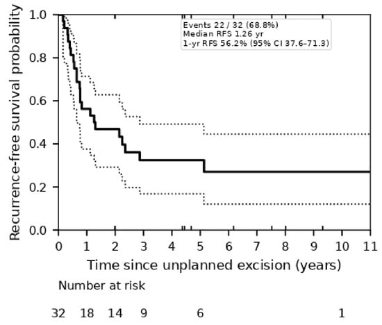
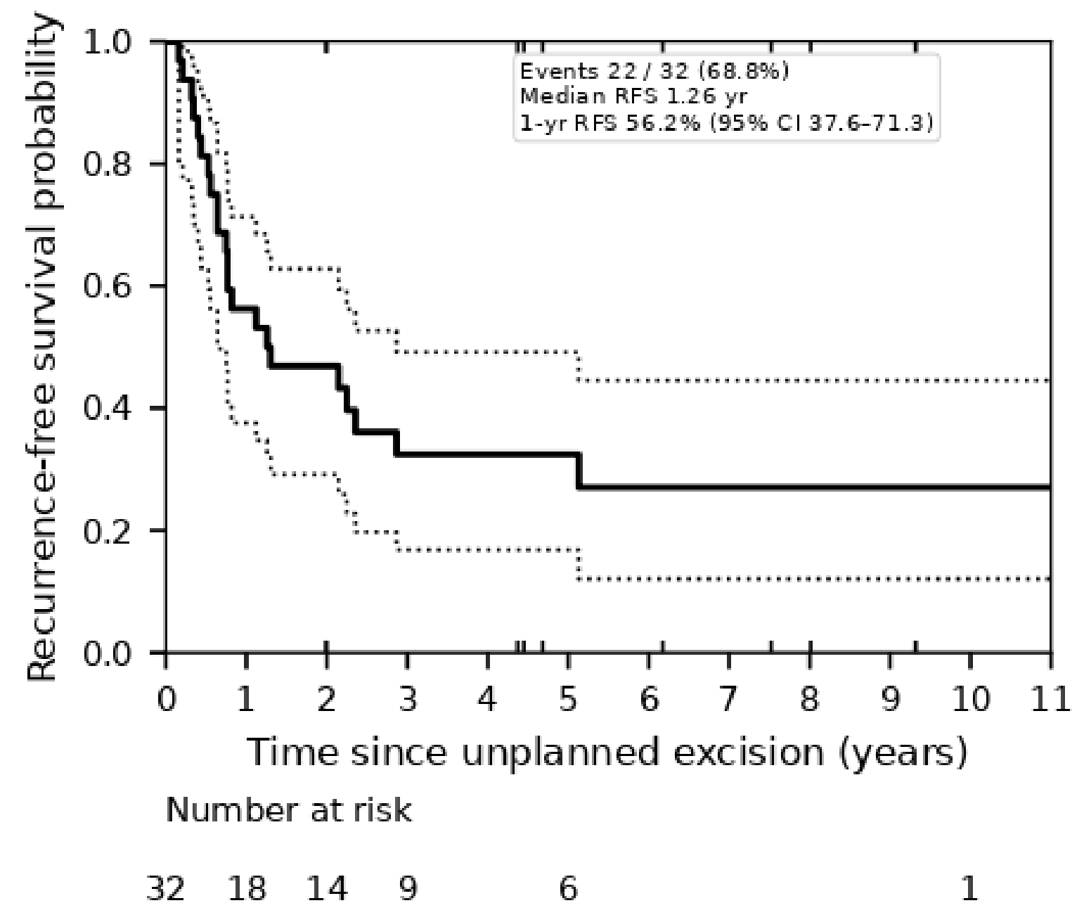
The median follow-up duration was 65.3 months (range, 4.5–343.1 months), and local recurrence was detected in 22 of the 32 patients (68.8%) during the observation period. The Kaplan–Meier RFS rates were 56.2% (95% confidence interval [CI] 37.6–71.3) at 1 year, 46.9% (95% CI 29.1–62.8) at 2 years, 32.5% (95% CI 16.8–49.1) at 3 years, 32.5% (95% CI 16.8–49.1) at 5 years, and 27.0% (95% CI 12.1–44.6) at 10 years, with a median RFS of 1.26 years (Table 2 and Figure 1).
Table 2.
Recurrence-free survival of the entire cohort (n = 32; 22 events, 10 censored).
Figure 1.
Kaplan–Meier curve of recurrence-free survival after unplanned excision in patients followed without additional wide excision (n = 32). Dotted lines indicate the 95% confidence interval. Numbers at risk are shown below the curve.
Among the 22 patients with recurrence, the median time to recurrence was 9.1 months (range, 1.9–61.6 months). Recurrence occurred within 12 months in 63.6% (14/22) and within 60 months in 95.5% (21/22) of cases, indicating that recurrences were concentrated in the early period after UE. However, late recurrence beyond 5 years was also observed in one patient (61.6 months).
3.3. Exploratory Histology-Stratified Analysis
In an exploratory analysis, patients were classified into three histological groups: UPS/MFS (n = 17), MLS (n = 6), and other histologies (LMS, SS, FS, ES, and MPNST; n = 9). Age differed significantly among the three groups (median 67.8, 37.1, and 52.5 years, respectively; Kruskal–Wallis p = 0.0036), reflecting the known age distribution of these subtypes, whereas no significant differences were observed in sex, tumor size, margin status, tumor location, or follow-up duration (all p > 0.05) (Table 3).
Table 3.
Patient characteristics stratified by histological group (UPS/MFS, MLS, and other histologies).
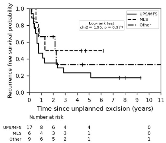
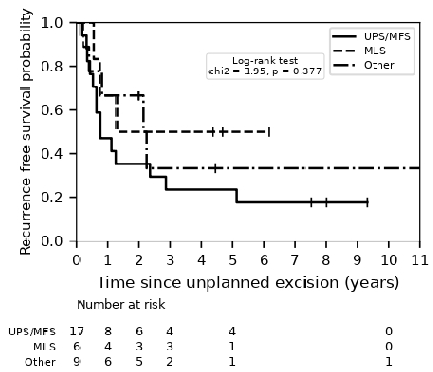
The 1-year RFS rates were 47.1% (95% CI 23.0–68.0) for UPS/MFS, 66.7% (95% CI 19.5–90.4) for MLS, and 66.7% (95% CI 28.2–87.8) for other histologies, and the corresponding 3-year rates were 23.5%, 50.0%, and 33.3%, respectively. The median RFS was 0.76 years in the UPS/MFS group and 2.15 years in the other histology group, whereas it was not reached in the MLS group. No statistically significant difference in RFS was observed among the three groups (overall log-rank test, χ2 = 1.95, p = 0.377), and all pairwise comparisons remained non-significant after Bonferroni adjustment (UPS/MFS vs. MLS, p = 0.685; UPS/MFS vs. other, p = 1.000; MLS vs. other, p = 1.000) (Table 4; Figure 2). Given the small subgroup sizes and confounding by age, these results should be interpreted as hypothesis generating. Within the combined UPS/MFS group, the local recurrence rate did not differ significantly between UPS and MFS (84.6% [11/13] vs. 75.0% [3/4]; Fisher’s exact test, p = 1.000), and the RFS curves were comparable (log-rank test, p = 0.874); UPS and MFS were therefore analyzed together.
Table 4.
Recurrence-free survival by histological group.
Figure 2.
Kaplan–Meier curves of recurrence-free survival stratified by histological group (UPS/MFS, solid line; MLS, dashed line; other histologies, dash-dotted line). Overall log-rank test, p = 0.377.
Regarding overall survival (OS), only one patient died during the follow-up (NED, 27; AWD, 4; DOD, 1). The 5-year OS rate was 96.0% (95% CI 74.8–99.4), and the median OS was not reached. Thus, although local recurrence was frequent after UE followed by observation, it was rarely fatal in this cohort, underscoring that the principal benefit of prompt additional wide excision lies in durable local control rather than in an improvement in survival.
3.4. Sensitivity Analyses
Because the interval between the initial unplanned excision and the first visit to our institution varied widely (median, 41 days; interquartile range, 21–134 days; range, 7–1839 days), two sensitivity analyses were performed to assess the potential influence of referral timing on the survival estimates. First, after excluding the 12 patients who were referred only after local recurrence had already developed, local recurrence occurred in 10 of the remaining 20 patients (50.0%), and the 1-, 2-, and 3-year RFS rates were 60.0%, 55.0%, and 48.9%, respectively, with a median RFS of 34.4 months. Second, when the analysis was restricted to the 22 patients who were referred within 90 days of the unplanned excision, local recurrence occurred in 13 patients (59.1%), and the 1-, 2-, and 3-year RFS rates were 54.5%, 45.5%, and 39.8%, respectively, with a median RFS of 13.4 months (Figure S1). In both analyses, local recurrence developed in approximately half of the patients, and recurrences remained concentrated in the first postoperative year.
4. Discussion
In the present study, we evaluated the oncological outcomes of patients with localized STS who underwent UE and were subsequently managed with observation alone, without immediate additional wide excision. Our primary finding was a high overall local recurrence rate of 68.8% (22/32 patients), with a 1-year RFS rate of only 56.2% and a median RFS of 1.26 years. Importantly, 63.6% (14/22) of all local recurrences occurred within the first 12 months following UE, indicating that local treatment failure in this setting occurs early as well as frequently.
These outcomes contrast with the local control rates achieved by standard primary planned excision or immediate re-excision. For extremity and truncal STS, primary planned resection generally results in 5-year local recurrence rates of 8.7–11.7% [8,13,14]. In UPS, a biologically aggressive high-grade histology, the 1-year RFS rate after primary curative resection reaches 84.4% (1-year local recurrence rate, 15.6%) [15]. For MFS, an adequate negative surgical margin results in a 2-year RFS rate of 91.7% [16]. The 1-year RFS rate of 56.2% observed in our observation cohort is markedly lower than these benchmark figures. This disparity indicates that observation after UE carries a high risk of early local treatment failure, even when initial postoperative imaging appears clear.
Our results also question the acceptability of a “watch and wait” approach following UE. Decanter et al. reported a 5-year RFS rate of 63.8% in a subgroup of 251 post-UE patients managed with observation, suggesting that delayed re-excision upon overt clinical recurrence might be a safe alternative in selected cases without compromising OS or limb salvage rates [17]. In our cohort, however, the 5-year RFS fell further to 32.5%, and most recurrences occurred within the first year. Sacchetti et al., in a systematic review and meta-analysis, reported that the development of a local recurrence significantly increases overall mortality (OR = 2.32) and the risk of distant metastasis (OR = 1.55) in STS patients [18]. A wait-and-see strategy therefore exposes patients to an avoidable local relapse, which may in turn affect long-term oncological outcome.
Why local recurrence occurred so often despite “negative” post-UE imaging is explained in part by the limitations of MRI in the early postoperative period. Postsurgical tissue distortion, extensive edema, hematoma, and seroma frequently mask microscopic tumor deposits along the surgical bed. Previous studies have reported microscopic residual sarcoma in the re-excision specimens of 50% to 70% of patients despite radiologically negative or equivocal post-UE MRI findings [1,2,19,20]. A radiologically “clear” post-UE status therefore cannot be equated with the absence of malignant cells and should not by itself justify withholding re-excision.
An important consideration in the interpretation of these results is the influence of surgical margin status. Positive margins were common in this cohort (22 of 32 patients, 68.8%) and plausibly contributed to the high rate of local recurrence. Margin status was not, however, significantly associated with recurrence (p = 0.196; Table 1): recurrence developed in 13 of 22 patients (59.1%) with a positive margin, in all 3 patients (100%) with a negative margin, and in 6 of 7 patients (85.7%) with an indeterminate margin, so that recurrence occurred in every margin category. The absence of a statistically significant association most likely reflects the small size of the cohort and the small number of patients with a negative margin (n = 3), rather than a lack of biological importance of margin status. The histological diagnosis and the assessment of the surgical margin were, moreover, made at our institution by pathologists with expertise in soft tissue sarcoma in 23 of the 32 patients (71.9%), so that our findings are unlikely to be explained by the way in which the margins were assessed. These observations reinforce the recommendation for re-excision at a sarcoma center: re-excision remains the most reliable means of achieving a confirmed R0 margin [5,21,22]. In this context, plastic and reconstructive techniques are of value because they allow wide resection with safe margins to be achieved in the extremities and trunk, even when the resection is extensive [22].
The risk of local recurrence after surgical treatment differs substantially among histological subtypes, which has direct implications for post-UE management. MFS, characterized by its infiltrative growth pattern along fascial planes (often visualized as the “tail sign” on MRI), and UPS, a high-grade malignancy, both carry intrinsically high local recurrence rates even after wide resection. Reported 5-year RFS rates range from 60% to 78% for MFS [16,21,23,24], while omission of radiotherapy in UPS significantly compromises local control [15,25]. In contrast, MLS is highly radiosensitive and generally exhibits favorable local control, with 5-year RFS rates exceeding 90% in standard surgical series [26,27,28,29]. In our observation cohort, the subgroup estimates were in line with these biological behaviors, although the comparison among histological groups did not reach statistical significance (log-rank p = 0.377). The UPS/MFS group had the worst course, with a 1-year RFS of 47.1% (recurrence in 14/17 patients) and a median RFS of 0.76 years. The MLS group had a higher 1-year RFS of 66.7%, and the median RFS was not reached. The lack of statistical significance is likely explained by the small sample size (MLS, n = 6) and the difference in age between groups (median 37.1 years in MLS vs. 67.8 years in UPS/MFS; p = 0.0036). Even in MLS, a subtype typically associated with low local failure rates after standard surgery [26,27,28,29], 3 of 6 patients (50%) developed local recurrence with observation alone. These findings indicate that withholding immediate re-excision after UE carries a risk of local failure across histological subtypes, and that the risk is highest for infiltrative or high-grade sarcomas such as MFS and UPS.
Despite the high early recurrence rate, OS in our cohort remained favorable (5-year rate, 96.0%), even though no additional therapy had been administered until local recurrence was detected. This favorable survival should be interpreted with caution for two reasons. First, patients treated with UE are known to be biased toward smaller, more superficial, and intrinsically lower-risk tumors, which may inflate survival estimates relative to a planned-excision comparison group; Smolle et al. reported that the initially better OS of patients after UE in a cohort of 728 patients with STS was no longer present after propensity-score weighting [30]. In our cohort, the median tumor sizes (35.0 mm in Group A and 37.5 mm in Group B) are consistent with this selection bias. The tumors in this cohort were, however, predominantly deep rather than superficial: among the 23 patients in whom depth could be determined, 19 (82.6%) were located deep to, or invaded, the muscular fascia. The present cohort therefore consists of small but deeply situated tumors that were excised without a preoperative suspicion of malignancy, a combination that is unfavorable with respect to local control. Second, Funovics et al. proposed that the final achievement of negative margins (R0), rather than the unplanned nature of the initial procedure, determines survival [5]; this also fits our findings, as additional wide excision was ultimately performed in all patients who developed local recurrence. The favorable OS therefore appears to reflect effective salvage resection rather than oncological equivalence between observation and immediate re-excision. Deferring re-excision nevertheless has a cost in local control: local recurrence developed in 22 of 32 patients (68.8%) after a median interval of 9.1 months.
These observations have two clinical implications. First, immediate additional wide excision should remain the standard of care for all eligible patients following UE, regardless of initial radiological findings [3,4,5]. Second, for the minority of patients in whom immediate re-excision cannot be performed because of severe comorbidities, advanced age, or patient refusal, close surveillance is required. Because nearly two-thirds of local recurrences occur within the first year, clinical and MRI examinations should be scheduled at short intervals, for example every 3 months during the first year after UE, so that local failure is detected as early as possible.
This study has several limitations. It was a retrospective analysis with a small sample size (n = 32), reflected by the wide confidence intervals around the survival estimates. The cohort lacked an internal control group treated with immediate additional wide excision during the same period, precluding direct intra-institutional comparison. Additionally, although UPS and MFS were merged based on their shared aggressive clinical behavior, the MFS subgroup was relatively small (n = 4); thus, the absence of statistical significance between subtypes should be interpreted with caution. Tumor grade and depth could not be determined in 9 patients each (28.1%), as some archived specimens and imaging studies were no longer available. The interval between the initial excision and referral also varied widely; although the two sensitivity analyses described above yielded consistent results, the possibility of residual selection bias cannot be entirely excluded. Despite these limitations, the early clustering of local recurrences observed here describes the clinical course of post-UE STS managed without immediate re-excision in routine practice.
5. Conclusions
UE of STS carries a high risk of early local recurrence, with most recurrences occurring within the first year. Because recurrence also developed in tumors of low histological grade (3 of 6 grade 1 tumors), aggressive additional wide excision should be actively recommended for all patients after UE, whenever feasible.
Supplementary Materials
The following supporting information can be downloaded at: https://www.mdpi.com/article/10.3390/curroncol33110673/s1. Figure S1: Sensitivity analyses of recurrence-free survival.
Author Contributions
Conceptualization, R.T., T.S., S.T. and K.T.; formal analysis, N.M., Y.K. and R.N.; investigation, R.T., T.S., Y.M. and S.S.; data curation, R.T.; writing—original draft preparation, R.T.; writing—review and editing, all authors; supervision, K.T., S.T. and T.S. All authors have read and agreed to the published version of the manuscript.
Funding
This study received no external funding.
Institutional Review Board Statement
This study was conducted in accordance with the Declaration of Helsinki and was approved by the Institutional Review Board of the University Hospital, Kyoto Prefectural University of Medicine (IRB No. ERB-C-2837, 12 June 2023, ethics approval).
Informed Consent Statement
The requirement for patient consent was waived due to the retrospective nature of the study and the use of anonymized data.
Data Availability Statement
The data presented in this study are available on request from the corresponding author due to privacy restrictions.
Acknowledgments
During the preparation of this manuscript, the authors used Genspark (Genspark AI, Super Agent) and ChatGPT 5.6 (OpenAI) for the purposes of English translation of the manuscript from Japanese and language editing, including improvement of readability of the text. The authors have reviewed and edited the output and take full responsibility for the content of this publication.
Conflicts of Interest
R.T. and S.S. are affiliated with an endowed chair funded by donations from medical corporations, which had no role in the conduct of this study or in the preparation of this manuscript. The authors declare no other conflicts of interest.
Abbreviations
UE, unplanned excision; STS, soft tissue sarcoma; RFS, recurrence-free survival; OS, overall survival; MRI, magnetic resonance imaging; CT, computed tomography; UPS, undifferentiated pleomorphic sarcoma; MFS, myxofibrosarcoma; MLS, myxoid liposarcoma; LMS, leiomyosarcoma; SS, synovial sarcoma; FS, fibrosarcoma; ES, Ewing sarcoma; MPNST, malignant peripheral nerve sheath tumor; IQR, interquartile range; CI, confidence interval; NED, no evidence of disease; AWD, alive with disease; DOD, died of disease; FNCLCC, Fédération Nationale des Centres de Lutte Contre le Cancer.
References
- Gingrich, A.A.; Elias, A.; Lee, C.Y.M.; Nakache, Y.P.N.; Li, C.S.; Shah, D.R.; Boutin, R.D.; Canter, R.J. Predictors of Residual Disease after Unplanned Excision of Soft Tissue Sarcomas. J. Surg. Res. 2017, 208, 26–32. [Google Scholar] [CrossRef] [Scilit] [PubMed]
- Grimer, R.; Parry, M.; James, S. Inadvertent Excision of Malignant Soft Tissue Tumours. EFORT Open Rev. 2019, 4, 321–329. [Google Scholar] [CrossRef] [Scilit] [PubMed]
- Pretell-Mazzini, J.; Barton, M.D., Jr.; Conway, S.A.; Temple, H.T. Unplanned Excision of Soft-Tissue Sarcomas: Current Concepts for Management and Prognosis. J. Bone Jt. Surg. Am. 2015, 97, 597–603. [Google Scholar]
- Koulaxouzidis, G.; Schwarzkopf, E.; Bannasch, H.; Stark, G.B. Is Revisional Surgery Mandatory When an Unexpected Sarcoma Diagnosis Is Made Following Primary Surgery? World J. Surg. Oncol. 2015, 13, 306. [Google Scholar] [CrossRef] [Scilit] [PubMed]
- Funovics, P.T.; Vaselic, S.; Panotopoulos, J.; Kotz, R.I.; Dominkus, M. The Impact of Re-Excision of Inadequately Resected Soft Tissue Sarcomas on Surgical Therapy, Results, and Prognosis: A Single Institution Experience with 682 Patients. J. Surg. Oncol. 2010, 102, 626–633. [Google Scholar] [CrossRef] [Scilit] [PubMed]
- Qureshi, Y.A.; Huddy, J.R.; Miller, J.D.; Strauss, D.C.; Thomas, J.M.; Hayes, A.J. Unplanned Excision of Soft Tissue Sarcoma Results in Increased Rates of Local Recurrence Despite Full Further Oncological Treatment. Ann. Surg. Oncol. 2012, 19, 871–877. [Google Scholar] [CrossRef] [Scilit] [PubMed]
- Lewis, J.J.; Leung, D.; Espat, J.; Woodruff, J.M.; Brennan, M.F. Effect of Reresection in Extremity Soft Tissue Sarcoma. Ann. Surg. 2000, 231, 655–663. [Google Scholar] [CrossRef] [Scilit] [PubMed]
- Nakamura, T.; Kawai, A.; Hagi, T.; Asanuma, K.; Sudo, A. A Comparison of Clinical Outcomes between Additional Excision after Unplanned and Planned Excisions in Patients with Soft-Tissue Sarcoma of the Limb: A Propensity Matching Cohort Study. Bone Jt. J. 2021, 103-B, 1809–1814. [Google Scholar]
- Nakamura, T.; Kawai, A.; Sudo, A. Analysis of the Patients with Soft Tissue Sarcoma Who Received Additional Excision after Unplanned Excision: Report from the Bone and Soft Tissue Tumor Registry in Japan. Jpn. J. Clin. Oncol. 2017, 47, 1055–1059. [Google Scholar] [CrossRef] [Scilit] [PubMed]
- Nakamura, T.; Hasegawa, M. Unplanned Excision in Soft Tissue Sarcoma: Current Knowledge and Remaining Gaps. Diagnostics 2025, 15, 453. [Google Scholar] [CrossRef] [Scilit] [PubMed]
- Widemann, B.C.; Italiano, A. Biology and Management of Undifferentiated Pleomorphic Sarcoma, Myxofibrosarcoma, and Malignant Peripheral Nerve Sheath Tumors: State of the Art and Perspectives. J. Clin. Oncol. 2018, 36, 160–167. [Google Scholar] [CrossRef] [Scilit] [PubMed]
- Brennan, M.F.; Antonescu, C.R.; Moraco, N.; Singer, S. Lessons Learned from the Study of 10,000 Patients with Soft Tissue Sarcoma. Ann. Surg. 2014, 260, 416–422. [Google Scholar] [CrossRef] [Scilit] [PubMed]
- Alsina, A.C.; Sacchetti, F.; Kaya, H.; Yaman, B.; Tamsel, İ.; Sabah, D. Impact of the Unplanned Excision on the Oncological Outcomes of Patients with Soft Tissue Sarcomas: A Single-Center Retrospective Review of 490 Patients. Acta Orthop. Traumatol. Turc. 2022, 56, 272–277. [Google Scholar] [CrossRef] [Scilit] [PubMed]
- Charoenlap, C.; Imanishi, J.; Tanaka, T.; Slavin, J.; Ngan, S.Y.; Chander, S.; Dowsey, M.M.; Goyal, C.; Choong, P.F. Outcomes of Unplanned Sarcoma Excision: Impact of Residual Disease. Cancer Med. 2016, 5, 980–988. [Google Scholar] [CrossRef] [Scilit] [PubMed]
- Goertz, O.; Pieper, A.; von der Lohe, L.; Stricker, I.; Dadras, M.; Behr, B.; Lehnhardt, M.; Harati, K. The Impact of Surgical Margins and Adjuvant Radiotherapy in Patients with Undifferentiated Pleomorphic Sarcomas of the Extremities: A Single-Institutional Analysis of 192 Patients. Cancers 2020, 12, 362. [Google Scholar] [CrossRef] [Scilit] [PubMed]
- Yurtbay, A.; Coşkun, H.S.; Say, F.; Dabak, N. Is the Thickness of the Margin Associated with Local Recurrence and Survival in Patients with Myxofibrosarcoma? Clin. Orthop. Relat. Res. 2023, 481, 2125–2136. [Google Scholar] [CrossRef] [Scilit] [PubMed]
- Decanter, G.; Stoeckle, E.; Honoré, C.; Meeus, P.; Mattei, J.C.; Dubray-Longeras, P.; Ferron, G.; Carrere, S.; Causeret, S.; Guilloit, J.M.; et al. Watch and Wait Approach for Re-Excision After Unplanned Yet Macroscopically Complete Excision of Extremity and Superficial Truncal Soft Tissue Sarcoma Is Safe and Does Not Affect Metastatic Risk or Amputation Rate. Ann. Surg. Oncol. 2019, 26, 3526–3534. [Google Scholar] [CrossRef] [Scilit] [PubMed]
- Sacchetti, F.; Alsina, A.C.; Morganti, R.; Innocenti, M.; Andreani, L.; Muratori, F.; Scoccianti, G.; Totti, F.; Campanacci, D.A.; Capanna, R. Re-Excision after Unplanned Excision of Soft Tissue Sarcoma: A Systematic Review and Meta-Analysis. The Rationale of Systematic Re-Excision. J. Orthop. 2021, 25, 244–251. [Google Scholar] [CrossRef] [Scilit] [PubMed]
- Davies, A.M.; Mehr, A.; Parsonage, S.; Evans, N.; Grimer, R.J.; Pynsent, P.B. MR Imaging in the Assessment of Residual Tumour Following Inadequate Primary Excision of Soft Tissue Sarcomas. Eur. Radiol. 2004, 14, 506–513. [Google Scholar] [CrossRef] [Scilit] [PubMed]
- Goodlad, J.R.; Fletcher, C.D.; Smith, M.A. Surgical Resection of Primary Soft-Tissue Sarcoma: Incidence of Residual Tumour in 95 Patients Needing Re-Excision after Local Resection. J. Bone Jt. Surg. Br. 1996, 78-B, 658–661. [Google Scholar]
- Boughzala-Bennadji, R.; Stoeckle, E.; Le Péchoux, C.; Méeus, P.; Honoré, C.; Attal, J.; Duffaud, F.; De Pinieux, G.; Bompas, E.; Thariat, J.; et al. Localized Myxofibrosarcomas: Roles of Surgical Margins and Adjuvant Radiation Therapy. Int. J. Radiat. Oncol. Biol. Phys. 2018, 102, 399–406. [Google Scholar] [CrossRef] [Scilit] [PubMed]
- Samà, L.; Binder, J.P.; Darrigues, L.; Boura, B.; Courtois, C.; Rimareix, F.; Honoré, C.; Le Cesne, A.; Faron, M. Safe-margin surgery by plastic reconstruction in extremities or parietal trunk soft tissue sarcoma: A tertiary single centre experience. Eur. J. Surg. Oncol. 2022, 48, 526–532. [Google Scholar] [CrossRef] [Scilit] [PubMed]
- Dadrass, F.; Gusho, C.; Yang, F.; Culvern, C.; Bloom, J.; Fillingham, Y.; Colman, M.; Gitelis, S.; Blank, A. A Clinicopathologic Examination of Myxofibrosarcoma. Do Surgical Margins Significantly Affect Local Recurrence Rates in This Infiltrative Sarcoma Subtype? J. Surg. Oncol. 2021, 123, 489–496. [Google Scholar] [CrossRef] [Scilit] [PubMed]
- Rhee, I.; Spazzoli, B.; Stevens, J.; Hansa, A.; Spelman, T.; Pang, G.; Guiney, M.; Powell, G.; Choong, P.; Di Bella, C. Oncologic Outcomes in Myxofibrosarcomas: The Role of a Multidisciplinary Approach and Surgical Resection Margins. ANZ J. Surg. 2023, 93, 577–584. [Google Scholar] [CrossRef] [Scilit] [PubMed]
- Makris, E.A.; Tran, T.B.; Delitto, D.J.; Lee, B.; Ethun, C.G.; Grignol, V.; Howard, J.H.; Bedi, M.; Gamblin, T.C.; Tseng, J.; et al. Natural History of Undifferentiated Pleomorphic Sarcoma: Experience from the US Sarcoma Collaborative. J. Surg. Oncol. 2024, 129, 1354–1363. [Google Scholar] [CrossRef] [Scilit] [PubMed]
- Masunaga, T.; Tsukamoto, S.; Nitta, Y.; Honoki, K.; Fujii, H.; Akahane, M.; Takeda, M.; Tanaka, Y.; Mavrogenis, A.F.; Errani, C.; et al. Is Perioperative Chemotherapy Effective in Patients with Localized Myxoid Liposarcoma? Jpn. J. Clin. Oncol. 2024, 54, 297–304. [Google Scholar] [CrossRef] [Scilit] [PubMed]
- Potkrajcic, V.; Zschiegner, M.; Niyazi, M.; Warm, V.; Thiel, J.T.; Frantz, S.; Deinzer, C.K.W.; Szelat, F.; Hoffmann, E.; Paulsen, F.; et al. Myxoid Liposarcoma: Treatment Outcomes, Metastatic Pattern and Volumetric Analysis. Strahlenther. Onkol. 2025, 201, 1239–1248. [Google Scholar] [CrossRef] [Scilit] [PubMed]
- Roohani, S.; Chung, P.W.M.; Dickson, B.C.; Ferguson, P.C.; Griffin, A.M.; Kirsch, D.G.; Krcek, R.; O’Sullivan, B.; Shultz, D.B.; Tsoi, K.M.; et al. Refining Surveillance in Myxoid Liposarcoma: Long-Term Recurrence Patterns and Functional Outcomes After Surgery ± Radiotherapy in 186 Patients. Radiother. Oncol. 2026, 214, 111275. [Google Scholar] [CrossRef] [Scilit] [PubMed]
- Baxter, K.J.; Govsyeyev, N.; Namm, J.P.; Gonzalez, R.J.; Roggin, K.K.; Cardona, K. Is Multimodality Therapy Necessary for the Management of Pure Myxoid Liposarcomas? A Multi-Institutional Series of Pure Myxoid Liposarcomas of the Extremities and Torso. J. Surg. Oncol. 2015, 111, 146–151. [Google Scholar] [CrossRef] [Scilit] [PubMed]
- Smolle, M.A.; Tunn, P.-U.; Goldenitsch, E.; Posch, F.; Szkandera, J.; Bergovec, M.; Liegl-Atzwanger, B.; Leithner, A. The Prognostic Impact of Unplanned Excisions in a Cohort of 728 Soft Tissue Sarcoma Patients: A Multicentre Study. Ann. Surg. Oncol. 2017, 24, 1596–1605. [Google Scholar] [CrossRef] [Scilit] [PubMed]

Disclaimer/Publisher’s Note: The statements, opinions and data contained in all publications are solely those of the individual author(s) and contributor(s) and not of MDPI and/or the editor(s). MDPI and/or the editor(s) disclaim responsibility for any injury to people or property resulting from any ideas, methods, instructions or products referred to in the content. |
© 2026 by the authors. Licensee MDPI, Basel, Switzerland. This article is an open access article distributed under the terms and conditions of the Creative Commons Attribution (CC BY) license.