


Summary
Principles: Newly developed qualitative rapid bedside immunoassays for markers of cardiac injury represent a promising tool for emergency decision making in patients with chest pain, but they have been tested mainly in conditions of clinical investigation. We prospectively evaluated in ordinary clinical settings the accuracy of two of these tests, the Cardiac StatusTM and the TROPT sensitive, for diagnosing acute myocardial infarction.
Methods: 83 patients who presented to the emergency department of a large public teaching hospital, within 12 hours of chest pain, were evaluated. Bedside tests and their corresponding plasma quantitative assays (myoglobin, CK-MB [mass], and troponin I) were routinely performed on admission and three times during the eight following hours. Handling of the bedside tests and interpretation of their results were achieved by ordinary nurses of the emergency department staff, that had been trained for it.
Results: 43 (52%) patients had a final diagnosis of acute myocardial infarction, based on electrographic and enzymatic criteria defined by the World Health Organization; unstable angina pectoris was diagnosed in 6 (7%) other patients, chronic ischemic heart disease in 15 (18%), pericarditis in 2 (2.5%) and non-cardiac chest pain in 17 (20.5%). Mean ± SD time between the onset of symptoms and arrival at the emergency department was 7 ± 11 hours and median time 3 hours. Upon arrival, the sensitivities of the bedside tests for diagnosing myocardial infarction were 35% for myoglobin, 14% for CK-MB mass and 9% for troponin T, while they were 72% (p <0.01), 42% (p <0.004), and 28% (p = 0.03) for corresponding plasma quantitative assays; specificities were similar for both methods: 80—90% for myoglobin; 100% for CK-MB mass; and 90—100% for troponin I and T. Eight hours after admission, sensitivities of the bedside tests were 54% for myoglobin, 40% for CK-MB mass, and 20% for troponin T, while they were 81% (p <0.05), 81% (p <0.01) and 73% (p <0.001), respectively, for their laboratory counterparts. Then, at all times, sensitivities of the bedside tests were mostly significantly lower than their laboratory counterparts, whereas their specificities were comparable.
Conclusions: Qualitative bedside tests were of little help for ruling out acute myocardial injury under usual clinical conditions. A new generation of such tests, allowing quantitative measurements, may improve their diagnostic accuracy.
Résumé
Principes: Les nouveaux tests immunologiques rapides au lit du patient pour la mesure des marqueurs de lésion cardiaque représentent un outil décisionnel prometteur chez les patients consultant pour douleurs thoraciques, mais leur validité a été essentiellement démontrée en conditions d'investigation clinique. Nous avons donc évalué deux de ces tests parmi les plus récents, le Cardiac StatusTM et le TROPT sensitive, pour le diagnostic d'infarctus aigu du myocarde en conditions cliniques habituelles.
Méthodes: nous avons évalué 83 patients qui se sont présentés au service des urgences d'un grand hôpital universitaire dans les 12 heures suivant la survenue de douleurs thoraciques. Les tests rapides qualitatifs au lit du patient et leurs équivalents quantitatifs de laboratoire (myoglobine, CK-MB massiques et troponine I) ont été effectués de routine ä l'admission, ainsi qu'à trois reprises dans les huit heures suivantes, et leurs résultats comparés. La manipulation des tests rapides ainsi que l'interprétation de leurs résultats ont été effectuées par les infirmières du service des urgences, spécialement entrainées cet effet.
Résultats: le diagnostic d'infarctus aigu du myocarde a été retenu chez 43 patients (52%), sur la base des criteres électrocardiographiques et enzymatiques définis par l'Organisation Mondiale de la Santé, alors que 6 des patients (7%) ont présenté un angor instable, 15 (18%) une cardiopathie ischémique chronique, 2 (2,5%) une péricardite et 17 (20,5%) une douleur thoracique d'origine non-cardiaque. La durée moyenne (± déviation standard) séparant l'apparition des symptômes de l'arrivée au service des urgences était de 7 ± 11 heures (durée médiane: 3 heures). A l'admission, les sensibilités des tests rapides au lit du patient étaient de 35% pour la myoglobine, 14% pour la CKMB massique et 9% pour la troponine T, alors que celles des tests quantitatifs de laboratoire correspondants étaient de 72% (p <0,01), 42% (p <0,004) et 28% (p = 0,03), respectivement; par contre, les spécificités étaient similaires pour les deux méthodes: 80—90% pour la myoglobine, 40% pour les CK-MB massiques et 90—100% pour les troponines T et I. Huit heures apres l'admission, les sensibilités des tests rapides au lit du patient étaient de 54% pour la myoglobine, 40% pour les CK-MB massiques et 20% pour la troponine T, contre 81% (p <0,05), 81% (p <0,01) et 73% (p <0,01) pour les tests de laboratoires respectifs correspondants. A chaque étape, les sensibilités des test rapides au lit du patient se sont donc révélées significativement inférieures à celles des tests quantitatifs de laboratoire correspondant, alors leurs sensibilités se sont avérées similaires.
Conclusions: les tests rapides qualitatifs au lit du patient sont de peu d'utilité pour écarter le diagnostic de nécrose myocardique en conditions ordinaires d'urgence. Une nouvelle génération de ces tests, permettant une mesure quantitative de ces différents marqueurs, devrait améliorer leur efficacité.
Introduction
Ischaemic heart disease remains the principal cause of death in industrialized countries and its consequences account for the consumption of vast economic ressources [1]. The goal of emergency therapy is to salvage myocardium, and this objective is best achieved when interventions occur early relative to the onset of acute clinical symptoms [2].
The ECG has remained the unquestioned standard for initiation of thrombolytic therapy and management is well established when typical ECG changes are present [3, 4]. In many cases, however, the ECG alone is not sufficient for decision making, because of atypical changes, presence of paced rhythms or bundle-branch blocks [1, 3, 5]. It is in these situations that results of cardiac enzymes levels are the most helpful [1, 6]. For several years, creatine kinase and its MB isoenzyme have been regarded as reference standards for biochemical diagnosis of acute myocardial infarction (AMI), with improvements in both sensitivity and specificity by the recent development of mass monoclonal antibody assays [1, 6, 7]. Measurement of serum myoglobin, a new highly sensitive, but non-specific marker, had been widely advocated, especially because of the excellent predictive value of a negative result [1]. A new generation of serodiagnostic tests simultaneously appeared with the measurement of the cardiac-specific contractile proteins troponin T and troponin I, that show superior diagnostic accuracy [1, 8]. Troponins have been shown to not only have a prognostic value in patients with acute coronary syndromes [4, 6, 7], but also to allow identification of a high risk subgroup of patients most likely to benefit from early and more aggressive interventions [9].
Thus, techniques allowing fast results of the serum levels of each marker seem very appealing.
Rapid bedside immunoassays have been developed for Troponins T and I, as well as for creatine kinase MB mass and myoglobin. The use of these diagnostic kits seems to be easier and faster than the conventional analytic techniques, and could represent a promizing tool for emergency decision making [4, 7, 10].
However, most studies having demonstrated their validity have been performed in conditions of clinical investigation, using specially trained nurses [4, 7, 10, 11]. Thus, we decided to prospectively evaluate in ordinary clinical settings two recent qualitative, whole blood, bedside, rapid immunological assays, the Cardiac Status™ and the TROPT sensitive, for their diagnostic accuracy in patients presenting to the emergency department with chest pain.
Methods
Patients
The study population consisted of 83 patients who presented between July 1996 and February 1997, at the emergency department of a 1200-bed public teaching hospital. To be eligible, patients had to have experienced acute chest pain within 12 hours of the emergency department visit, that was not consecutive to trauma (x-ray confirmed) or that was not associated with acute pulmonary edema on admission.
Study protocol
The study protocol was approved by the ethics committee of the hospital. The study was conducted in a single blinded fashion by the nurses of the emergency room staff that had been trained in the handling of the bedside tests. Test procedures including interpretation of results were taught to the nurses of the staff by a trained physician during four general lessons, and individually later on; they performed them as part of their ordinary duties, and no specific nurses were assigned to this task. Neither the physician nor the biochemist involved had access to the individual values of the cardiac marker tested on an individual patient. Markers' results did not alter the management of the patients, which was provided similarly as for all patients with acute coronary syndromes.
Each patient underwent a 12-lead ECG on admission. Patients were followed until discharge from the hospital, and clinical data including history, findings of physical examination, discharge diagnoses, electrocardiograms and biochemical tests were recorded.
Analytic techniques
For qualitative determination of serum cardiac troponin T, we used the TROPT sensitive, a whole blood, bedside, rapid immunological assay device (Boehringer Mannheim, Germany). This assay uses a cardiospecific monoclonal antibody for troponin T, which is labeled with biotin, and a second, gold-labeled cardiospecific monoclonal antibody, which binds to a different epitope of troponin T. Immunocomplexes are then immobilized in a streptavidin microtiter plate that constitutes the reading zone. After addition of 120 to 150 μl of whole, venous, anticoagulated blood, the turnaround time of the assay is 20 minutes. Positivity is detected by the eye as a colored band that develops in the reading zone of the kit at troponin T levels >0.1 μg/l. Results are then classified as positive, negative or uncertain.
For simultaneous qualitative determination of serum myoglobin and CK-MB (mass), we used the Cardiac Status™, a whole blood, bedside, rapid immunochromatographic assay device (Spectral Diagnostics, Toronto, Canada). This device incorporates a collection-separation membrane system to separate erythrocytes from plasma, followed by a dye-based immunochromatographic assay using the plasma after a run over filtration paper incorporated in the cartridge. A volume of 150 μl of heparinized whole blood was applied with a micropipet according to the instructions of the manufacturer. After three minutes, the separation membrane system was removed and the elution buffer was added to the cartridge sample area. At abnormal concentrations of a marker, antibody-marker complexes bind to matching immobilized antibodies on the test strip, forming brown-red bands that are visible in a result window. This window contains an area for CK-MB, one for myoglobin and a control area to indicate successful transfer of plasma to the result window and development of the colored band. The result has to be read qualitatively, as either normal, abnormal or uncertain, after exactly 15 minutes. According to the manufacturer, the test reads positive when CK-MB is elevated above 5 μg/l and when myoglobin is elevated above 50 μg/l.
For the quantitative determination of myoglobin, we used a Stratus II Dade analyser, based on a sandwich-tape immunodosage, with an upper reference limit of 90 μg/l, which is the upper reference limit in our laboratory.
For the quantitative determination of total creatine kinase and its MB isoenzyme activity, we used a BM/Hitachi 717 colorimeter, with an upper reference limit of 270 international units per liter (U/l) in men and 150 international units per liter (U/l) in women.
For quantitative determination of serum CK-MB (mass), we used a Stratus II Analyser (Dade, Miami), based on a immuno-enzymo-fluorimetric method, with an upper reference limit of 7 μg/l, which is the upper reference limit in our laboratory.
For quantitative detection of serum troponin I, we used a Stratus II analyser too, with an upper reference limit of 0.6 μg/l.
The Cardiac Status™ (CK-MB [mass]/myoglobin) panel and the TROPT sensitive (troponin T) were routinely performed on admission and repeated four times after two, four and eight hours, respectively, if both tests were not simultaneously positive. If they were, the sampling stopped. At the same times, and for each newly performed rapid test, quantitative measurements of plasma total CK (enzymatic activity), CK-MB (mass), myoglobin, troponin I and creatinin were achieved. The Cardiac Status™ was considered positive only if both CK-MB (mass) and myoglobin were simultaneously positive. Blood samples for quantitative measurements were collected in standard test tubes containing heparin (Li⁺ or Na⁺), and most of them were analyzed immediately. If specimens were to be stored, the plasma obtained following centrifugation was refrigerated at 2–8 °C for storage periods shorter than 24 hours. For longer periods of time, samples were kept at temperatures below –20 °C.
Definitions
Unstable angina pectoris was defined as type IB (new onset of severe angina or accelerated angina without rest pain, that develops in absence of extracardiac conditions) or IIIB (angina at rest within 48 hours, that develops in absence of extracardiac conditions) according to Braunwald's classification [12]. Acute myocardial infarction was considered to be present if patients met the electrographic and/or enzymatic criteria defined by the World Health Organization [13] (creatine kinase activity >270 U/l in men and >150 U/l in women, or creatine kinase-MB >10% of creatine kinase activity, or creatine kinase (mass) >7 μg/l).
Statistical analysis
Sensitivities and specificities between bedside tests and laboratory tests were compered using a chi-square test or a Fisher's exact test, when appropriate. P values of less than 0.05 were considered to indicate statistical significance.
Results
Characteristics of the patients and final diagnoses
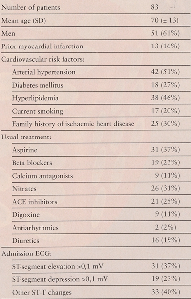
The clinical characteristics of the 83 study patients, their usual medication and their ECG findings upon admission are described in Table 1. They were predominantly male, with a mean age of 70 years. On admission, 31 (37%) of them presented with significant ST segment elevation on the ECG, 19 (23%) with significant ST segment depression and 11 (13%) with conduction disturbances; 13 (16%) of them had a prior history of myocardial infarction.
Table 1.
Characteristics of the study patients.
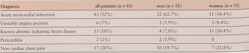
Table 2 shows the discharge diagnoses of the study patients. Of 83 consecutive patients, 43 (52%) had a final diagnosis of acute myocardial infarction. Among the other 40 patients, unstable angina pectoris was diagnosed in 6 (7%), known chronic ischaemic heart disease in 15 (18%), pericarditis in 2 (2%) and non-cardiac chest pain in 17 (20%).
Table 2.
Discharge diagnoses.
Patients with confirmed AMI had the following mean ± SD peak values of serum enzymes levels: total CK: 840 ± 789 U/l; CK MB: 117 ± 95 U/l; CK MB mass: 120 ± 178 μg/l; myoglobin: 906 ± 1129 μg/l; troponin I: 28 ± 53 μg/l.
For patients with unstable angina, the corresponding levels were: total CK: 97 ± 77 U/l; CK MB mass: 0.5 ± 0.9 μg/l; myoglobin: 58 ± 32 μg/l; troponin I: 0.2 ± 0.3 μg/l (CK MB enzymatic activity was not measured for these patients, because of normal total CK values).
Laboratory test results
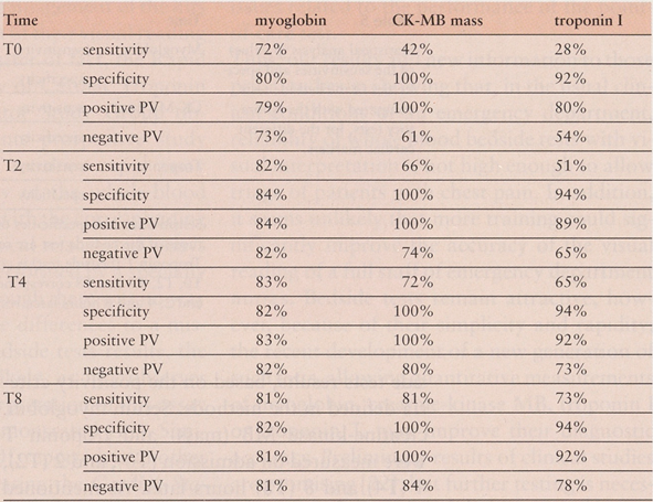
Table 3 shows the sensitivity, specificity, positive and negative predictive values of the laboratory tests results, based on the positivity criteria defined in the methods. Serum myoglobin, creatine-kinase MB (mass) and troponin I were measured on admission (T0), and 2 (T2), 4 (T4) and 8 (T8) hours later. The mean time between onset of symptoms and arrival at the emergency room for patients with chest pain in our hospital was 7 ± 11 hours and the median time 3 hours. For each marker, sensitivity for diagnosing acute myocardial infarction rose progressively along time, and was maximal 8 hours after admission for CK-MB mass and troponin I, with values of 81% and 73%, respectively. Myoglobin maximal sensitivity was reached earlier (4 hours after admission: 83%) and was the highest of the 3 markers on admission, as well as at 2 and 4 hours; 8 hours after admission, myoglobin sensitivity, however, was comparable to CK-MB mass sensitivity (81%). Specificity was 100% for CK-MB mass at each measurement but ranged from 92 to 94% for troponin I. As expected, myoglobin specificity was lower than the 2 other cardiac specific markers's, ranging from 72% to 84%.
Table 3.
Sensitivity, specificity, positive and negative predictive values (PV) of laboratory tests:
Bedside tests results
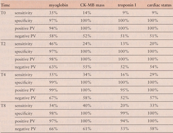
Table 4 shows the sensitivity, specificity, positive and negative predictive values of the bedside tests results, based on the positivity criteria defined in the methods. Serum myoglobin, creatine-kinase MB (mass) and troponin T were measured on admission (T0), and 2 (T2), 4 (T4) and 8 (T8) hours later. As mentioned earlier, the Cardiac Status™ panel was considered as positive only if both myoglobin and CK-MB mass were simultaneously positive. Sensitivity of myoglobin, CK-MB mass and troponin T rose progressively along time, from 35%, 14% and 9%, respectively, on admission, to 54%, 40% and 20%, respectively, eight hours later. At all times, myoglobin sensitivity was higher than the one of the other markers. Specificities were high for all markers and at all times, particularly so for CK-MB mass and troponin T. As a result, the positive predictive values of the bedside tests were very high, while their negative predictive values were low.
Table 4.
Sensitivity, specificity, positive and negative predictive values (PV) of bedside tests.
Comparison between laboratory and bedside tests results
In comparison with corresponding laboratory tests results (laboratory troponin I was compared with bedside troponin T), all sensitivities of the bedside tests were most of the time significantly lower than their laboratory counterparts. On the other hand, specificities of the bedside tests were closer to the ones of the laboratory tests for each marker, at each measurement, and even higher for CK-MB mass, troponin T and myoglobin.
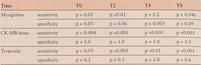
Table 5 shows the p values corresponding to comparison of the sensitivities and specificities of the bedside tests with those of the laboratory tests, for the different cardiac markers. For each marker except troponin, and at each time period, the bedside tests results were compared with the corresponding laboratory tests results; troponin T bedside test results were compared with troponin I laboratory test results.
Table 5.
Statistical analysis (p value) of the sensitivities and specificities of the bedside tests, compared with the laboratory tests, for the different cardiac markers.
P values reflect significantly lower sensitivities for most results of the bedside tests compared with their corresponding laboratory tests. There was only one exception: the sensitivities of myoglobin 4 hours after admission were not significantly different between bedside and laboratory tests. On the other hand, there were no statistically significant differences for specificities between bedside tests and laboratory tests results, with the only exception of myoglobin, on admission and 4 hours later, whose specificities were significantly higher than their laboratory counterparts.
Discussion
Our results are expressed in relation to time of arrival at the hospital. The mean +SD time between onset of symptoms and arrival at the emergency room department for patients with chest pain in our hospital was 7 + 11 hours (median time 3 hours). Accordingly, our results for the laboratory tests are roughly similar to those published in the general literature, at least when comparable parameters are explicitly mentioned [14, 16].
Concerning bedside tests, our results showed no statistically different or worse specificities compared with those of the laboratory tests, but their sensitivities were significantly different and dramatically lower than expected. Several experimental studies [4, 7, 10, 17, 18] had clearly established the accuracy of bedside tests; in these studies, however, specific research nurses had been assigned to their handling and reading. In our study, we chose to evaluate these tests under ordinary clinical settings, i.e when read by ordinary nurses of an emergency department, who had been exposed to the training of the handling of the kits, as recommended by the manufacturer. Thus, the poor accuracy of the bedside tests in our study very likely reflected mismanagement of the kits and/or misinterpretation of their visual reading by such nurses. As a matter of fact, the Rapid Evaluation by the Assay of Cardiac Troponin T (REACTT) Investigator Study Group observed, in a 721 patients prospective study evaluating the TROPT sensitive rapid assay [11], a lower sensitivity of the whole-blood bedside tests compared with the corresponding serum assay. In this clinical trial, rapid assays had nevertheless been performed by a specially trained study staff. Although there was no way to clearly attribute these differences to a misinterpretation of the bedside tests results, the authors considered it likely, as interobserver variability in visual interpretation of this assay had previously been demonstrated [19]. Similarly, Schouten, et al. [20], reported, in another prospective study evaluating the Cardiac Status™ rapid assay, a high false-negative rate for both bedside CK-MB mass and myoglobin results, compared with those of the corresponding quantitative assays, and consequently, low sensitivities for these markers. Finally, in a recent study evaluating risk stratification with a point-of-care qualitative troponin T bedside test in AMI patients [21], as part of the GUSTO III trial, 257/807 (32%) hospitals did not participate in the substudy because of logistical issues related to the performance of the point-of-care test.
Thus, our results add new information to those prior studies in showing that, in the usual clinical conditions of an emergency department, reliability of whole blood bedside tests with visual interpretation is not high enough to allow triage of patients with chest pain. In addition, it seems unlikely that more training could significantly improve the accuracy of the visual reading of a full staff of emergency department nurses. Bedside tests remain attractive, however, because of their simplicity and rapidity; the recent development of a new generation of such tests, allowing quantitative measurements of myoglobin, creatine-kinase MB, troponin I or troponin T, may improve their diagnostic accuracy. Preliminary results of clinical studies are promising [22], but further testing is necessary to confirm their validity.
A limitation of our study resulted from the fact that the accuracy of the bedside test for troponin T was assessed by comparison with serum quantitative troponin I levels. The sensitivity and the specificity of serum troponin I and troponin T levels have been shown to be very close for the diagnosis of AMI in emergency room patients with chest pain [4].
References
- Keffer JH. Myocardial markers of injury. Evolution and insights. Am J Clin Pathol 1996;105:305–20.
- Apple FS. Acute myocardial infarction and coronary reperfusion. Serum cardiac markers for the 1990s. Am J Clin Pathol 1992;97:217–26.
- Rottbauer W, Greten T, Muller-Bardorff M, Remppis A, Zehelein J, Grünig E, et al. Troponin T: a diagnostic marker for myocardial infarction and minor cardiac cell damage. Eur Heart J 1996;17(suppl F):3–8.
- Hamm CW, Goldmann BU, Heeschen C, Kreymann G, Berger J, Meinertz T. Emergency room triage of patients with acute chest pain by means of rapid testing for cardiac troponin T or troponin I. N Engl J Med 1997;337:1648–53.
- Ohman EM, Armstrong PW, Christenson RH, Granger CB, Katus HA, Hamm CW, et al. Cardiac troponin T levels for risk stratification in acute myocardial ischemia. GUSTO IIA Investigators. N Engl J Med 1996;335:1333–41.
- Katus HA, Remppis A, Neumann FJ, Scheffold T, Diederich KW, Vinar G, et al. Diagnostic efficiency of troponin T measurements in acute myocardial infarction. Circulation 1991; 83:902–12.
- Mach F, Lovis C, Chevrolet JC, Urban P, Unger PF, Bouillie M, et al. Rapid bedside whole blood cardiospecific troponin T immunoassay for the diagnosis of acute myocardial infarction. Am J Cardiol 1995;75:842–5.
- Lindhal B, Venge P, Wallentin L. Relation between troponin T and the risk of subsequent cardiac events in unstable coronary artery disease. The FRISC study group. Circulation 1996;93:1651–7.
- Lindhal B, Venge P, Wallentin L. Troponin T identifies patients with unstable coronary artery disease who benefit from long-term antithrombotic protection. Framing in Unstable Coronary Artery Disease (FRISC) Study Group. J Am Coll Cardiol 1997;29:43–8.
- Antman EM, Grudzien C, Sacks DB. Evaluation of a rapid bedside assay for detection of serum cardiac troponin T. JAMA 1995;273:1279–82.
- Baxter MS, Brogan Jr. GX, Harcherload FP, Knoop KJ, Zackowski SW, Ryan RJ, et al. Evaluation of a bedside whole-blood rapid troponin T assay in the emergency department. Rapid Evaluation by Assay of Cardiac Troponin T (REACTT) Investigators Study Group. Acad Emerg Med 1997;4:1018–24.
- Braunwald E. Unstable angina. A classification. Circulation 1989;80:410–14.
- Nomenclature and criteria for diagnosis of ischemic heart disease. Report of the Joint International Society and Federation of Cardiology / World Health Organization task force on standardization of clinical nomenclature. Circulation 1979;59:607–09.
- de Winter RJ, Koster RW, Sturk A, Sanders GT. Value of myoglobin, troponin T, and CK-Mb mass in ruling out an acute myocardial infarction in the emergency room. Circulation 1995;92:3401–07.
- Bakker AJ, Koelemay MJ, Gorgels JP, Van Vlies B, Smits R, Tijssen JG, et al. Failure of new biochemical markers to exclude acute myocardial infarction at admission. Lancet 1993;342:1220–2.
- Zimmerman J, Fromm R, Meyer D, Boudreaux A, Wun CC, Smalling R, et al. Diagnostic marker cooperative study for the diagnosis of myocardial infarction. Circulation 1999; 99:1671–7.
- Luscher MS, Ravkilde J, Thygesen K. Clinical application of two novel rapid bedside tests for the detection of cardiac troponin T and creatine kinase-MB mass/myoglobin in whole blood in acute myocardial infarction. Cardiology 1998; 89:222–8.
- Panteghini M, Cuccia C, Pagani F, Turla C. Comparison of the diagnostic performance of two rapid bedside biochemical assays in early detection of acute myocardial infarction. Clin Cardiol 1998;21:394–8.
- Muller-Bardorff M, Freitag H, Scheffold T, Remppis A, Kubler W, Katus HA. Development and characterization of a rapid assay for bedside determinations of cardiac troponin T. Circulation 1995;92:2869–75.
- Schouten Y, de Winter RJ, Gorgels JP, Koster RW, Adams R, Sanders GT. Clinical evaluation of the CARDIAC STATUS, a rapid immunochromatographic assay for simultaneous detection of elevated concentrations of CK-MB and myoglobin in whole blood. Clin Chem Lab Med 1998;36:469–73.
- Ohman EM, Armstrong PW, White HD, Granger CB, Wilcox RG, Weaver WD, et al. Risk stratification with a point-of-care cardiac troponin T test in acute myocardial infarction. GUSTO III Investigators. Global Use of Strategies To Open Occluded Coronary Arteries. Am J Cardiol 1999; 84:1281–6.
- Apple FS, Christenson RH, Valdes Jr. R, Andriak AJ, Berg A, Duh SH, et al. Simultaneous rapid measurement of whole blood myoglobin, creatine kinase MB, and cardiac troponin I by the triage cardiac panel for detection of myocardial infarction. Clin Chem 1999;45:199–205.

© 2001 by the author. Attribution - Non-Commercial - NoDerivatives 4.0.