


Abstract
Aortic stenosis (AS) is increasingly recognized as a complex myocardial disease rather than an isolated valvular disorder. Emerging translational and clinical evidence suggests that diastolic dysfunction in AS is not merely an epiphenomenon of aging or comorbidity burden but develops early and may represent a central mechanistic link between valvular obstruction and heart failure. Chronic left ventricular pressure overload increases wall stress and myocardial oxygen demand, triggering a cascade of structural, metabolic, and microvascular alterations that culminate in left ventricular hypertrophy, interstitial fibrosis, impaired relaxation, and reduced ventricular compliance. The severity of diastolic dysfunction before aortic valve replacement significantly influences clinical outcomes. Persistent myocardial fibrosis and impaired diastolic reserve may remain despite successful valve intervention, helping explain why a substantial proportion of patients continue to experience heart failure symptoms after valve replacement. Despite its clinical relevance, diastolic dysfunction remains insufficiently integrated into current guideline-directed decision-making, raising the question of whether it should assume a more central role in determining the optimal timing of aortic valve replacement. Beyond correction of valvular obstruction alone, diastolic dysfunction may therefore represent both a marker of advanced myocardial remodeling and a potential therapeutic target in patients with AS.
1. Introduction
Aortic stenosis (AS) is the most common valvular heart disease in developed countries and represents a prototypical model of chronic pressure overload [1]. Although the clinical focus traditionally centers on valve area, gradients, and systolic function, the natural history of AS is profoundly influenced by progressive alterations in left ventricular (LV) structure and diastolic properties [2].
Diastolic dysfunction and aortic stenosis frequently coexist in elderly patients. This raises the question of whether this association merely reflects age-related myocardial changes or represents a direct pathophysiological relationship [3,4,5]. While aging itself promotes structural and functional myocardial changes that predispose to diastolic dysfunction, growing evidence suggests that the hemodynamic burden of aortic stenosis plays a significant causal role in accelerating these processes. Consequently, the interaction between these conditions is likely bidirectional and multifactorial [3,4,5].
2. Pathophysiological Relationship Between Aortic Stenosis and Diastolic Dysfunction
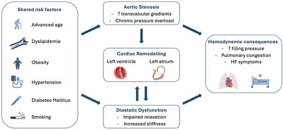
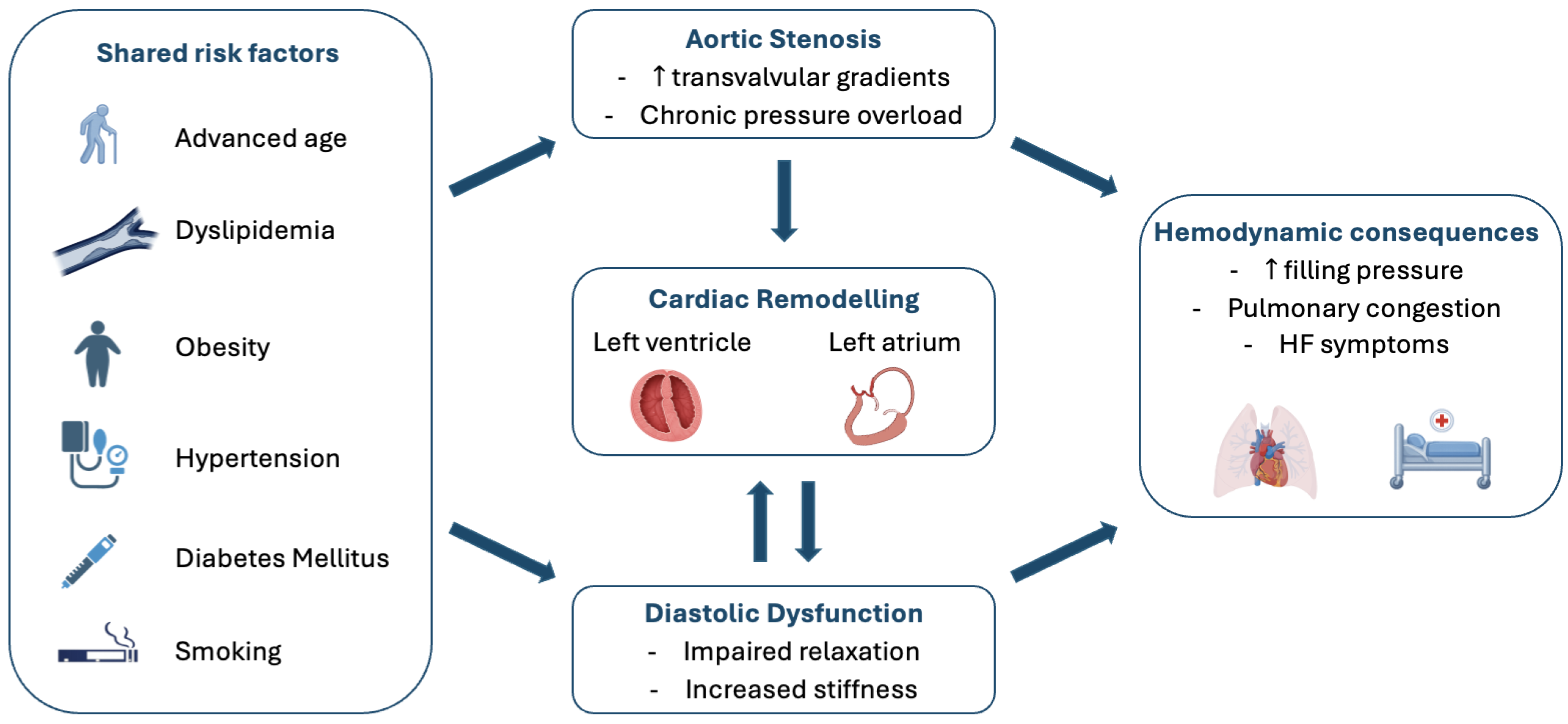
Long before left ventricular ejection fraction declines, the pressure overload imposed by a stenotic aortic valve induces concentric hypertrophy, interstitial fibrosis, and impaired myocardial relaxation (Figure 1). Diastolic dysfunction therefore constitutes an early and central component of AS pathophysiology and plays a key role in symptom development and outcomes after valve replacement [6].
Figure 1.
Pathophysiological relationship between aortic stenosis and diastolic dysfunction. Aortic stenosis (AS) and diastolic dysfunction (DD) frequently coexist and share several common risk factors. Chronic pressure overload caused by AS promotes left ventricular hypertrophy, impaired relaxation, and increased myocardial stiffness, leading to progressive diastolic dysfunction. In turn, DD contributes to left atrial dilation and further adverse cardiac remodeling. These pathophysiological changes result in elevated filling pressures, pulmonary congestion, exercise intolerance, and heart failure symptoms. Therefore, AS and DD interact in a self-reinforcing manner, whereby AS worsens diastolic impairment, while DD contributes substantially to symptom development, functional limitation, and adverse clinical outcomes.
Diastolic function encompasses the sequence of mechanical events that allow the left ventricle to fill adequately without an abnormal rise in filling pressures. Its main determinants are active myocardial relaxation and the passive diastolic properties of the LV chamber, particularly chamber stiffness. Diastolic dysfunction is characterized by impaired relaxation and/or increased chamber stiffness, which may lead to elevated LV diastolic pressures and, in more advanced stages, elevated LV filling pressures.
In AS, progressive narrowing of the aortic valve orifice leads to a chronic increase in LV systolic pressure. In response to this pressure overload, the ventricle undergoes compensatory concentric left ventricular hypertrophy (LVH), which normalizes wall stress and preserves systolic performance according to the Laplace relationship [1].
However, while concentric remodeling does not directly reduce ejection fraction, it adversely affects diastolic properties [1].
Indeed, it is well established that coronary endothelial function is significantly impaired in patients with left ventricular hypertrophy and aortic stenosis, resulting in reduced coronary flow reserve and an imbalance between myocardial oxygen supply and demand [7]. In addition, capillary rarefaction, increased extravascular compressive forces, and endothelial dysfunction contribute to impaired myocardial perfusion, particularly during stress. These mechanisms promote subendocardial ischemia and stimulate the development of myocardial fibrosis, both of which increase myocardial stiffness and impair ventricular relaxation [7,8].
Increased wall thickness impairs early diastolic relaxation and reduces ventricular compliance, requiring higher filling pressures to obtain a normal end-diastolic volume.
The fibrotic remodeling further increases myocardial stiffness and contributes to worsening diastolic dysfunction. These structural changes are notoriously resistant to immediate reversal, while muscular hypertrophy may regress within months of successful aortic valve replacement, the regression of interstitial fibrosis is a protracted process that may take years, which explains the common clinical observation that diastolic dysfunction often persists or only partially improves long after the valvular obstruction is removed [4,9].
As AS progresses, the left atrium (LA) undergoes compensatory remodeling to maintain ventricular filling in the presence of impaired diastolic function. This transition results in a greater reliance on atrial contraction to achieve complete ventricular filling. Consequently, the rise in left atrial and pulmonary venous pressures leads to pulmonary congestion, which manifests clinically as exertional dyspnea and, in advanced stages, overt heart failure [4].
Subendocardial ischemia, due to elevated wall stress and reduced coronary reserve, further impairs active relaxation. In addition, several comorbid conditions highly prevalent in patients with AS, such as chronic kidney disease, coronary artery disease, and diabetes, can directly contribute to diastolic dysfunction.
In AS, diastolic dysfunction typically progresses from impaired relaxation with initially normal filling pressures to a gradual increase in LV filling pressures. Over time, this increased pressure burden promotes LA remodeling and dysfunction and may ultimately contribute to the development of pulmonary hypertension.
Importantly, these changes often precede systolic impairment, underscoring the need for careful diastolic evaluation even when ejection fraction is preserved.
Since this pathophysiological progression has important implications for clinical outcomes, a staging system of cardiac damage in aortic stenosis has been proposed. This classification describes a stepwise progression from stage 0 (no cardiac damage), to stage 1 (left ventricular damage), stage 2 (left atrial and/or mitral valve involvement), stage 3 (tricuspid valve damage), and stage 4 (right ventricular dysfunction) Table 1 [10,11]. Although the staging of cardiac damage influences outcomes in patients with aortic stenosis, it does not correlate directly with the severity of the valvular disease. Indeed, cardiac damage beyond stage 1 is present in up to 80% of patients even with non-severe aortic stenosis. The extent of cardiac damage is more strongly influenced by comorbidities such as hypertension, ischemic heart disease, renal dysfunction, and pulmonary disease [10,11]. Differentiating diastolic dysfunction primarily driven by aortic stenosis from that related to concomitant comorbidities remains challenging, as both conditions frequently coexist and share common pathophysiological mechanisms. Rather than relying on a single parameter, a multiparametric approach should be adopted.
Table 1.
Staging of Cardiac Damage in Aortic Stenosis.
Several findings may suggest that pressure overload from aortic stenosis is the predominant mechanism, including severe valve obstruction, concentric left ventricular hypertrophy disproportionate to the degree of systemic hypertension, progressive impairment of global longitudinal strain despite preserved ejection fraction, and the presence of diffuse myocardial fibrosis on cardiovascular magnetic resonance in the absence of extensive ischemic scar. Improvement in diastolic parameters following aortic valve replacement also supports a major contribution of valvular afterload [12].
Conversely, when diastolic dysfunction appears disproportionate to the severity of aortic stenosis, clinicians should actively search for alternative or concomitant causes, including longstanding hypertension, diabetes mellitus, chronic kidney disease, obesity, coronary artery disease, infiltrative cardiomyopathies, or atrial fibrillation. Features such as eccentric remodeling, previous myocardial infarction, severe diffuse vascular disease, or persistent elevation of filling pressures despite successful valve replacement may suggest that myocardial dysfunction is predominantly related to these comorbidities [12].
Ultimately, distinguishing these entities is clinically relevant because it may influence therapeutic priorities. Patients in whom pressure overload is the principal driver are expected to derive the greatest benefit from timely aortic valve replacement, whereas optimization of guideline-directed medical therapy targeting hypertension, diabetes, heart failure, and other comorbidities becomes particularly important when myocardial disease extends beyond the valvular lesion. In most patients, however, diastolic dysfunction reflects the combined effects of aortic stenosis and systemic comorbidities, highlighting the need for an individualized, multimodality assessment [12]. In particular, cardiac amyloidosis represents an important infiltrative cause of disproportionate diastolic dysfunction in patients with AS and has been reported in up to 16% of patients undergoing transcatheter aortic valve replacement (TAVR) [13]. Transthyretin cardiac amyloidosis (ATTR-CA), particularly wild-type ATTR, may coexist with AS in older patients and can contribute independently to left LV wall thickening, increased myocardial stiffness, impaired relaxation, and elevated filling pressures. The coexistence of AS and ATTR-CA is clinically relevant because both conditions share several phenotypic features, including concentric LV hypertrophy, preserved left ventricular ejection fraction, low-flow states, and advanced diastolic dysfunction, potentially leading to an overestimation of the myocardial consequences attributable solely to valvular afterload [14,15]. The presence of marked LV wall thickening that appears disproportionate to the severity of AS, low-flow/low-gradient AS, reduced global longitudinal strain with relative apical sparing, increased wall thickness-to-voltage ratio, conduction abnormalities, or extracardiac red flags should therefore raise suspicion of concomitant cardiac amyloidosis [14,15]. In selected patients, cardiovascular magnetic resonance may provide additional evidence of infiltrative myocardial disease through diffuse late gadolinium enhancement and increased native T1 and extracellular volume, whereas bone scintigraphy with bone-seeking tracers, after appropriate exclusion of light-chain amyloidosis, can establish the diagnosis of ATTR-CA [14,15]. Recognition of concomitant amyloidosis is particularly important when interpreting diastolic dysfunction and myocardial damage in AS, as persistent myocardial abnormalities after aortic valve replacement may partly reflect an underlying infiltrative cardiomyopathy rather than irreversible consequences of valvular pressure overload [12].
3. Contribution of Diastolic Dysfunction to Stroke Volume Reduction in Aortic Stenosis
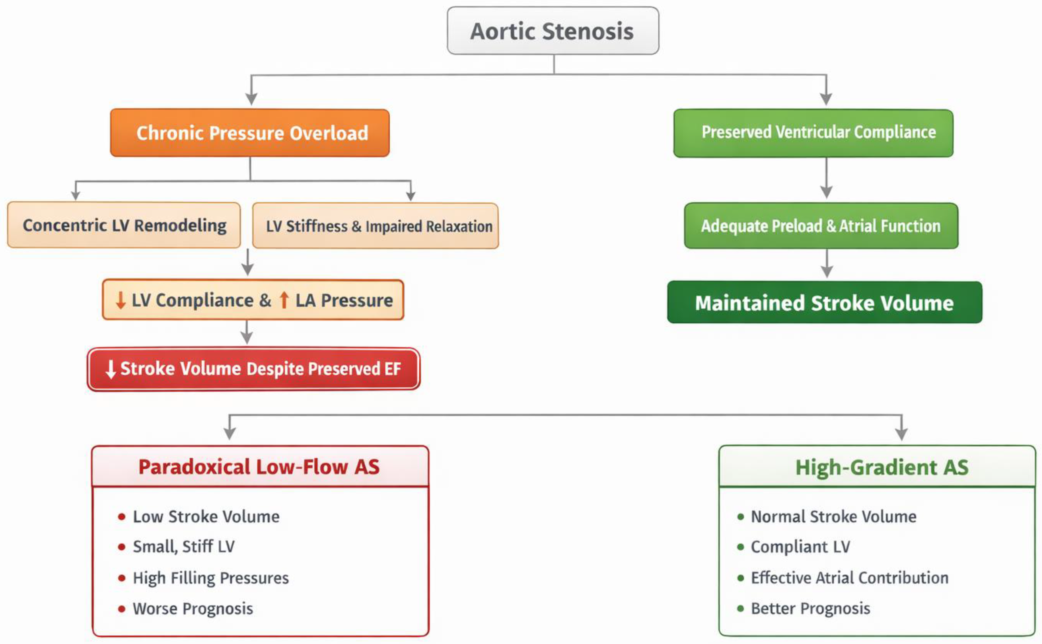
Diastolic dysfunction is a determinant of stroke volume reduction in patients with AS [16]. Chronic pressure overload induces concentric left ventricular remodeling, increased myocardial stiffness, and impaired relaxation, all of which limit ventricular filling during diastole. As left ventricular compliance progressively decreases, filling becomes increasingly dependent on higher left atrial pressures and atrial contraction. When these compensatory mechanisms are lacking, the reduction in effective ventricular preload leads to a decrease in stroke volume despite preserved systolic function. This mechanism is particularly relevant in the development of the paradoxical low-flow aortic stenosis phenotype observed in 25–35% of patients with severe AS, a small, concentrically remodeled and stiff ventricle [17,18,19]. In these cases, the stroke volume is low because the ventricle cannot fill adequately—a condition often referred to as “underfilling”. Diastolic assessment in this group frequently reveals advanced (Grade II or III) dysfunction and high filling pressures, which are the primary drivers of the low-flow state and the associated poor prognosis.
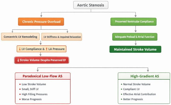
However, not all patients with AS develop a low-flow paradoxical phenotype. Patients who maintain relatively preserved ventricular compliance, adequate preload, and effective atrial contribution may preserve stroke volume despite significant valvular obstruction (Figure 2).
Figure 2.
Diastolic dysfunction as a determinant of stroke volume in aortic stenosis.
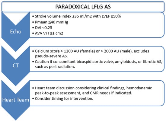
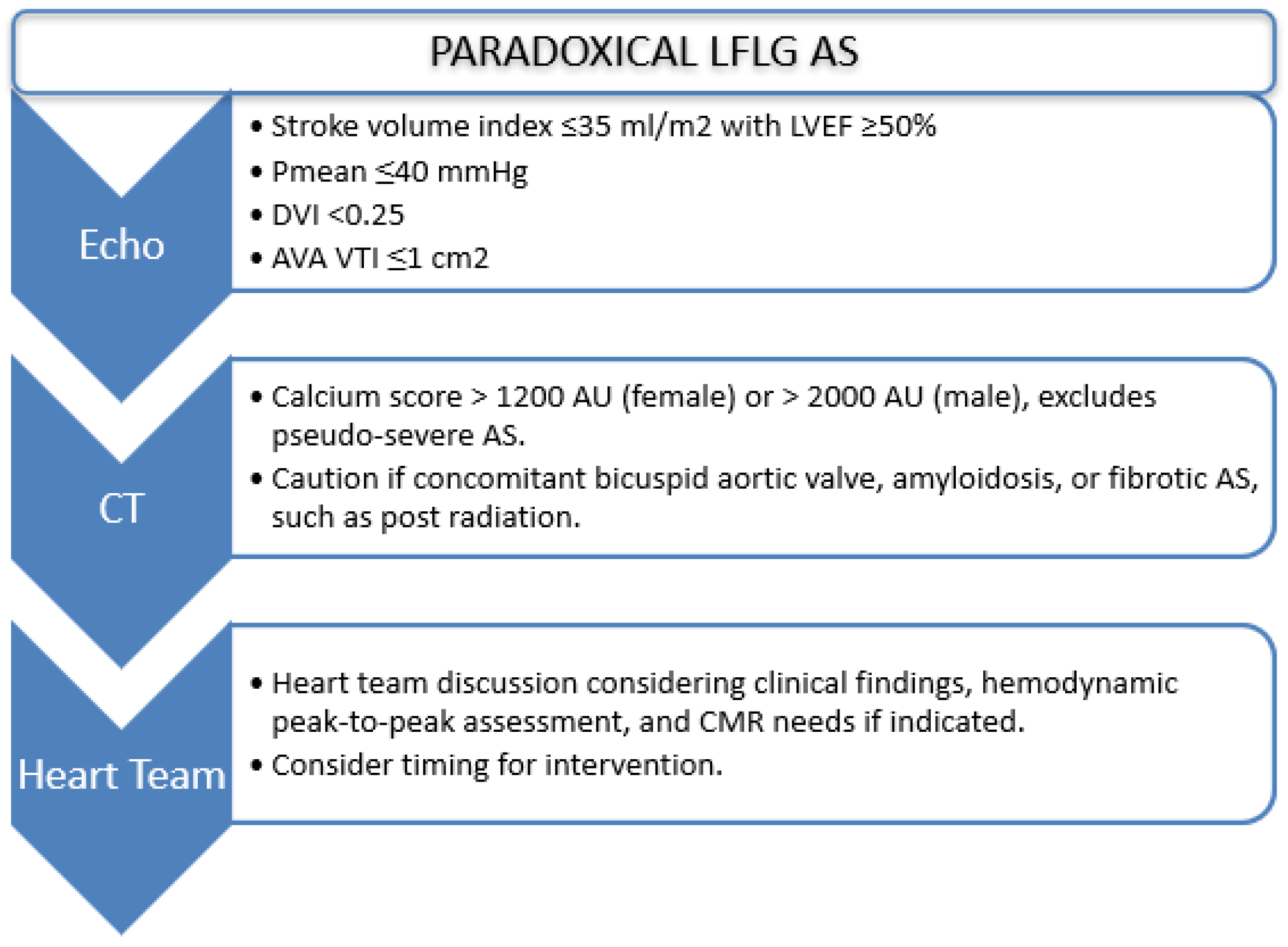
Moreover, patients with paradoxical low-flow, low-gradient aortic stenosis have a worse prognosis compared with those with high-gradient severe aortic stenosis and those with pseudo-severe aortic stenosis, but a more favorable outcome than patients with classical low-flow, low-gradient aortic stenosis [20,21]. In this setting, comprehensive multiparametric imaging integrating Doppler echocardiography, myocardial deformation imaging, and, when appropriate, multimodality imaging is essential to accurately assess disease severity and guide clinical decision-making (Figure 3).
Figure 3.
Proposed multiparametric approach for the evaluation and management of patients with paradoxical low-flow, low-gradient aortic stenosis. DVI: Doppler Velocity Index. AVA VTI: Aortic Valve Area Velocity–Time Integral. AU: Agatston Units; AS: Aortic Stenosis. CMR: Cardiac Magneetic Resonance.
4. Echocardiographic Assessment of Left Ventricular Diastolic Function in Aortic Stenosis
Transthoracic echocardiography remains the definitive non-invasive tool for evaluating diastolic function in patients with aortic stenosis. A multiparametric approach is required to integrate findings from mitral inflow Doppler, tissue Doppler imaging, and left atrial morphology.
4.1. Mitral Inflow Patterns in the Pressure-Overloaded Ventricle
Pulse-wave Doppler evaluation of mitral inflow provides immediate insights into the phases of diastole. The E-wave represents early diastolic filling, which is driven by the pressure gradient between the left atrium and the relaxing left ventricle. In the early stages of aortic stenosis, impaired relaxation causes a decrease in the E-wave velocity and an extension of the deceleration time (DT) as the ventricle takes longer to drop its pressure below that of the atrium [22].
The A-wave, representing atrial contraction, becomes more prominent as the ventricle’s reliance on the late filling phase increases. Consequently, a low E/A ratio (<0.8) is often the first identifiable sign of diastolic impairment in this population. However, as the disease progresses and filling pressures rise, the left atrial pressure increases to force blood into the stiff ventricle. This can artificially increase the E-wave velocity and shorten the DT, leading to a “pseudonormal” pattern that masks the underlying pathology [22].
In advanced stages, restrictive filling (E/A ≥ 2 with shortened deceleration time) reflects markedly increased chamber stiffness and carries adverse prognostic implications.
A critical consideration in AS is load dependence. The elevated afterload and altered LV–LA interaction can influence transmitral velocities, making isolated reliance on E/A patterns insufficient [22].
Moreover, atrial fibrillation presents several challenges, including tachycardia, the absence of an A wave, and variable cycle lengths. To assess filling pressures in atrial fibrillation, it is necessary to average measurements over several cycles that reflect the average heart rate. Key indicators of elevated LAP in atrial fibrillation include an E velocity ≥100 cm/s, a septal E/e′ > 11, a TR velocity >2.8 m/s, and a short DT of the E wave (≤160 ms) [22].
4.2. Tissue Doppler and E/e′: Interpretative Nuances in LV Hypertrophy
Tissue Doppler imaging measures the velocity of myocardial motion, typically at the septal and lateral aspects of the mitral annulus. The early diastolic annular velocity (e′) is a relatively load-independent marker of myocardial relaxation. In aortic stenosis, the e′ velocity is characteristically reduced, often falling below 7 cm/s at the septum or 10 cm/s at the lateral wall, reflecting the intrinsic impairment of the longitudinal myocardial fibers caused by hypertrophy and fibrosis [23].
The average E/e′ ratio has emerged as one of the most robust non-invasive estimates of left ventricular filling pressure. An average E/e′ ratio exceeding 14 is highly suggestive of elevated left atrial pressure in the general population. In the context of aortic stenosis, however, the high-afterload environment and the presence of significant hypertrophy may necessitate the use of higher thresholds or additional parameters to ensure accuracy [23].
Moreover, Mitral annular calcification (MAC) can restrict annular motion, falsely reducing the e′ velocity and elevating the E/e′ ratio. In patients with moderate to severe MAC, an E/A ratio <0.8 suggests normal pressures, while a ratio >1.8 suggests elevated pressures. If the ratio is intermediate (0.8–1.8), an IVRT < 80 ms is used as a marker for elevated LAP [22].
4.3. The Left Atrium: Chronic Pressure Sensor and Functional Marker
The left atrium serves as a chronic barometer of left ventricular filling pressures. Chronic exposure to elevated pressures leads to left atrial stretching and structural dilation. The left atrial volume index (LAVI), calculated using the biplane area-length method and indexed to body surface area, is a critical marker of the chronicity and severity of diastolic dysfunction. A LAVI greater than 34 mL/m2 is a major criterion for diagnosing diastolic dysfunction and is associated with an increased risk of adverse cardiovascular events, heart failure readmissions, and the development of atrial fibrillation [24].
However, LA enlargement is often a late finding [24].
4.4. Pulmonary Pressures and Advanced Disease
The peak velocity of the tricuspid regurgitation jet allows for the estimation of pulmonary artery systolic pressure. In the absence of primary pulmonary disease, an elevated TR velocity, typically defined as >2.8 m/s, often reflects a “backwards” transmission of high left-sided filling pressures through the pulmonary circulation. In patients with aortic stenosis, an elevated TR velocity is a potent indicator of advanced diastolic dysfunction and is strongly linked to a poor prognosis, particularly in the perioperative phase of valve replacement [25].
Peak tricuspid regurgitation velocity provides an estimate of pulmonary artery systolic pressure and reflects the hemodynamic consequences of sustained elevation of LV filling pressures. In AS, the development of pulmonary hypertension signals advanced diastolic burden and is associated with worse surgical and transcatheter outcomes.
Thus, TR velocity serves as both a diagnostic adjunct and a prognostic marker in the diastolic evaluation of AS [25].
4.5. Advanced Imaging: The Integration of Left Atrial Strain, Global Longitudinal Strain and Stress Echo
The traditional assessment of left atrial size via LAVI provides a view of structural remodeling but often fails to capture the earlier functional changes. Left atrial strain (LAS), specifically left atrial reservoir strain (LARS), has emerged as a superior and more sensitive marker of early diastolic impairment.
The left atrium performs three distinct functional roles during the cardiac cycle, each of which can be quantified using speckle-tracking echocardiography:
- Reservoir Function: Occurring during ventricular systole, the atrium expands to receive blood from the pulmonary veins. This is quantified by LARS, which reflects atrial compliance and intrinsic myocardial function.
- Conduit Function: Representing the passive flow of blood from the atrium into the ventricle during early diastole.
- Contractile Function: Representing the active contraction of the atrium during late diastole to complete ventricular filling.
Left atrial strain represents the longitudinal deformation of the atrial myocardium and is largely independent of volume, making it an early marker of dysfunction.
In patients across the spectrum of aortic stenosis, from moderate to severe stages, LARS has been shown to be an independent predictor of cardiac mortality and heart failure hospitalization. Notably, LARS often outperforms traditional measures like LAVI and even left ventricular global longitudinal strain (GLS) in its predictive power. A reduction in LARS is indicative of advanced atrial remodeling and fibrosis, signaling a significantly higher risk of adverse outcomes even after successful aortic valve replacement [26].
A Systematic review involving over 7000 patients have confirmed that for each unit increase in LA Peak Longitudinal Strain, the risk of primary endpoint events decreases by 7%. This underscores the importance of functional atrial assessment as a component of the comprehensive echocardiographic report for aortic stenosis patients [27].
The most recent recommendations for the assessment of diastolic function propose a stepwise approach [22].
In the first step, the following parameters should be evaluated: reduced e′ velocity (septal ≤ 6 cm/s, lateral ≤ 7 cm/s, or average ≤ 6.5 cm/s); increased E/e′ ratio (septal ≥ 15, lateral ≥ 13, or average ≥ 14); and increased tricuspid regurgitation (TR) velocity (≥2.8 m/s) or pulmonary artery systolic pressure (PASP ≥ 35 mmHg) [22].
If all three criteria are met, left atrial pressure is considered elevated, and diastolic dysfunction is present. In this case, grading between grade II and grade III diastolic dysfunction is based on the E/A ratio (E/A < 2 vs. ≥ 2).
If none of these criteria are met, diastolic dysfunction is excluded.
If only e′ velocity is reduced and the E/A ratio is ≤0.8, this is consistent with grade I diastolic dysfunction.
If two criteria are present, or if E/e′ is increased, or if PASP is elevated, further evaluation is required. In this setting, additional parameters should be assessed, including left atrial volume index (LAVI ≥ 34 mL/m2), left atrial reservoir strain (LARS < 18%), and isovolumetric relaxation time (IVRT < 70 ms). The presence of at least one of these supports the diagnosis of grade II or III diastolic dysfunction, depending on the E/A ratio [22].
Speckle-tracking echocardiography has expanded our understanding of myocardial mechanics in AS. GLS is frequently reduced despite preserved ejection fraction, reflecting subendocardial dysfunction and fibrosis.
Reduced GLS often parallels worsening diastolic stiffness, reinforcing the concept of systolic–diastolic coupling.
In AS, the combination of impaired GLS and abnormal LA strain may represent a phenotype of advanced myocardial remodeling, even before overt systolic failure [28].
Stress echocardiography is a critical tool for the functional assessment of aortic stenosis, particularly when symptoms are equivocal or absent. By reproducing the physiological demands of daily activity, stress testing can unmask “latent” disease. This is often achieved through the provocation of exertional symptoms, the detection of an abnormal blood pressure response, or the identification of an excessive rise in the mean transvalvular gradient.
Furthermore, stress echocardiography allows for the evaluation of the “diastolic reserve.” A failure of the diastolic reserve is characterized by a significant increase in the E/e′ ratio and pulmonary artery pressures during exercise, even when the resting values appear within normal limits [29,30].
5. Cardiovascular Magnetic Resonance: Myocardial Fibrosis and Risk Stratification
Cardiovascular magnetic resonance (CMR) has emerged as an important complementary imaging modality for the assessment of myocardial remodeling in patients with aortic stenosis. While echocardiography remains the cornerstone for evaluating diastolic function and hemodynamic severity, CMR provides unique tissue characterization, enabling non-invasive assessment of myocardial fibrosis, a key determinant of left ventricular decompensation and adverse clinical outcomes [31,32].
Late gadolinium enhancement (LGE) identifies focal replacement fibrosis, which represents irreversible myocardial injury and has consistently been associated with increased all-cause mortality, cardiovascular mortality, and major adverse cardiovascular events. Conversely, native T1 mapping and extracellular volume (ECV) quantification detect diffuse interstitial fibrosis, an earlier stage of myocardial remodeling that is more closely related to increased myocardial stiffness and diastolic dysfunction. Unlike replacement fibrosis, diffuse fibrosis appears to be at least partially reversible following aortic valve replacement, highlighting the importance of identifying myocardial damage before irreversible remodeling occurs [31,32].
Growing evidence indicates that CMR-derived markers of fibrosis improve risk stratification beyond conventional measures of aortic stenosis severity. Indeed, myocardial fibrosis may develop before symptom onset and cannot be reliably predicted by valve severity alone. Therefore, the combined assessment of LGE, native T1, and ECV may help identify patients with early left ventricular decompensation who could benefit from earlier aortic valve intervention. Moreover, regression of native T1 values after valve replacement has been associated with left ventricular mass regression and functional recovery, suggesting that CMR may also be useful for monitoring myocardial reverse remodeling following intervention [31,32].
Although routine CMR is not currently recommended for all patients with aortic stenosis, it represents a valuable complementary tool in selected patients, providing incremental prognostic information and supporting individualized timing of aortic valve replacement [31,32].
6. Medical Therapeutic Approach
Diastolic dysfunction in patients with severe aortic valve stenosis is recognized as a marker of increased cardiovascular risk and is associated with higher mortality [33,34,35]. Recently, a paradigm shift has occurred in the understanding of aortic stenosis, which is no longer considered only a disease of the valve but also a disease of the myocardium [36,37,38]. Although aortic valve replacement remains the only established disease-modifying treatment for severe aortic stenosis, increasing attention has been directed toward adjunctive medical therapies aimed at attenuating myocardial remodeling, improving diastolic function, and reducing residual cardiovascular risk. Among these, inhibition of the renin–angiotensin system has been associated with regression of left ventricular hypertrophy, attenuation of adverse myocardial remodeling, and potential slowing of valvular calcification. Observational studies have also reported lower cardiovascular mortality in patients receiving angiotensin-converting enzyme inhibitors or angiotensin receptor blockers before or after TAVR, although randomized evidence is currently lacking and the optimal timing of therapy remains uncertain [12].
Mineralocorticoid receptor antagonists (MRAs) represent another mechanistically attractive strategy because of their antifibrotic properties and their ability to attenuate neurohormonal activation. While MRAs have shown beneficial effects on diffuse interstitial fibrosis in heart failure, TAVR-specific data remain scarce, and their long-term efficacy and safety in this elderly population—particularly regarding renal dysfunction and hyperkalemia—have yet to be established. Using non-steroidal MRS, Finrenone before TAVR was associated with fewer hospitalizations at 1 year. However, 3-year outcomes after TAVR were not superior among patients receiving Finrenone compared with other MRAs [12,39].
Similarly, beta-blockers may provide benefits beyond heart rate control through modulation of sympathetic activation, reduction in myocardial oxygen demand, and prevention of arrhythmias. Contemporary observational studies suggest that continuation of beta-blocker therapy during the TAVR pathway is generally safe and may be associated with lower rates of heart failure and cardiovascular mortality in selected patients. However, evidence derives almost exclusively from registry data, and randomized trials evaluating their routine use in patients with aortic stenosis are still unavailable [12].
Among currently available pharmacological strategies, sodium–glucose cotransporter-2 (SGLT2) inhibitors have emerged as one of the most promising therapeutic options [12,40]. This class of drugs may act as a disease-modifying therapy in aortic stenosis, supported by emerging evidence suggesting that SGLT2 inhibitor use is associated with a slower progression of non-severe aortic valve stenosis; however, it requires confirmation in further powered randomized trials [41] (Table 2).
Table 2.
Potential mechanisms by which SGLT2 Inhibitors may slow aortic stenosis progression.
Beyond the aforementioned benefits in slowing aortic stenosis progression, SGLT2 inhibitors have demonstrated significant myocardial reverse remodeling. This comes with beneficial effects on diastolic dysfunction in heart failure with preserved or reduced ejection fraction, regardless of diabetes status [48]. Although SGLT2 is not expressed in cardiomyocytes [49], several pathophysiological mechanisms have been proposed to explain the cardioprotective effects of this drug class. These include improvements in myocardial energetics, reductions in systemic inflammation and oxidative stress, attenuation of myocardial fibrosis, modulation of intracellular sodium and calcium handling, and favorable effects on microvascular function and ventricular loading conditions [50,51,52,53,54].
Specifically, dapagliflozin has been shown to promote myocardial reverse remodeling by reducing left ventricular hypertrophy [55]. Regression of left ventricular hypertrophy has been associated with a lower risk of mortality and heart failure–related hospitalizations at one-year follow-up [56]. Despite effective treatment of aortic stenosis, heart failure hospitalizations remain frequent after TAVR, reaching rates of up to 20% within the first year [57,58]. In the DAPA-TAVI trial, treatment with the SGLT2 inhibitor dapagliflozin, initiated after TAVR, in patients with previous heart failure hospitalization and chronic kidney disease, heart failure with reduced ejection fraction, or diabetes, was associated with a significant reduction in adverse clinical outcomes. Compared with standard care alone, dapagliflozin therapy led to an approximate 28% relative reduction in the risk of the composite endpoint of all-cause mortality or worsening heart failure, highlighting the potential role of SGLT2 inhibition as an adjunctive strategy to improve outcomes following valve intervention [59]. Importantly, the observed clinical benefit was achieved without an excess of safety concerns. These findings suggest that dapagliflozin may favorably modulate post-procedural ventricular remodeling and diastolic function, thereby improving short- and mid-term outcomes [59]. In addition, in a study by Paolisso et al. [40], the use of SGLT2 inhibitors in diabetic patients with ejection fraction lower than 50%, undergoing TAVR for severe aortic stenosis and established cardiac damage, was associated with improved clinical outcomes. Compared with patients not treated with SGLT2 inhibitors, SGLT2i therapy was linked to a lower risk of major adverse cardiovascular events, all-cause mortality, and heart failure–related hospitalizations over a median follow-up of 24 months [40].
Moreover, SGLT2 inhibitor therapy has been associated with a lower incidence of acute kidney injury following transcatheter aortic valve implantation, further supporting their potential role in improving peri-procedural outcomes in this patient population [60].
Despite the observed cardiovascular and renal benefits, the addition of SGLT2 inhibitors to TAVR did not translate into further improvements in patient-reported health status, as assessed by KCCQ scores, compared with TAVR combined with standard care [61]. Dapagliflozin seems to primarily serve a disease-modifying role by reducing clinical events, rather than improving patient-perceived health status. After valve replacement, any additional symptomatic relief from SGLT2 inhibition may be minimal or undetectable, particularly when significant residual congestion or volume overload is absent [61]. Despite the encouraging evidence available to date, several important questions remain unanswered. In particular, while several mechanisms potentially underlying the effects of SGLT2 inhibitors on the aortic valve have been proposed, the available evidence is largely derived from preclinical and experimental studies, and their direct effects on human aortic valve disease remain to be established. Moreover, the optimal timing for initiating SGLT2 inhibitors in patients with aortic stenosis has yet to be established. It remains unclear whether these agents should be routinely introduced before TAVR, initiated early after valve implantation, or reserved for selected patients with established heart failure, diabetes mellitus, or chronic kidney disease. Furthermore, whether their clinical benefit varies according to heart failure phenotype (HFrEF versus HFpEF), the burden of residual myocardial fibrosis, or the extent of extravalvular cardiac damage remains to be determined. Future prospective randomized studies are also needed to evaluate the efficacy and tolerability of SGLT2 inhibitors in underrepresented populations, particularly frail older adults and patients with advanced chronic kidney disease. Overall, SGLT2 inhibitors represent one of the most promising adjunctive pharmacological strategies currently under investigation for patients with aortic stenosis and concomitant myocardial dysfunction. Nevertheless, the available evidence is still largely based on observational studies and a limited number of randomized trials. Therefore, although preliminary findings are encouraging, larger randomized studies are required before these agents can be routinely recommended specifically for patients with aortic stenosis [59].
7. Impact of Aortic Valve Replacement on Diastolic Function
TAVR or surgical aortic valve replacement (SAVR) effectively interrupts the vicious cycle of chronic left ventricular pressure overload and myocardial fibrosis induced by severe AS. Preoperative DD has also been associated with increased morbidity and mortality after AVR [4]. Table 3 summarizes the changes induced by aortic valve replacement, schematically categorizing them into short-, intermediate-, and long-term effects.
Table 3.
Impact of AVR on Diastolic Function: Short- vs. Long-Term Effects.
AVR rapidly reduces transvalvular gradients and systolic wall stress, halting the progression of concentric LV hypertrophy and enabling reverse remodeling. Serial studies demonstrate LV mass regression at 1-year follow-up, alongside improved global longitudinal strain reflecting decreased fibrosis burden [62,63]. As a consequence, diastolic parameters benefit substantially, including increased septal/lateral e′ velocity and reduced E/e′ ratio. Several studies have suggested that recovery of left ventricular diastolic function may occur earlier after TAVR than after SAVR. This observation is likely multifactorial rather than solely attributable to the avoidance of cardiopulmonary bypass and ischemia–reperfusion injury. Compared with SAVR, TAVR is associated with a less invasive procedural approach, avoids myocardial manipulation, has lower systemic inflammatory activation, and less peri-procedural myocardial injury, all of which may facilitate earlier improvement in myocardial relaxation and left ventricular filling pressures. Moreover, the absence of cardioplegic arrest may contribute to better preservation of myocardial function in the immediate postoperative period. Nevertheless, the available comparative evidence remains heterogeneous, with differences in study design, patient selection, and follow-up duration. Therefore, although current data suggest a more rapid early recovery of diastolic function after TAVR, further prospective comparative studies are required to clarify the underlying mechanisms and determine whether these early differences translate into sustained long-term clinical benefit [64,65,66]. Moreover, even after TAVR the baseline severity of diastolic dysfunction has a significant impact on prognosis and survival. Indeed, patients with grade 2 or 3 diastolic dysfunction at baseline exhibit worse outcomes [67]. More severe diastolic dysfunction is associated with a higher one-year mortality rate and prevalence of pulmonary hypertension and atrial fibrillation, as well as with higher circulating levels of N-terminal pro–B-type natriuretic peptide. These findings have important clinical implications, as such patient subgroups may benefit from earlier intervention for aortic valve disease, even in the absence of symptoms [67,68]. Following TAVR, LARS shows a strong association with the development of new-onset atrial fibrillation. Importantly, post-procedural LARS may represent a more sensitive marker of intrinsic left atrial structural and functional impairment, capturing atrial remodeling and dysfunction that persist despite correction of the valvular obstruction [69].
A similar pattern has been observed in patients undergoing SAVR. In this population, preoperative diastolic dysfunction has likewise been linked to increased morbidity and mortality [4,70,71]. Moreover, baseline diastolic dysfunction was associated with greater difficulty in weaning from cardiopulmonary bypass [72]. In the study by Krayenbuehl et al., myocardial mass was reduced 18 months after SAVR, largely as a result of regression of cardiomyocyte hypertrophy; however, a relative increase in myocardial fibrosis led to a deterioration in diastolic function [73].
In the myocardium of patients with aortic stenosis, histological examination reveals an increase in cardiomyocyte cell volume accompanied by interstitial fibrosis. These structural changes lead to diastolic dysfunction, characterized by reduced ventricular compliance and increased left atrial and left ventricular end-diastolic pressures [74].
At longer-term follow-up (6 years after surgery), both left ventricular mass and fibrotic content declined, although complete normalization was never achieved [73].
The extent of reverse myocardial remodeling after AVR is highly variable and depends on the stage of myocardial disease at the time of intervention. Patients undergoing valve replacement before the development of advanced myocardial damage generally experience greater regression of left ventricular hypertrophy, more pronounced improvement in diastolic function, and better recovery of myocardial deformation. Conversely, advanced age, longstanding pressure overload, atrial fibrillation, pulmonary hypertension, chronic kidney disease, and diabetes mellitus have all been associated with less favorable reverse remodeling. Imaging biomarkers also provide important prognostic information: severe baseline diastolic dysfunction, impaired global longitudinal strain, left atrial dysfunction, and the presence of replacement fibrosis on cardiovascular magnetic resonance are associated with persistent myocardial dysfunction and worse clinical outcomes after valve replacement. Optimization of medical therapy before and after valve intervention may further influence the reverse remodeling process. Therefore, treatment of aortic valve disease should ideally be undertaken before the onset of cardiomyocyte loss, myocardial fibrosis, and advanced diastolic dysfunction, a stage at which the benefits of intervention are likely to be attenuated [68]. Consequently, although current guidelines recommend intervention for aortic stenosis in asymptomatic patients when systolic dysfunction is present (left ventricular ejection fraction <50%), diastolic dysfunction is far more prevalent in this population and carries important prognostic implications. The traditional watchful-waiting strategy for other asymptomatic patients has recently been challenged by several randomized trials evaluating earlier aortic valve intervention. The RECOVERY trial demonstrated that early SAVR reduced the risk of the composite of operative mortality or cardiovascular death compared with conservative management in patients with asymptomatic very severe aortic stenosis [75]. More recently, the AVATAR trial extended these findings to patients with asymptomatic severe aortic stenosis, preserved left ventricular ejection fraction, and negative exercise testing, showing a lower incidence of the composite of all-cause death, acute myocardial infarction, stroke, or unplanned heart failure hospitalization with early SAVR during extended follow-up [76]. The EARLY TAVR trial further expanded this concept to transcatheter therapy, demonstrating that early TAVR was superior to clinical surveillance in reducing the composite of death, stroke, or unplanned hospitalization for cardiovascular causes in patients with asymptomatic severe aortic stenosis. However, these findings are not entirely consistent across patient populations [77]. In the EVOLVED trial, early aortic valve intervention did not significantly reduce the risk of all-cause death or unplanned aortic stenosis-related hospitalization in asymptomatic patients with severe aortic stenosis and CMR-detected myocardial fibrosis [78,79]. Taken together, these studies suggest a potential shift toward earlier intervention in selected asymptomatic patients, while also highlighting that myocardial fibrosis alone may not be sufficient to identify those who derive clinical benefit from early valve replacement. Further integration of clinical status, valve severity, myocardial remodeling, fibrosis, ventricular function, and multimodality imaging may therefore be required to refine patient selection and determine the optimal timing of intervention [78,79].
8. Future Perspectives
Beyond AVR, persistent diastolic dysfunction may represent a relevant therapeutic target in patients who remain symptomatic after TAVR or SAVR despite successful relief of valvular obstruction. In recent years, several transcatheter and device-based strategies have been investigated in HFpEF, aiming to reduce left-sided filling pressures, improve haemodynamic monitoring, modulate autonomic imbalance, or favorably influence ventricular–arterial coupling. These approaches include interatrial shunt devices for left atrial decompression, implantable haemodynamic monitors to guide decongestion, and renal sympathetic denervation [80,81]. In patients with persistent symptoms after TAVR or SAVR, future trials should evaluate whether a phenotype-guided approach, based on residual diastolic dysfunction and objectively elevated filling pressures, may identify subgroups who could benefit from adjunctive transcatheter therapies.
In addition, advances in artificial intelligence and deep learning applied to echocardiography may further improve the identification of patients with AS at higher risk of progressive myocardial remodeling and adverse outcomes. Recent data suggest that deep learning–based assessment of diastolic dysfunction can stratify the latent risk associated with the progression of early-stage AS, supporting the potential role of artificial intelligence in future risk stratification and therapeutic decision-making [82].
9. Conclusions
Aortic stenosis is not merely a valvular disease, but also a myocardial disorder driven by chronic pressure overload. Diastolic dysfunction develops early and frequently precedes overt systolic impairment. Early recognition of myocardial involvement is crucial, as it may refine risk stratification and help optimize the timing of aortic valve intervention.
Echocardiography provides a comprehensive, noninvasive framework for this assessment; however, in the hypertrophied ventricle of aortic stenosis, Doppler indices require careful interpretation. A multiparametric approach integrating transmitral flow, tissue Doppler imaging, left atrial size and function, pulmonary pressures, and myocardial deformation offers the most physiologically sound evaluation.
Emerging therapies targeting myocardial fibrosis, inflammation, and filling pressures represent a promising adjunctive strategy. Future studies are warranted to determine whether the use of these therapies before or after AVR can translate into improved clinical outcomes.
Author Contributions
Conceptualization, V.M.D.L., K.-P.K., K.-P.R., S.B. and S.C.; methodology, K.-P.K. and K.-P.R.; resources, A.S., F.C. and F.F.; writing—original draft preparation, V.M.D.L., S.C. and S.B.; writing—review and editing, K.-P.K., K.-P.R., S.B. and S.C.; visualization A.S., P.L., F.C. and F.F.; supervision: S.B., P.L., A.S., F.C. and F.F. All authors have read and agreed to the published version of the manuscript.
Funding
This research received no external funding.
Data Availability Statement
No new data were created or analyzed in this study. Data sharing is not applicable to this article.
Conflicts of Interest
All authors have reported that they have no relationships relevant to the contents of this paper to disclose.
Abbreviations
| AS | Aortic Stenosis |
| LV | Left Ventricular |
| LVH | Left Ventricular Hypertrophy |
| AVR | Aortic Valve Replacement |
| LA | Left Atrium |
| DT | Deceleration Time |
| MAC | Mitral Annular Calcification |
| LAVI | Left Atrial Volume Index |
| LAS | Left Atrial Strain |
| LARS | Left Atrial Reservoir Strain |
| GLS | Global Longitudinal Strain |
| TR | Tricuspid Regurgitation |
| PASP | Pulmonary Artery Systolic Pressure |
| IVRT | Isovolumetric Relaxation Time |
| SGLT2 | sodium–glucose cotransporter 2 |
| TAVR | Transcatheter Aortic Valve Replacement |
| SAVR | Surgical Aortic Valve Replacement |
References
- Clavel, M.A.; Tastet, L.; Pibarot, P. Cardiac remodelling in aortic stenosis. Arch. Cardiovasc. Dis. 2026, 119, 32–38. [Google Scholar] [CrossRef] [Scilit]
- Fukui, M.; Généreux, P.; Cavalcante, J.L. Assessment of Cardiac Damage in Aortic Stenosis. Cardiol. Clin. 2020, 38, 23–31. [Google Scholar] [CrossRef] [Scilit]
- Klein, A.L.; Ramchand, J.; Nagueh, S.F. Aortic Stenosis and Diastolic Dysfunction: Partners in Crime. J. Am. Coll. Cardiol. 2020, 76, 2952–2955. [Google Scholar] [CrossRef] [Scilit]
- Ong, G.; Pibarot, P.; Redfors, B.; Weissman, N.J.; Jaber, W.A.; Makkar, R.R.; Lerakis, S.; Gopal, D.; Khalique, O.; Kodali, S.K.; et al. Diastolic Function and Clinical Outcomes after Transcatheter Aortic Valve Replacement: PARTNER 2 SAPIEN 3 Registry. J. Am. Coll. Cardiol. 2020, 76, 2940–2951. [Google Scholar] [CrossRef] [Scilit]
- Barletta, G.; Del Bene, M.R. Diastolic dysfunction in aortic stenosis: Old pathophysiologic observations made a new tool. Echocardiography 2024, 41, e15816. [Google Scholar] [CrossRef] [Scilit]
- Hess, O.M.; Villari, B.; Krayenbuehl, H.P. Diastolic dysfunction in aortic stenosis. Circulation 1993, 87, IV73–IV76. [Google Scholar] [CrossRef] [Scilit]
- Bozbas, H.; Pirat, B.; Yildirir, A.; Şimşek, V.; Sade, E.; Eroglu, S.; Atar, I.; Altin, C.; Demirtas, S.; Ozin, B.; et al. Coronary flow reserve is impaired in patients with aortic valve calcification. Atherosclerosis 2008, 197, 846–852. [Google Scholar] [CrossRef] [Scilit]
- Ma, L.N.; Zhao, S.P.; Gao, M.; Zhou, Q.C.; Fan, P. Endothelial dysfunction associated with left ventricular diastolic dysfunction in patients with coronary heart disease. Int. J. Cardiol. 2000, 72, 275–279. [Google Scholar] [CrossRef] [Scilit]
- Villari, B.; Vassalli, G.; Monrad, E.S.; Chiariello, M.; Turina, M.; Hess, O.M. Normalization of diastolic dysfunction in aortic stenosis late after valve replacement. Circulation 1995, 91, 2353–2358. [Google Scholar] [CrossRef] [Scilit]
- Tastet, L.; Tribouilloy, C.; Maréchaux, S.; Vollema, E.M.; Delgado, V.; Salaun, E.; Shen, M.; Capoulade, R.; Clavel, M.-A.; Arsenault, M.; et al. Staging Cardiac Damage in Patients with Asymptomatic Aortic Valve Stenosis. J. Am. Coll. Cardiol. 2019, 74, 550–563. [Google Scholar] [CrossRef] [Scilit]
- Vollema, E.M.; Amanullah, M.R.; A Prihadi, E.; Ng, A.C.T.; van der Bijl, P.; Sin, Y.K.; Marsan, N.A.; Ding, Z.P.; Généreux, P.; Leon, M.B.; et al. Incremental value of left ventricular global longitudinal strain in a newly proposed staging classification based on cardiac damage in patients with severe aortic stenosis. Eur. Heart J.-Cardiovasc. Imaging 2020, 21, 1248–1258. [Google Scholar] [CrossRef] [Scilit]
- Onishi, T.; Tang, G.H.L.; Moyal, A.; Vemuri, K.S.; Swaminathan, S.; Contreras, J.; Moss, N.; Khera, S.; Hooda, A.; Dangas, G.; et al. Heart Failure Pharmacotherapy Across the Transcatheter Aortic Valve Replacement Continuum. J. Card. Fail. 2026, in press. [Google Scholar] [CrossRef] [Scilit] [PubMed]
- Nitsche, C.; Aschauer, S.; Kammerlander, A.A.; Schneider, M.; Poschner, T.; Duca, F.; Binder, C.; Koschutnik, M.; Stiftinger, J.; Goliasch, G.; et al. Light-chain and transthyretin cardiac amyloidosis in severe aortic stenosis: Prevalence, screening possibilities, and outcome. Eur. J. Heart Fail. 2020, 22, 1852–1862. [Google Scholar] [CrossRef] [Scilit]
- Castaño, A.; Narotsky, D.L.; Hamid, N.; Khalique, O.K.; Morgenstern, R.; DeLuca, A.; Rubin, J.; Chiuzan, C.; Nazif, T.; Vahl, T.; et al. Unveiling transthyretin cardiac amyloidosis and its predictors among elderly patients with severe aortic stenosis undergoing transcatheter aortic valve replacement. Eur. Heart J. 2017, 38, 2879–2887. [Google Scholar] [CrossRef] [Scilit]
- Mattig, I.; Wrede-Wihl, K.; Sökmen, S.; Huscher, D.; Wetz, C.; Heidecker, B.; Barzen, G.; Pernice, H.; Klein, C.; Unbehaun, A.; et al. Screening for amyloidosis before aortic valve elective replacement: Results from the SAVER study. Eur. J. Prev. Cardiol. 2026, zwag158. [Google Scholar] [CrossRef] [Scilit] [PubMed]
- Clavel, M.A.; Magne, J.; Pibarot, P. Low-gradient aortic stenosis. Eur. Heart J. 2016, 37, 2645–2657. [Google Scholar] [CrossRef] [Scilit]
- Hachicha, Z.; Dumesnil, J.G.; Bogaty, P.; Pibarot, P. Paradoxical low-flow, low-gradient severe aortic stenosis despite preserved ejection fraction is associated with higher afterload and reduced survival. Circulation 2007, 115, 2856–2864. [Google Scholar] [CrossRef] [Scilit]
- Minners, J.; Allgeier, M.; Gohlke-Baerwolf, C.; Kienzle, R.P.; Neumann, F.J.; Jander, N. Inconsistent grading of aortic valve stenosis by current guidelines: Haemodynamic studies in patients with apparently normal left ventricular function. Heart 2010, 96, 1463–1468. [Google Scholar] [CrossRef] [Scilit]
- Lancellotti, P.; Magne, J.; Donal, E.; Davin, L.; O’COnnor, K.; Rosca, M.; Szymanski, C.; Cosyns, B.; Piérard, L.A. Clinical outcome in asymptomatic severe aortic stenosis: Insights from the new proposed aortic stenosis grading classification. J. Am. Coll. Cardiol. 2012, 59, 235–243. [Google Scholar] [CrossRef] [Scilit]
- Tribouilloy, C.; Rusinaru, D.; Maréchaux, S.; Castel, A.-L.; Debry, N.; Maizel, J.; Mentaverri, R.; Kamel, S.; Slama, M.; Lévy, F. Low-gradient, low-flow severe aortic stenosis with preserved left ventricular ejection fraction: Characteristics, outcome, and implications for surgery. J. Am. Coll. Cardiol. 2015, 65, 55–66. [Google Scholar] [CrossRef] [Scilit]
- Mehrotra, P.; Jansen, K.; Flynn, A.W.; Tan, T.C.; Elmariah, S.; Picard, M.H.; Hung, J. Differential left ventricular remodelling and longitudinal function distinguishes low flow from normal-flow preserved ejection fraction low-gradient severe aortic stenosis. Eur. Heart J. 2013, 34, 1906–1914. [Google Scholar] [CrossRef] [Scilit]
- Nagueh, S.F.; Sanborn, D.Y.; Oh, J.K.; Anderson, B.; Billick, K.; Derumeaux, G.; Klein, A.; Koulogiannis, K.; Mitchell, C.; Shah, A.; et al. Recommendations for the Evaluation of Left Ventricular Diastolic Function by Echocardiography and for Heart Failure with Preserved Ejection Fraction Diagnosis: An Update from the American Society of Echocardiography. J. Am. Soc. Echocardiogr. 2025, 38, 537–569. [Google Scholar] [CrossRef] [Scilit]
- Bruch, C.; Stypmann, J.; Grude, M.; Gradaus, R.; Breithardt, G.; Wichter, T. Tissue Doppler imaging in patients with moderate to severe aortic valve stenosis: Clinical usefulness and diagnostic accuracy. Am. Heart J. 2004, 148, 696–702. [Google Scholar] [CrossRef] [Scilit]
- Rusinaru, D.; Bohbot, Y.; Kowalski, C.; Ringle, A.; Maréchaux, S.; Tribouilloy, C. Left Atrial Volume and Mortality in Patients with Aortic Stenosis. J. Am. Heart Assoc. 2017, 6, e006615. [Google Scholar] [CrossRef] [Scilit]
- Maeder, M.T.; Weber, L.; Rickli, H. Pulmonary hypertension in aortic valve stenosis. Trends Cardiovasc. Med. 2022, 32, 73–81. [Google Scholar] [CrossRef] [Scilit]
- Bak, M.; Lee, S.Y.; Park, S.-J.; Shin, H.; Kim, J.; Kim, E.K.; Chang, S.-A.; Lee, S.-C.; Park, S.W. Left Atrial Strain Predicts Cardiac Outcomes in Moderate Aortic Stenosis. Circ. Cardiovasc. Imaging 2026, 19, e018451. [Google Scholar] [CrossRef] [Scilit]
- Chen, N.; Gu, W.; Wu, J. Prognostic value of left atrial strain in significant aortic valve disease: A systematic review and meta-analysis. Front. Cardiovasc. Med. 2025, 12, 1667871. [Google Scholar] [CrossRef] [Scilit]
- Cramariuc, D.; Gerdts, E.; Davidsen, E.S.; Segadal, L.; Matre, K. Myocardial deformation in aortic valve stenosis: Relation to left ventricular geometry. Heart 2010, 96, 106–112. [Google Scholar] [CrossRef] [Scilit]
- Ha, J.W.; Andersen, O.S.; Smiseth, O.A. Diastolic Stress Test: Invasive and Noninvasive Testing. JACC Cardiovasc. Imaging 2020, 13, 272–282. [Google Scholar] [CrossRef] [Scilit]
- Clavel, M.-A.; Ennezat, P.V.; Maréchaux, S.; Dumesnil, J.G.; Capoulade, R.; Hachicha, Z.; Mathieu, P.; Bellouin, A.; Bergeron, S.; Meimoun, P.; et al. Stress echocardiography to assess stenosis severity and predict outcome in patients with paradoxical low-flow, low-gradient aortic stenosis and preserved LVEF. JACC Cardiovasc. Imaging 2013, 6, 175–183. [Google Scholar] [CrossRef] [Scilit]
- Zhang, C.; Liu, J.; Qin, S. Prognostic value of cardiac magnetic resonance in patients with aortic stenosis: A systematic review and meta-analysis. PLoS ONE 2022, 17, e0263378. [Google Scholar] [CrossRef] [Scilit] [PubMed] [PubMed Central]
- Papanastasiou, C.A.; Kokkinidis, D.G.; Kampaktsis, P.N.; Bikakis, I.; Cunha, D.K.; Oikonomou, E.K.; Greenwood, J.P.; Garcia, M.J.; Karamitsos, T.D. The Prognostic Role of Late Gadolinium Enhancement in Aortic Stenosis: A Systematic Review and Meta-Analysis. JACC Cardiovasc. Imaging 2020, 13, 385–392. [Google Scholar] [CrossRef] [Scilit] [PubMed]
- Ladeiras-Lopes, R.; Araújo, M.; Sampaio, F.; Leite-Moreira, A.; Fontes-Carvalho, R. The impact of diastolic dysfunction as a predictor of cardiovascular events: A systematic review and meta-analysis. Rev. Port. Cardiol. 2019, 38, 789–804. [Google Scholar] [CrossRef] [Scilit]
- Sorop, O.; Heinonen, I.; van Kranenburg, M.; van de Wouw, J.; de Beer, V.J.; Nguyen, I.T.N.; Octavia, Y.; van Duin, R.W.B.; Stam, K.; van Geuns, R.-J.; et al. Multiple common comorbidities produce left ventricular diastolic dysfunction associated with coronary microvascular dysfunction, oxidative stress, and myocardial stiffening. Cardiovasc. Res. 2018, 114, 954–964. [Google Scholar] [CrossRef] [Scilit]
- Ladeiras-Lopes, R.; Moreira, H.T.; Bettencourt, N.; Fontes-Carvalho, R.; Sampaio, F.; Ambale-Venkatesh, B.; Wu, C.; Liu, K.; Bertoni, A.G.; Ouyang, P.; et al. Metabolic Syndrome Is Associated with Impaired Diastolic Function Independently of MRI-Derived Myocardial Extracellular Volume: The MESA Study. Diabetes 2018, 67, 1007–1012. [Google Scholar] [CrossRef] [Scilit]
- Généreux, P.; Pibarot, P.; Redfors, B.; Mack, M.J.; Makkar, R.R.; A Jaber, W.; Svensson, L.G.; Kapadia, S.; Tuzcu, E.M.; Thourani, V.H.; et al. Staging classification of aortic stenosis based on the extent of cardiac damage. Eur. Heart J. 2017, 38, 3351–3358. [Google Scholar] [CrossRef] [Scilit]
- Dweck, M.R.; Loganath, K.; Bing, R.; A Treibel, T.; McCann, G.P.; E Newby, D.; Leipsic, J.; Fraccaro, C.; Paolisso, P.; Cosyns, B.; et al. Multi-modality imaging in aortic stenosis: An EACVI clinical consensus document. Eur. Heart J.-Cardiovasc. Imaging 2023, 24, 1430–1443. [Google Scholar] [CrossRef] [Scilit]
- Marsan, N.A.; Delgado, V.; Shah, D.J.; Pellikka, P.; Bax, J.J.; Treibel, T.; Cavalcante, J.L. Valvular heart disease: Shifting the focus to the myocardium. Eur. Heart J. 2023, 44, 28–40. [Google Scholar] [CrossRef] [Scilit]
- Massad, F.; Badwan, O.; Rosenzveig, A.; Zghyer, F.; Shahrori, Z.; Motairek, I.; Hatab, T.; Alkhatib, R.; Kapadia, S. Finerenone in Patients Undergoing TAVR for Nonrheumatic Aortic Stenosis: Insights from a Real-World Cohort. Struct. Heart 2025, 10, 100751. [Google Scholar] [CrossRef] [Scilit] [PubMed] [PubMed Central]
- Paolisso, P.; Belmonte, M.; Gallinoro, E.; Scarsini, R.; Bergamaschi, L.; Portolan, L.; Armillotta, M.; Esposito, G.; Moscarella, E.; Benfari, G.; et al. SGLT2-inhibitors in diabetic patients with severe aortic stenosis and cardiac damage undergoing transcatheter aortic valve implantation (TAVI). Cardiovasc. Diabetol. 2024, 23, 420. [Google Scholar] [CrossRef] [Scilit]
- Shah, T.; Zhang, Z.; Shah, H.; Fanaroff, A.C.; Nathan, A.S.; Parise, H.; Lutz, J.; Sugeng, L.; Bellumkonda, L.; Redfors, B.; et al. Effect of Sodium-Glucose Cotransporter-2 Inhibitors on the Progression of Aortic Stenosis. JACC Cardiovasc. Interv. 2025, 18, 738–748. [Google Scholar] [CrossRef] [Scilit]
- Elrakaybi, A.; Laubner, K.; Zhou, Q.; Hug, M.J.; Seufert, J. Cardiovascular protection by SGLT2 inhibitors—Do anti-inflammatory mechanisms play a role? Mol. Metab. 2022, 64, 101549. [Google Scholar] [CrossRef] [Scilit]
- Mone, P.; Varzideh, F.; Jankauskas, S.S.; Pansini, A.; Lombardi, A.; Frullone, S.; Santulli, G. SGLT2 Inhibition via Empagliflozin Improves Endothelial Function and Reduces Mitochondrial Oxidative Stress: Insights from Frail Hypertensive and Diabetic Patients. Hypertension 2022, 79, 1633–1643. [Google Scholar] [CrossRef] [Scilit]
- Lu, J.; Xie, S.; Deng, Y.; Xie, X.; Liu, Y. Blocking the NLRP3 inflammasome reduces osteogenic calcification and M1 macrophage polarization in a mouse model of calcified aortic valve stenosis. Atherosclerosis 2022, 347, 28–38. [Google Scholar] [CrossRef] [Scilit]
- Manduteanu, I.; Simionescu, D.; Simionescu, A.; Simionescu, M. Aortic valve disease in diabetes: Molecular mechanisms and novel therapies. J. Cell Mol. Med. 2021, 25, 9483–9495. [Google Scholar] [CrossRef] [Scilit]
- Chen, X.; Yang, Q.; Bai, W.; Yao, W.; Liu, L.; Xing, Y.; Meng, C.; Qi, P.; Dang, Y.; Qi, X. Dapagliflozin Attenuates Myocardial Fibrosis by Inhibiting the TGF-β1/Smad Signaling Pathway in a Normoglycemic Rabbit Model of Chronic Heart Failure. Front. Pharmacol. 2022, 13, 873108. [Google Scholar] [CrossRef] [Scilit]
- Goerg, J.; Sommerfeld, M.; Greiner, B.; Lauer, D.; Seckin, Y.; Kulikov, A.; Ivkin, D.; Kintscher, U.; Okovityi, S.; Kaschina, E. Low-Dose Empagliflozin Improves Systolic Heart Function after Myocardial Infarction in Rats: Regulation of MMP9, NHE1, and SERCA2a. Int. J. Mol. Sci. 2021, 22, 5437. [Google Scholar] [CrossRef] [Scilit]
- Connelly, K.; Cai, S. Softening the Stiff Heart: SGLT2 Inhibition and Diastolic Function. JACC Cardiovasc. Imaging 2021, 14, 408–410. [Google Scholar] [CrossRef] [Scilit]
- Chen, J.; Williams, S.; Ho, S.; Loraine, H.; Hagan, D.; Whaley, J.M.; Feder, J.N. Quantitative PCR tissue expression profiling of the human SGLT2 gene and related family members. Diabetes Ther. Res. Treat. Educ. Diabetes Relat. Disord. 2010, 1, 57–92. [Google Scholar] [CrossRef] [Scilit]
- Toyama, T.; Neuen, B.L.; Jun, M.; Ohkuma, T.; Neal, B.; Jardine, M.J.; Heerspink, H.L.; Wong, M.G.; Ninomiya, T.; Wada, T.; et al. Effect of SGLT2 inhibitors on cardiovascular, renal and safety outcomes in patients with type 2 diabetes mellitus and chronic kidney disease: A systematic review and meta-analysis. Diabetes Obes. Metab. 2019, 21, 1237–1250. [Google Scholar] [CrossRef] [Scilit]
- Connelly, K.A.; Zhang, Y.; Visram, A.; Advani, A.; Batchu, S.N.; Desjardins, J.-F.; Thai, K.; Gilbert, R.E. Empagliflozin Improves Diastolic Function in a Nondiabetic Rodent Model of Heart Failure with Preserved Ejection Fraction. JACC Basic. Transl. Sci. 2019, 4, 27–37. [Google Scholar] [CrossRef] [Scilit]
- Santos-Gallego, C.G.; Requena-Ibanez, J.A.; Antonio, R.S.; Garcia-Ropero, A.; Ishikawa, K.; Watanabe, S.; Picatoste, B.; Vargas-Delgado, A.P.; Flores-Umanzor, E.J.; Sanz, J.; et al. Empagliflozin Ameliorates Diastolic Dysfunction and Left Ventricular Fibrosis/Stiffness in Nondiabetic Heart Failure: A Multimodality Study. JACC Cardiovasc. Imaging 2021, 14, 393–407. [Google Scholar] [CrossRef] [Scilit]
- Singh, J.S.; Mordi, I.R.; Vickneson, K.; Fathi, A.; Donnan, P.T.; Mohan, M.; Choy, A.M.J.; Gandy, S.; George, J.; Khan, F.; et al. Dapagliflozin Versus Placebo on Left Ventricular Remodeling in Patients with Diabetes and Heart Failure: The REFORM Trial. Diabetes Care 2020, 43, 1356–1359. [Google Scholar] [CrossRef] [Scilit]
- Verma, S.; Mazer, C.D.; Yan, A.T.; Mason, T.; Garg, V.; Teoh, H.; Zuo, F.; Quan, A.; Farkouh, M.E.; Fitchett, D.H.; et al. Effect of Empagliflozin on Left Ventricular Mass in Patients with Type 2 Diabetes Mellitus and Coronary Artery Disease: The EMPA-HEART CardioLink-6 Randomized Clinical Trial. Circulation 2019, 140, 1693–1702. [Google Scholar] [CrossRef] [Scilit]
- Brown, A.J.M.; Gandy, S.; McCrimmon, R.; Houston, J.G.; Struthers, A.D.; Lang, C.C. A randomized controlled trial of dapagliflozin on left ventricular hypertrophy in people with type two diabetes: The DAPA-LVH trial. Eur. Heart J. 2020, 41, 3421–3432. [Google Scholar] [CrossRef] [Scilit]
- Chau, K.H.; Douglas, P.S.; Pibarot, P.; Hahn, R.T.; Khalique, O.K.; Jaber, W.A.; Cremer, P.; Weissman, N.J.; Asch, F.M.; Zhang, Y.; et al. Regression of Left Ventricular Mass After Transcatheter Aortic Valve Replacement: The PARTNER Trials and Registries. J. Am. Coll. Cardiol. 2020, 75, 2446–2458. [Google Scholar] [CrossRef] [Scilit]
- Nombela-Franco, L.; del Trigo, M.; Morrison-Polo, G.; Veiga, G.; Jimenez-Quevedo, P.; Altisent, O.A.-J.; Campelo-Parada, F.; Biagioni, C.; Puri, R.; DeLarochellière, R.; et al. Incidence, Causes, and Predictors of Early (≤30 Days) and Late Unplanned Hospital Readmissions After Transcatheter Aortic Valve Replacement. JACC Cardiovasc. Interv. 2015, 8, 1748–1757. [Google Scholar] [CrossRef] [Scilit]
- Cid-Menéndez, A.; López-Otero, D.; González-Ferreiro, R.; Iglesias-Álvarez, D.; Álvarez-Rodríguez, L.; Antúnez-Muiños, P.J.; Cid-Álvarez, B.; Sanmartin-Pena, X.; Trillo-Nouche, R.; González-Juanatey, J.R. Predictors and outcomes of heart failure after transcatheter aortic valve implantation using a self-expanding prosthesis. Rev. Esp. Cardiol. 2020, 73, 383–392. [Google Scholar] [CrossRef] [Scilit]
- Raposeiras-Roubin, S.; Amat-Santos, I.J.; Rossello, X.; Ferreiro, R.G.; Bermúdez, I.G.; Otero, D.L.; Nombela-Franco, L.; Gheorghe, L.; Diez, J.L.; Zorita, C.B.; et al. Dapagliflozin in Patients Undergoing Transcatheter Aortic-Valve Implantation. N. Engl. J. Med. 2025, 392, 1396–1405. [Google Scholar] [CrossRef] [Scilit]
- Paolisso, P.; Belmonte, M.; Gallinoro, E.; Scarsini, R.; Bergamaschi, L.; Portolan, L.; Armillotta, M.; Esposito, G.; Moscarella, E.; Montalto, C.; et al. Impact of SGLT2-inhibitors on acute kidney injury in diabetic patients with severe aortic stenosis undergoing transcatheter aortic valve implantation (TAVI). Cardiovasc. Diabetol. 2025, 24, 221. [Google Scholar] [CrossRef] [Scilit]
- Bonanad-Lozano, C.; Blas, S.G.; Amat-Santos, I.; Gonzalez-Manzanares, R.; Sanchís, J.; Otero, D.L.; Franco, L.N.; Gheorge, L.; Sanz-Sánchez, J.; Zorita, C.B.; et al. Effect of Dapagliflozin on Quality of Life of Patients with Aortic Stenosis Undergoing Transcatheter Aortic Valve Implantation. J. Am. Coll. Cardiol. 2025, 86, 1128–1138. [Google Scholar] [CrossRef] [Scilit]
- Lindman, B.R.; Stewart, W.J.; Pibarot, P.; Hahn, R.T.; Otto, C.M.; Xu, K.; Devereux, R.B.; Weissman, N.J.; Enriquez-Sarano, M.; Szeto, W.Y.; et al. Early Regression of Severe Left Ventricular Hypertrophy After Transcatheter Aortic Valve Replacement Is Associated with Decreased Hospitalizations. JACC Cardiovasc. Interv. 2014, 7, 662–673. [Google Scholar] [CrossRef] [Scilit]
- Grayburn, P.A. “Cherry-on-Top” Pattern of Left Ventricular Longitudinal Strain in Aortic Stenosis Is Common and Resolves After Transcatheter Aortic Valve Replacement. Am. J. Cardiol. 2023, 198, 133–134. [Google Scholar] [CrossRef] [Scilit]
- Ha, S.J.; Yoo, S.Y.; Hong, M.K.; Hong, G.R. Immediate and Evolutionary Recovery of Left Ventricular Diastolic Function after Transcatheter Aortic Valve Replacement: Comparison with Surgery. Yonsei Med. J. 2020, 61, 30. [Google Scholar] [CrossRef] [Scilit]
- Shim, J.K.; Choi, Y.S.; Chun, D.H.; Hong, S.W.; Kim, D.H.; Kwak, Y.L. Relationship between echocardiographic index of ventricular filling pressure and intraoperative haemodynamic changes during off-pump coronary bypass surgery. Br. J. Anaesth. 2009, 102, 316–321. [Google Scholar] [CrossRef] [Scilit]
- Guarracino, F.; Talini, E.; Landoni, G.; Petronio, S.; Giannini, C.; Di Bello, V. Effect of aortic valve surgery on left ventricular diastole assessed by echocardiography and neuroendocrine response: Percutaneous versus surgical approach. J. Cardiothorac. Vasc. Anesth. 2010, 24, 25–29. [Google Scholar] [CrossRef] [Scilit]
- Koifman, E.; Medvedofsky, D.; Didier, R.; Torguson, R.; Jerusalem, Z.; Kiramijyan, S.; Ben-Dor, I.; Wang, Z.; Goldstein, S.A.; Xu, L.; et al. Impact of Baseline Left Ventricular Diastolic Dysfunction in Patients with Severe Aortic Stenosis Undergoing Transcatheter Aortic Valve Implantation. Am. J. Cardiol. 2020, 125, 258–263. [Google Scholar] [CrossRef] [Scilit]
- Pellikka, P.A.; Padang, R. Diastolic Dysfunction Pre-Transcatheter Aortic Valve Replacement: Is it Too Late? JACC Cardiovasc. Interv. 2018, 11, 602–604. [Google Scholar] [CrossRef] [Scilit]
- Butcher, S.C.; Hirasawa, K.; Meucci, M.C.; Stassen, J.; Kuneman, J.H.; Pereira, A.R.; van der Kley, F.; de Weger, A.; van Rosendael, P.J.; Marsan, N.A.; et al. Prognostic implications and alterations in left atrial deformation following transcatheter aortic valve implantation. Eur. Heart J.-Cardiovasc. Imaging 2024, 25, 1638–1648. [Google Scholar] [CrossRef] [Scilit]
- Gjertsson, P.; Caidahl, K.; Farasati, M.; Odén, A.; Bech-Hanssen, O. Preoperative moderate to severe diastolic dysfunction: A novel Doppler echocardiographic long-term prognostic factor in patients with severe aortic stenosis. J. Thorac. Cardiovasc. Surg. 2005, 129, 890–896. [Google Scholar] [CrossRef] [Scilit]
- Dahl, J.S.; Videbæk, L.; Poulsen, M.K.; Pellikka, P.A.; Veien, K.; Andersen, L.I.; Haghfelt, T.; Møller, J.E. Noninvasive assessment of filling pressure and left atrial pressure overload in severe aortic valve stenosis: Relation to ventricular remodeling and clinical outcome after aortic valve replacement. J. Thorac. Cardiovasc. Surg. 2011, 142, e77–e83. [Google Scholar] [CrossRef] [Scilit]
- Denault, A.Y.; Couture, P.; Buithieu, J.; Haddad, F.; Carrier, M.; Babin, D.; Levesque, S.; Tardif, J.-C. Left and right ventricular diastolic dysfunction as predictors of difficult separation from cardiopulmonary bypass. Can. J. Anaesth. 2006, 53, 1020–1029. [Google Scholar] [CrossRef] [Scilit]
- Krayenbuehl, H.P.; Hess, O.M.; Monrad, E.S.; Schneider, J.; Mall, G.; Turina, M. Left ventricular myocardial structure in aortic valve disease before, intermediate, and late after aortic valve replacement. Circulation 1989, 79, 744–755. [Google Scholar] [CrossRef] [Scilit]
- Vizzardi, E.; D’aLoia, A.; Fiorina, C.; Bugatti, S.; Parrinello, G.; De Carlo, M.; Giannini, C.; Di Bello, V.; Petronio, A.S.; Curello, S.; et al. Early regression of left ventricular mass associated with diastolic improvement after transcatheter aortic valve implantation. J. Am. Soc. Echocardiogr. 2012, 25, 1091–1098. [Google Scholar] [CrossRef] [Scilit]
- Kang, D.H.; Park, S.J.; Lee, S.A.; Lee, S.; Kim, D.H.; Kim, H.K.; Yun, S.C.; Hong, G.R.; Song, J.M.; Chung, C.H.; et al. Early Surgery or Conservative Care for Asymptomatic Aortic Stenosis. N. Engl. J. Med. 2020, 382, 111–119. [Google Scholar] [CrossRef] [Scilit] [PubMed]
- Banovic, M.; Putnik, S.; Penicka, M.; Doros, G.; Deja, M.A.; Kockova, R.; Kotrc, M.; Glaveckaite, S.; Gasparovic, H.; Pavlovic, N.; et al. Aortic Valve Replacement Versus Conservative Treatment in Asymptomatic Severe Aortic Stenosis: The AVATAR Trial. Circulation 2022, 145, 648–658, Erratum in Circulation 2022, 145, e761. https://doi.org/10.1161/CIR.0000000000001057. PMID: 34779220. [Google Scholar] [CrossRef] [Scilit]
- Généreux, P.; Schwartz, A.; Oldemeyer, J.B.; Pibarot, P.; Cohen, D.J.; Blanke, P.; Lindman, B.R.; Babaliaros, V.; Fearon, W.F.; Daniels, D.V.; et al. Transcatheter Aortic-Valve Replacement for Asymptomatic Severe Aortic Stenosis. N. Engl. J. Med. 2025, 392, 217–227. [Google Scholar] [CrossRef] [Scilit] [PubMed]
- Loganath, K.; Craig, N.J.; Everett, R.J.; Bing, R.; Tsampasian, V.; Molek, P.; Botezatu, S.; Aslam, S.; Lewis, S.; Graham, C.; et al. Early Intervention in Patients with Asymptomatic Severe Aortic Stenosis Myocardial Fibrosis: The EVOLVED Randomized Clinical Trial. JAMA 2025, 333, 213–221. [Google Scholar] [CrossRef] [Scilit] [PubMed] [PubMed Central]
- Lancellotti, P.; Coisne, A.; Cosyns, B.; Dulgheru, R.; Garbi, M.; Hong, G.R.; Hanzevacki, J.S.; Moscarelli, M.; Sugimoto, T.; Donal, E.; et al. Early aortic valve intervention in asymptomatic severe aortic stenosis: A clinical dilemma in evolution. Eur. Heart J. Open. 2025, 5, oeaf114. [Google Scholar] [CrossRef] [Scilit] [PubMed] [PubMed Central]
- Wu, Y.; Song, M.; Wu, M.; Lin, L. Advances in device-based treatment of heart failure with preserved ejection fraction: Evidence from clinical trials. ESC. Heart Fail. 2024, 11, 13–27. [Google Scholar] [CrossRef] [Scilit]
- Kresoja, K.P.; Rommel, K.P.; Fengler, K.; von Roeder, M.; Besler, C.; Lücke, C.; Gutberlet, M.; Desch, S.; Thiele, H.; Böhm, M.; et al. Renal sympathetic denervation in patients with heart failure with preserved ejection fraction. Circ. Heart Fail. 2021, 14, e007421. [Google Scholar] [CrossRef] [Scilit]
- Tokodi, M.; Shah, R.; Jamthikar, A.; Craig, N.; Hamirani, Y.; Casaclang-Verzosa, G.; Hahn, R.T.; Dweck, M.R.; Pibarot, P.; Yanamala, N.; et al. Deep learning model of diastolic dysfunction risk stratifies the progression of early-stage aortic stenosis. JACC Cardiovasc. Imaging 2025, 18, 150–165. [Google Scholar] [CrossRef] [Scilit]

Disclaimer/Publisher’s Note: The statements, opinions and data contained in all publications are solely those of the individual author(s) and contributor(s) and not of MDPI and/or the editor(s). MDPI and/or the editor(s) disclaim responsibility for any injury to people or property resulting from any ideas, methods, instructions or products referred to in the content. |
© 2026 by the authors. Licensee MDPI, Basel, Switzerland. This article is an open access article distributed under the terms and conditions of the Creative Commons Attribution (CC BY) license.