


Abstract
Neuropathic pain is a chronic, difficult-to-treat condition arising from injury or disease of the somatosensory nervous system. Current pharmacological treatments provide limited relief for many patients and are often associated with adverse effects, highlighting the need for complementary mechanism-based strategies. Neuroinflammation and oxidative stress are pivotal and interdependent drivers of neuropathic pain, promoting glial activation, cytokine release, mitochondrial dysfunction, redox imbalance, ion channel sensitization, and peripheral and central sensitization. Nutraceuticals and dietary bioactive compounds have gained attention because of their pleiotropic actions, i.e., multiple pharmacological activities, on inflammatory, oxidative, metabolic, mitochondrial, and neuroimmune pathways. This review summarizes the mechanistic roles of neuroinflammation and oxidative stress in neuropathic pain and discusses evidence supporting nutraceutical-based interventions, including polyphenols, terpenoids, alkaloids, fatty acids and lipid-derived compounds, antioxidant nutraceuticals, vitamins, and micronutrients. Preclinical studies indicate that these compounds can attenuate pain-related behaviors by modulating redox-sensitive inflammatory signaling, glial reactivity, mitochondrial function, nociceptive ion channels, and endogenous antioxidant defenses. Although clinical evidence remains limited, selected compounds, including alpha-lipoic acid, palmitoylethanolamide, omega-3 fatty acids, B-complex vitamins, vitamin D, and capsaicin, show translational promise. Future studies should prioritize standardized formulations, bioavailability, dosing, safety, mechanistic biomarkers, and robust randomized trials to clarify their clinical role as complementary, multi-target strategies for neuropathic pain management.
1. Introduction
Neuropathic pain is defined as “pain arising as a direct consequence of a lesion or disease affecting the somatosensory nervous system” [1,2]. Unlike nociceptive pain, which results from the activation of peripheral nociceptors by tissue injury or inflammation, neuropathic pain originates from pathological alterations within the peripheral or central nervous system and is characterized by maladaptive changes in neuronal signaling [3,4]. Clinically, neuropathic pain presents with a range of sensory abnormalities, including spontaneous pain, burning sensations, electric shock-like pain, paresthesia, dysesthesia, mechanical allodynia, and thermal or mechanical hyperalgesia [4,5]. These symptoms often persist long after the initial injury or disease has resolved and are frequently accompanied by significant emotional, cognitive, and functional impairments [6].
Neuropathic pain affects approximately 7–10% of the general population and represents a major public health challenge worldwide [7]. Neuropathic pain is prevalent among middle-aged and older adults with diverse underlying etiologies and is associated with a greater health burden than non-neuropathic pain [8]. It is associated with a broad spectrum of conditions, including diabetic peripheral neuropathy, chemotherapy-induced peripheral neuropathy, postherpetic neuralgia, traumatic nerve injuries, spinal cord injury, multiple sclerosis, and stroke [4,9]. The chronic nature of neuropathic pain imposes a substantial socioeconomic burden through increased healthcare utilization, reduced productivity, disability, sleep disturbances, anxiety, depression, and diminished quality of life [6,10]. Consequently, neuropathic pain remains one of the most debilitating and difficult-to-manage chronic pain conditions [4].
Current pharmacological management relies primarily on antidepressants, anticonvulsants, topical agents, and, in some cases, opioids [11,12]. However, therapeutic outcomes remain unsatisfactory for many patients. Available treatments often provide only partial pain relief, with less than half of treated individuals achieving clinically meaningful reductions in pain intensity [11,13]. Furthermore, dose-limiting adverse effects, poor tolerability, drug interactions, and concerns regarding long-term safety, particularly with opioid use, frequently compromise treatment adherence and effectiveness [11,13]. These limitations highlight the urgent need for novel therapeutic approaches capable of addressing the underlying biological mechanisms that sustain neuropathic pain.
The pathophysiology of neuropathic pain is highly complex and involves dynamic interactions between neurons, glial cells, immune cells, and multiple intracellular signaling pathways [14,15,16]. Peripheral nerve injury or disease initiates a cascade of molecular and cellular events that promote both peripheral and central sensitization [17]. Peripheral sensitization refers to the “increased responsiveness and reduced threshold of nociceptive neurons in the periphery to the stimulation of their receptive fields” [18,19], while central sensitization refers to the “increased responsiveness of nociceptive neurons in the central nervous system to their normal or subthreshold afferent input” [19,20]. These processes include alterations in ion channel expression and function, aberrant neurotransmitter release, activation of immune and glial cells, and extensive neuroplastic changes within pain-processing pathways [14,17,21]. Rather than being driven by a single pathological mechanism, neuropathic pain emerges from the convergence of multiple interconnected processes that collectively contribute to its initiation, maintenance, and chronification. Among these mechanisms, neuroinflammation and oxidative stress have emerged as central contributors to neuropathic pain pathogenesis [14,16,22,23]. Following nerve injury, activation of resident and infiltrating immune cells, together with microglia, astrocytes, Schwann cells, and satellite glial cells (SGCs), leads to the sustained production of pro-inflammatory cytokines, chemokines, growth factors, and other inflammatory mediators [14,24,25]. These molecules enhance neuronal excitability and facilitate central sensitization [14,16]. Simultaneously, excessive production of reactive oxygen species (ROS) and reactive nitrogen species (RNS), often driven by mitochondrial dysfunction and activation of enzymatic oxidant systems, promotes cellular damage and further amplifies inflammatory signaling [22,26]. Importantly, neuroinflammation and oxidative stress are tightly interconnected processes that reinforce one another through reciprocal positive feedback loops involving key signaling pathways such as nuclear factor-κB (NF-κB), mitogen-activated protein kinases (MAPKs), and the NLRP3 inflammasome [23,27,28]. This bidirectional interaction contributes substantially to the persistence of neuropathic pain and represents an attractive target for therapeutic intervention [23,28].
In recent years, growing attention has been directed toward nutraceuticals and dietary bioactive compounds as complementary or alternative approaches for the management of chronic pain disorders [29,30,31,32]. Nutraceuticals encompass a diverse group of naturally occurring compounds derived from foods and food sources, including polyphenols, flavonoids, terpenoids, alkaloids, fatty acids, vitamins, minerals, and other bioactive molecules [30,33]. Unlike conventional pharmacological agents that often target a single molecular pathway, many nutraceuticals exert pleiotropic actions and can simultaneously modulate multiple biological processes involved in neuropathic pain [30,31]. Experimental and emerging clinical evidence suggests that these compounds may attenuate neuroinflammation, reduce oxidative stress, improve mitochondrial function, regulate glial activation, modulate nociceptive ion channels, and enhance endogenous antioxidant defenses [29,31,32,33]. Such multi-target properties make nutraceuticals particularly attractive candidates for mechanism-based interventions in complex disorders such as neuropathic pain.
Several previous reviews have discussed nutritional interventions for chronic pain, dietary supplements in neuropathic pain, phytochemicals, or bioactive compounds with analgesic potential. However, most have focused primarily on clinical efficacy, selected supplement categories, preclinical pharmacological effects, or broad nutritional approaches. In contrast, the present review specifically integrates the reciprocal mechanistic relationship between neuroinflammation and oxidative stress in neuropathic pain and links this crosstalk to nutraceutical-based interventions across multiple compound classes. Particular emphasis is placed on redox-sensitive inflammatory pathways, glial activation, mitochondrial dysfunction, inflammasome signaling, nociceptive ion channels, endogenous antioxidant defenses, and translational gaps.
Therefore, the aim of this review is to provide a comprehensive overview of the roles of neuroinflammation and oxidative stress in the development and maintenance of neuropathic pain and to discuss how these interconnected mechanisms may be targeted through nutraceutical interventions. Particular emphasis is placed on the molecular pathways underlying neuroimmune and redox dysregulation, as well as on the current evidence supporting the therapeutic potential of major classes of nutraceutical compounds. By integrating mechanistic insights with preclinical and clinical findings, this review seeks to evaluate the potential contribution of nutraceuticals as complementary and multi-target strategies for the management of neuropathic pain and to identify future directions for research and clinical translation.
2. Literature Search Strategy
This narrative review was based on a structured literature search of PubMed, Scopus, and Web of Science databases. The search covered references published from 1983 through 2026, corresponding to the publication years of the studies cited in this review. Searches were performed using combinations of the following terms: “neuropathic pain”, “neuroinflammation”, “oxidative stress”, “reactive oxygen species”, “mitochondrial dysfunction”, “nutraceuticals”, “dietary supplements”, “polyphenols”, “flavonoids”, “terpenoids”, “alkaloids”, “antioxidants”, “anti-inflammatory”, “vitamins”, and “micronutrients”. Preclinical studies, clinical trials, systematic reviews, meta-analyses, and mechanistic studies were considered when relevant to neuropathic pain mechanisms or nutraceutical interventions. Priority was given to studies addressing neuroinflammatory, oxidative, mitochondrial, glial, or nociceptive signaling pathways. Articles not directly related to neuropathic pain or lacking mechanistic or therapeutic relevance were excluded. Preference was also given to peer-reviewed studies providing mechanistic insights and/or evidence from well-established experimental models or clinical investigations. When multiple studies addressed similar mechanisms, representative and complementary publications were selected to provide a balanced overview of the available evidence. Because the objective of this work was to provide a comprehensive narrative synthesis of current knowledge rather than a systematic evidence synthesis, the review did not follow PRISMA guidelines and no formal risk-of-bias assessment was performed.
3. Neuroinflammation as a Key Driver of Neuropathic Pain
Neuroinflammation emerges as a primary driver of neuropathic pain following peripheral nerve injury or disease affecting the somatosensory system, arising from sustained activation of glial cells and intracellular inflammatory signaling cascades in the spinal cord and supraspinal regions, which ultimately promote neuronal hyperexcitability and pain chronification [34,35]. Unlike nociceptive pain, neuropathic pain is strongly associated with persistent neuroinflammatory processes that contribute to its initiation, maintenance, and progression [36]. These processes reflect tightly orchestrated yet ultimately maladaptive interactions between neurons, glial cells, and immune mediators across the peripheral and central nervous systems, where inflammatory signaling and oxidative stress reinforce each other in a self-perpetuating loop [37,38].
3.1. Immune Cells and Inflammatory Cascades Following Peripheral Nerve Injury
Following peripheral nerve injury, a complex inflammatory cascade is triggered. Damaged axons activate the extracellular signal-regulated MAPK pathway in Schwann cells, promoting immune cell recruitment and inflammatory mediator production [39]. Demyelination of injured peripheral nerves usually occurs through Wallerian degeneration, an inflammatory response that occurs after nerve injury, in which the distal portion of the axon degenerates after losing connection and trophic support from the neuronal cell body [40,41]. Resident immune cells, including mast cells, are rapidly activated and release histamine, serotonin (5-HT), prostaglandin E2 (PGE2), bradykinin, nerve growth factor (NGF), and leukotrienes, which directly sensitize nociceptors and promote neutrophil recruitment [42,43,44]. Neutrophils infiltrate the damaged tissue during the early inflammatory phase and facilitate the activation and recruitment of macrophages and T lymphocytes [45,46]. Macrophages play a key role in phagocytosing myelin debris and injured axons, thereby contributing to Wallerian degeneration, while simultaneously releasing pro-inflammatory cytokines that amplify nociceptive signaling [47,48,49,50]. T cells further modulate the inflammatory milieu by releasing both pro-inflammatory cytokines, such as interleukin (IL)-1β, tumor necrosis factor (TNF)-α, and IL-17, and anti-inflammatory cytokines, including IL-4 and IL-10 [51,52]. Notably, regulatory T cells (Tregs) have been shown to exert anti-nociceptive effects by suppressing effector immune responses and promoting the resolution of neuroinflammation. Furthermore, their depletion exacerbates neuropathic pain behaviors in preclinical models [51]. In parallel, sensory nerve terminals release vasoactive neuropeptides such as substance P and calcitonin gene-related peptide (CGRP) peripherally, increasing vascular permeability and enhancing immune cell infiltration at the site of injury through a mechanism known as neurogenic inflammation [53,54]. Within days after injury, immune cell infiltration into the dorsal root ganglion (DRG) promotes cytokine-mediated sensitization of primary sensory neurons [4,24,55,56,57]. Moreover, colony-stimulating factor 1 (CSF-1) released from injured afferents activates spinal microglia and astrocytes, leading to further production of neuroinflammatory mediators within the dorsal horn [4,47,55]. These mediators enhance excitatory synaptic transmission and may induce apoptosis of inhibitory GABAergic interneurons, thereby reducing spinal inhibitory control and facilitating central sensitization [4].
3.2. Pro-Inflammatory Cytokines in Neuropathic Pain Hypersensitivity
Pro-inflammatory cytokines are central mediators of neuropathic pain hypersensitivity. IL-1β enhances excitatory currents, reduces inhibitory synaptic transmission, and sensitizes nociceptors through direct activation of transient receptor potential vanilloid type 1 (TRPV1) channels and increased prostaglandin synthesis [58,59,60,61]. TNF-α acts as a potential initiator of neuropathic pain by sensitizing C-fibers and DRG neurons, and its signaling is closely associated with mechanical allodynia, a painful response to a stimulus that is normally non-noxious, and hyperalgesia, an exaggerated pain response to a noxious stimulus [62,63,64]. IL-6 has also been implicated in thermal hyperalgesia and mechanical hypersensitivity [65]. These cytokines activate intracellular signaling pathways such as MAPK, including p38 and c-Jun N-terminal kinase (JNK), and NF-κB, which regulate transcription of additional inflammatory mediators and modulate ion channel expression [61,66].
Inflammatory mediators directly alter ion channel function in nociceptors, lowering activation thresholds and increasing neuronal excitability. Histamine, prostaglandins, bradykinin, NGF, and substance P can directly activate or sensitize sensory terminals [41,67,68]. NGF, in particular, enhances thermal and mechanical hyperalgesia through modulation of TRPV1 and sodium channels [67,68]. Collectively, these molecular changes promote peripheral sensitization and facilitate synaptic transmission in the spinal dorsal horn, contributing to the development of persistent allodynia and hyperalgesia, both hallmarks of neuropathic pain.
3.3. Glial Cells as Orchestrators of Neuroinflammation
Microglia are the innate immune cells of the central nervous system, functioning as vigilant sentinels that quickly detect danger-associated molecular patterns (DAMP) released following nerve injury. In response, they proliferate, undergo morphological and functional changes, associated with a predominantly pro-inflammatory activation state, particularly within the dorsal horn of the spinal cord [69,70,71]. Activated microglia upregulate purinergic (e.g., P2X4, P2X7), chemokine, and Toll-like receptors (TLRs), leading to the production of TNF-α, IL-1β, IL-6, brain-derived neurotrophic factor (BDNF), and prostaglandins, which enhance synaptic transmission by increasing α-amino-3-hydroxy-5-methyl-4-isoxazolepropionic acid (AMPA)/N-methyl-D-aspartate (NMDA) receptor conductance and reduce inhibitory GABAergic tone, thereby contributing to mechanical allodynia and thermal hyperalgesia [27,71,72,73,74,75]. In the peripheral nervous system, however, microglia are absent; their functional role in the DRG is instead fulfilled by resident macrophages and SGCs, forming a macrophage-SGC-neuron triad in response to signals of nerve injury. This macrophage–SGC–neuron triad communicates through adenosine triphosphate (ATP) and other inflammatory mediators, contributing to the initiation, maintenance, and progression of inflammation-associated neuropathic pain [76]. In addition, following peripheral nerve injury, Schwann cells contribute to Wallerian degeneration by degrading myelin and releasing inflammatory signals [40].
Astrocytes, which normally maintain ion homeostasis, neurotransmitter uptake, and blood–brain barrier integrity, also undergo reactive astrogliosis following peripheral or central nerve injury, characterized by upregulation of glial fibrillary acidic protein (GFAP), hypertrophy of processes, and altered expression of connexins and transporters. Reactive astrocytes sustain and amplify neuroinflammation by releasing cytokines (TNF-α, IL-1β), chemokines (CCL2, CXCL1, CXCL12), ATP, and glutamate through hemichannels such as connexin 43 (Cx43), which activate microglia and neurons, and facilitate long-term maintenance of neuropathic pain states [77,78,79,80].
Beyond their individual contributions, microglia and astrocytes engage in a tightly regulated bidirectional signaling network that sustains chronic neuroinflammation. Following nerve injury, activated microglia release pro-inflammatory mediators such as TNF-α and IL-1β, which induce reactive astrogliosis and enhance the activity of astrocytic Cx43 and pannexin-1 (Panx1) hemichannels. Increased hemichannel opening promotes the extracellular release of ATP and glutamate, thereby amplifying neuron–glia communication [78,80,81,82]. Extracellular ATP activates purinergic receptors, particularly P2X7 receptors expressed by microglia [83,84], leading to further release of TNF-α, IL-1β, and other inflammatory mediators, as well as NLRP3 inflammasome activation and ROS production [83,84]. Concurrently, astrocyte-derived glutamate enhances excitatory neurotransmission and reduces inhibitory control within dorsal horn circuits, contributing to central sensitization [77,80]. Consequently, a self-perpetuating microglia–astrocyte amplification loop is established, representing a critical upstream mechanism linking neuroinflammation, oxidative stress, and persistent neuropathic pain [71,81]. Importantly, several nutraceuticals discussed in this review may interfere with components of this pathway through their ability to suppress glial activation, inhibit NF-κB/MAPK/NLRP3 signaling, reduce oxidative stress, and modulate purinergic and glutamatergic signaling [77,81].
Continuous bidirectional communication between microglia and astrocytes, mediated by soluble factors and type I interferon (IFN-I) signaling, establishes a self-sustaining inflammatory cycle. Experimental studies have shown that selective modulation or depletion of either astrocytes or microglia can reduce neuropathic pain [80,81,85,86]. Importantly, microglial pyroptosis, a highly inflammatory form of programmed cell death mediated by gasdermin D following caspase-1 activation, may have a dual role. Although it can locally enhance inflammation, it may also help limit neuroinflammation by removing excessively activated microglial cells. This complex effect should be considered when developing therapeutic strategies targeting glial cell activation.
3.4. Pro-Inflammatory Signaling Pathways: NF-κB, MAPKs, and the NLRP3 Inflammasome
A central node in glial-driven neuroinflammation is NF-κB, which is activated in microglia and astrocytes by pro-inflammatory cytokines, pattern-recognition receptor signaling, and oxidative stress, leading to nuclear translocation of p65 subunits and transcription of genes encoding TNF-α, IL-1β, IL-6, cyclooxygenase (COX)-2, and inducible nitric oxide synthase (iNOS). In neuropathic pain models, including chronic constriction injury (CCI) and spinal nerve ligation, pharmacological or genetic inhibition of NF-κB in the spinal cord reduces inflammatory mediator expression and alleviates mechanical allodynia, highlighting this pathway as a key mechanistic target [61,66].
MAPKs, including p38 MAPK, extracellular signal-regulated kinase (ERK), and JNK, are also activated in microglia and astrocytes following nerve injury. These kinases integrate upstream signals from cytokines, chemokines, and ROS, regulating both transcriptional and post-translational mechanisms involved in pain sensitization [87]. In CCI and spared nerve injury models, activation of p38 MAPK in spinal microglia correlates temporally with the onset of mechanical allodynia and thermal hyperalgesia, whereas intrathecal administration of p38 or ERK inhibitors reduces the production of TNF-α, IL-1β, and BDNF, thereby attenuating central sensitization and spontaneous pain behaviors [61,66].
Another pivotal signaling platform in neuropathic pain is the NLRP3 inflammasome, which functions as a sensor of mitochondrial dysfunction, oxidative stress, and ionic disturbances in microglia and, to a lesser extent, astrocytes [59]. Although NLRP3 inflammasome signaling has traditionally been associated with glial cells and other immune cells, growing evidence indicates that primary sensory neurons also express functional inflammasome components [88,89]. In sensory neurons, mitochondrial dysfunction, oxidative stress, metabolic stress, and disturbances in ionic homeostasis may promote NLRP3 activation, indicating that inflammasome signaling contributes not only to neuroimmune responses but also to neuronal dysfunction during neuropathic pain. Activation of the NLRP3 inflammasome occurs through two mechanistically distinct steps. An initial priming phase is triggered by inflammatory mediators and pattern-recognition receptor signaling, resulting in NF-κB-dependent transcriptional upregulation of NLRP3, pro-IL-1β, and pro-IL-18 [88,90,91]. A subsequent activation phase is induced by danger-associated signals, including extracellular ATP, ROS accumulation, mitochondrial dysfunction, lysosomal disruption, and ionic fluxes, promoting NLRP3 oligomerization, recruitment of the adaptor protein ASC (apoptosis-associated speck-like protein containing a CARD), and caspase-1 activation (NLRP3/ASC/Caspase-1 axis), leading to maturation of IL-1β and IL-18 [90,91,92]. Inflammasome activation may also trigger gasdermin D-mediated pyroptosis, thereby amplifying local inflammatory responses and promoting central sensitization and mechanical allodynia. Beyond cytokine maturation, neuronal NLRP3 activation has been implicated in axonal degeneration, neuronal dysfunction, and increased nociceptor excitability, suggesting a direct contribution to neuropathic pain initiation. Moreover, neuronal inflammasome activation may further amplify glial inflammatory responses, reinforcing the bidirectional crosstalk between stressed neurons and activated glial cells that underlies neuroimmune dysregulation in neuropathic pain [88,89]. This distinction between priming and activation is mechanistically important because therapeutic interventions may selectively target one or both phases of inflammasome signaling.
Importantly, neuroinflammation and oxidative stress are tightly interconnected processes that mutually reinforce neuropathic pain. Sustained production of ROS and RNS in injured sensory neurons and spinal cord microglia not only damages cellular macromolecules but also acts as a signaling mechanism [93]. ROS activate redox-sensitive pathways, including NF-κB and MAPKs, promoting the expression of pro-inflammatory cytokines and iNOS [94,95]. In turn, nicotinamide adenine dinucleotide phosphate (NADPH) oxidase (NOX)-derived and mitochondrial-derived ROS facilitate both NF-κB-dependent inflammasome priming and NLRP3 activation, leading to caspase-1 activation, IL-1β/IL-18 maturation, and, in some models, pyroptotic cell death, all of which contribute to central sensitization and mechanical allodynia [88]. Conversely, activation of NF-κB and the NLRP3 inflammasome further enhances ROS production through NOX2 activation and mitochondrial dysfunction, establishing a self-perpetuating feed-forward loop between oxidative stress and neuroinflammation that sustains neuropathic pain [38].
This mechanistic framework is particularly relevant for nutraceutical interventions because different bioactive compounds appear to interfere with distinct stages of the inflammatory cascade. Several nutraceuticals reviewed herein suppress NF-κB signaling and therefore are likely to predominantly inhibit the priming phase of NLRP3 inflammasome activation [96,97,98,99]. In contrast, others reduce mitochondrial ROS production, improve mitochondrial function, and/or attenuate purinergic signaling, thereby limiting inflammasome activation and downstream neuroinflammatory responses [100,101,102,103,104,105,106,107,108,109,110,111]. Because several nutraceuticals possess both antioxidant and anti-inflammatory properties, some compounds may simultaneously modulate NF-κB-dependent priming and activation signals required for NLRP3 inflammasome assembly, providing a mechanistic basis for their multi-target therapeutic actions [99,100]. This concept may extend beyond glial cells, as several nutraceuticals discussed in this review target upstream events known to promote neuronal NLRP3 activation, including mitochondrial dysfunction, ROS accumulation, redox imbalance, and NF-κB signaling [88,89]. Consequently, part of their neuroprotective and antinociceptive effects may result from the simultaneous modulation of neuronal and glial inflammasome signaling.
Although inflammation constitutes a physiological component of tissue repair following nerve injury, persistent immune activation and an imbalance between pro- and anti-inflammatory mediators promote maladaptive plasticity and chronic neuropathic pain [4,24,55,56,57,112]. Experimental studies demonstrate that targeting specific cytokines, stabilizing mast cells, reducing macrophage activation, or modulating autophagic pathways attenuates pain-related behaviors in preclinical models [44,49,59,64]. Nevertheless, the intricate interactions among neurons, immune cells, glial cells, redox signaling, and intracellular inflammatory pathways remain incompletely understood. Further elucidation of these neuroimmune mechanisms will be essential for developing more effective therapeutic strategies, including mechanism-based nutraceutical interventions, for neuropathic pain.
3.5. Sex Dimorphism in Neuroimmune Pain Signaling
Growing evidence indicates that neuroimmune mechanisms contributing to neuropathic pain differ substantially between males and females, with important implications for the development and translation of therapeutic strategies. Although neuroinflammation is recognized as a central driver of neuropathic pain in both sexes, the relative contribution of microglia, astrocytes, and adaptive immune cells appears to be sexually dimorphic [113,114].
In male rodents, spinal microglia play a dominant role in the initiation and maintenance of neuropathic pain. Following peripheral nerve injury, microglial activation is associated with increased expression of P2X4 receptors, activation of p38 MAPK, and release of pro-inflammatory mediators such as TNF-α, IL-1β, and BDNF, which contribute to central sensitization and pain hypersensitivity [14,27,71,73,74]. Pharmacological inhibition of microglial signaling or p38 MAPK effectively attenuates neuropathic pain behaviors in male animals, highlighting the importance of microglia-dependent pathways in males [115]. In contrast, female rodents appear to rely less on microglial-mediated signaling and more on adaptive immune responses. Sorge and co-workers [113] demonstrated that disruption of microglial signaling reversed mechanical allodynia in males but not in females, indicating that distinct neuroimmune mechanisms underlie pain hypersensitivity across sexes. In females, T lymphocytes appear to play a more prominent role in mediating nociceptive sensitization [113,114]. Furthermore, astrocytes, which are increasingly recognized as critical contributors to chronic pain maintenance [16,77,78,79,80], may have a greater influence on pain processing in females, although the underlying mechanisms remain incompletely understood [114,115].
These sex-dependent neuroimmune pathways have important implications for nutraceutical interventions targeting neuroinflammation and oxidative stress. Many compounds discussed in this review exert anti-neuropathic effects through modulation of microglial activation, MAPK signaling, cytokine production, and glia-neuron communication [96,100,101,102,110,111,116,117,118,119]. Because microglial p38 MAPK signaling appears to be particularly important in males, whereas T-cell- and astrocyte-dependent pathways may contribute more strongly in females, the efficacy and mechanisms of action of nutraceutical interventions may differ according to sex [113,114,115]. However, most preclinical studies investigating nutraceuticals in neuropathic pain have not systematically evaluated sex-specific responses. Consequently, future experimental and clinical studies should incorporate sex as a biological variable and determine whether nutraceutical interventions differentially modulate male- and female-specific neuroimmune pathways. Such an approach may improve translational relevance and support the development of more personalized nutraceutical strategies for neuropathic pain management.
4. The Role of Oxidative Stress in Neuropathic Pain
Oxidative stress or nitroxidative stress arises from a disruption in cellular oxidation-reduction (redox) homeostasis. This imbalance occurs when the production of ROS and/or RNS exceeds the capacity of endogenous antioxidant defense systems to neutralize and eliminate these reactive intermediates. Excessive ROS and RNS generation may result from mitochondrial dysfunction, altered cellular metabolism, environmental stressors, or impaired activity of enzymatic and non-enzymatic antioxidants [26]. Increased levels of ROS and RNS, and mitochondrial dysfunction have been reported to be involved in several types of pain [22,23,120]. In fact, in rodent models, the increased levels of ROS in the DRG and spinal cord were implicated in the development of neuropathic pain [121,122,123,124,125]. Changes in ROS levels have also been associated with the development of chronic pain in humans [126,127,128]. In neuropathic pain, the formation of ROS leads to significant damage to proteins, lipids, and DNA, eventually leading to mitochondrial dysfunction and cell death [129]. Furthermore, the oxidative environment can impair the function of ion channels and receptors involved in nociception, intensifying pain perception and perpetuating a continuous cycle of inflammation and pain [130,131,132,133].
4.1. Mitochondrial Dysfunction and ROS/RNS Production
Mitochondria are a major intracellular source of ROS, particularly during oxidative phosphorylation [134,135,136]. Under physiological conditions, electrons are transferred along the mitochondrial electron transport chain to support ATP synthesis. However, a small proportion of electrons can prematurely leak, mainly from complexes I and III, and react with molecular oxygen to generate superoxide radicals [134,135,137]. Superoxide can subsequently be converted into hydrogen peroxide (H2O2) by superoxide dismutases (SODs), while excessive superoxide may react with nitric oxide (NO) to form peroxynitrite, a highly RNS [138,139]. Thus, mitochondrial redox balance depends on the equilibrium between ROS/RNS generation and antioxidant defense systems, including manganese SOD (MnSOD), glutathione-related enzymes and proteins, catalase, and peroxiredoxins [138,140,141,142]. When mitochondrial function is impaired, electron leakage increases, antioxidant defenses may be overwhelmed, and ROS/RNS production is amplified [134,135,140].
In the context of neuropathic pain, injury-induced increases in ROS and RNS can impair mitochondrial function within nociceptive pathways, promoting further mitochondrial ROS release and the extrusion of DAMPs that activate inflammasomes and TLRs [23]. This bidirectional crosstalk establishes a self-perpetuating cycle of redox imbalance and neuroinflammation, driving the transcriptional upregulation of iNOS and NOXs that produce NO and ROS, further exacerbating oxidative and nitroxidative stress [23]. The resultant mitochondrial dysfunction contributes to neuronal hyperexcitability by impairing ATP synthesis and increasing superoxide production in the spinal cord [143,144]. The consequent energy deficiency disrupts the action of ion pumps, including the Na+/K+-ATPase, resulting in neuronal hyperexcitability and abnormal pain signaling [145]. Furthermore, dysfunctional mitochondria generate pro-apoptotic proteins, initiating neuronal apoptosis and increasing neuronal loss in pain pathways [89].
Impairment of the mitochondrial electron transport chain further promotes electron leakage, resulting in increased superoxide generation and triggering a broader cascade of ROS production. ROS generated in the spinal cord following nerve injury can enhance excitatory synaptic transmission in dorsal horn neurons via phosphorylation of NMDA receptors, phosphorylation and cell surface localization of AMPA receptors, and dysfunction or loss of gamma-aminobutyric acid neurons [125,146,147]. The production of superoxide via xanthine oxidase, together with superoxide and NO as precursors of peroxynitrite, has been shown to mediate central sensitization associated with NMDA receptor phosphorylation [148].
Peripheral nerve injury also promotes glutamate release, which activates NMDA receptors in the dorsal horn and contributes to the production of superoxide and NO. Superoxide reacts with NO to form peroxynitrite, which can perpetuate oxidative stress by promoting additional superoxide generation and inactivating intrinsic MnSOD through nitration. These processes contribute to spinal cord hypersensitivity and NMDA-mediated central sensitization [146,149]. Consistent with this mechanism, administration of SOD was shown to inhibit phosphorylation of spinal NMDA receptor subunit 1, thereby reducing mechanical allodynia and attenuating central sensitization in a rat spinal cord injury model [148].
Emerging nanozyme technologies, based on nanomaterials with enzyme-like activity, offer a promising strategy to counteract the complex mechanisms underlying neuropathic pain. By scavenging ROS, modulating inflammatory pathways, and reversing mitochondrial dysfunction, nanozymes may provide neuroprotection and pain relief [150].
Beyond impaired ATP production and increased electron leakage, mitochondrial dysfunction in neuropathic pain is closely linked to altered mitochondrial dynamics. Mitochondria continuously undergo fission and fusion, processes that regulate mitochondrial distribution, bioenergetic efficiency, calcium buffering, mitophagy, and cellular stress responses [151,152]. Mitochondrial fission is largely regulated by dynamin-related protein 1 (Drp1), which translocates from the cytosol to the outer mitochondrial membrane and promotes mitochondrial constriction and fragmentation through interactions with mitochondrial receptor/adaptor proteins, including Mff, Fis1, MiD49, and MiD51 [151,153]. Although physiological fission is necessary for mitochondrial turnover and quality control, excessive Drp1 activation after nerve injury may promote mitochondrial fragmentation, loss of mitochondrial membrane potential, impaired oxidative phosphorylation, and increased mitochondrial ROS production [154,155]. These events can contribute to nociceptor hyperexcitability, glial activation, and amplification of redox-sensitive inflammatory signaling pathways in neuropathic pain states [154,155]. In contrast, mitochondrial fusion, mediated primarily by mitofusin 1 and 2 (Mfn1/Mfn2) at the outer mitochondrial membrane and optic atrophy 1 (OPA1) at the inner mitochondrial membrane, preserves mitochondrial network integrity and supports mitochondrial function [151,152]. OPA1 is essential for cristae organization and respiratory chain efficiency, whereas Mfn2 also contributes to mitochondria–endoplasmic reticulum communication and calcium homeostasis [156,157,158]. Impairment of OPA1- or Mfn2-mediated fusion favors mitochondrial fragmentation, mitochondrial ROS generation, and mitochondrial DNA instability [151,156,157]. These mitochondrial alterations may facilitate the release of mitochondrial DAMPs, including oxidized mitochondrial DNA, which can activate pattern-recognition receptors and contribute to NLRP3 inflammasome priming and assembly in microglia and other immune-responsive cells [159,160,161]. Therefore, an imbalance favoring Drp1-mediated fission over OPA1/Mfn-mediated fusion may represent an important mechanistic bridge linking mitochondrial injury, ROS bursts, mitochondrial DNA release, inflammasome activation, and persistent neuroinflammation in neuropathic pain [154,155,159,160,161,162]. Restoration of mitochondrial fusion, improvement of mitophagy, or inhibition of excessive Drp1 activation may limit mitochondrial danger signaling and attenuate redox-driven neuroinflammatory pain mechanisms [155,162].
4.2. ROS/RNS in Neuropathic Pain: Major Species
ROS and RNS comprise a chemically diverse group of molecules that differ markedly in their reactivity, diffusion capacity, half-life, and biological functions [163]. A better understanding of the specific reactive species involved is essential for elucidating disease mechanisms and for developing targeted antioxidant therapies.
Among ROS, the superoxide anion (O2•−) is considered the primary radical generated following peripheral nerve injury. Superoxide originates predominantly from electron leakage at mitochondrial complexes I and III of the electron transport chain, NADPH oxidases (particularly NOX2 and NOX4), xanthine oxidase, and uncoupled NO synthase [136,164,165,166,167]. Although superoxide itself exhibits relatively limited membrane permeability and moderate reactivity, it serves as the precursor for several downstream reactive species implicated in neuronal dysfunction, neuroinflammation and neuropathic pain [168,169]. Superoxide is rapidly dismutated by SOD to form H2O2 [163]. Unlike superoxide, H2O2 is relatively stable, readily diffuses across biological membranes, and functions as an important intracellular signaling molecule [170]. Under physiological conditions, H2O2 participates in reversible redox signaling through selective oxidation of cysteine residues within regulatory proteins [170]. However, excessive H2O2 accumulation activates redox-sensitive pathways including NF-κB, and promotes microglia proliferation [171,172]. Additionally, in the presence of transition metals such as ferrous iron (Fe2+), H2O2 undergoes the Fenton reaction, producing the highly reactive hydroxyl radical (•OH) [163]. Owing to its extremely short half-life and high reactivity, the hydroxyl radical reacts almost immediately with nearby lipids, proteins, carbohydrates, and nucleic acids, causing irreversible oxidative damage [163]. Unlike H2O2, hydroxyl radicals cannot be detoxified enzymatically [173] and therefore represent one of the most destructive ROS contributing to mitochondrial dysfunction and neuronal degeneration during neuropathic pain [93].
Among RNS, NO is the principal signaling molecule involved in neuropathic pain. NO is synthesized by neuronal (nNOS), endothelial (eNOS), and iNOS. While constitutive NO production regulates neurotransmission and vascular homeostasis, excessive NO generated by iNOS during neuroinflammation contributes to pathological redox signaling [23,26]. In particular, NO rapidly reacts with superoxide at diffusion-limited rates to generate peroxynitrite (ONOO−), one of the most cytotoxic RNS [22,26]. Peroxynitrite promotes tyrosine nitration, lipid peroxidation, DNA damage, mitochondrial dysfunction, and nitration-mediated inactivation of mitochondrial MnSOD, thereby amplifying oxidative stress and sustaining central sensitization [22,23]. Experimental studies have consistently identified peroxynitrite as a major mediator linking oxidative stress to persistent neuropathic pain [23,169].
In addition to primary ROS and RNS, increasing attention has been directed toward secondary oxidation products that function as bioactive mediators rather than merely markers of oxidative damage. Lipid peroxidation generates highly reactive aldehydes such as 4-hydroxynonenal (4-HNE) and 4-oxo-2-nonenal (4-ONE), which covalently modify ion channels, mitochondrial proteins, transcription factors, and inflammatory signaling molecules involved in nociceptive processing [174,175]. Similarly, hypochlorous acid (HOCl), generated by myeloperoxidase in activated neutrophils and macrophages, contributes to oxidative protein modification and may participate in inflammatory pain signaling [176].
Because ROS and RNS are highly reactive and generally exhibit half-lives ranging from nanoseconds to milliseconds, their direct measurement remains technically challenging. Consequently, many studies investigating oxidative stress in neuropathic pain rely on indirect biomarkers, including malondialdehyde (MDA), 4-HNE, protein carbonyls, 3-nitrotyrosine, 8-hydroxy-2′-deoxyguanosine (8-OHdG), glutathione redox status (GSH/GSSG), and antioxidant enzyme activities (SOD, catalase and glutathione peroxidase). For more detailed information on approaches to measuring ROS/RNS and oxidative damage, see Ref. [177]. While these biomarkers provide valuable information regarding cumulative oxidative damage, they do not identify the specific reactive species responsible nor their temporal or subcellular distribution [177,178].
4.3. Oxidative Stress and Modulation of TRP and Voltage-Gated Ion Channels
The oxidative environment modulates the activity of ion channels critical for nociception such as transient receptor potential (TRP) ion channels, which are sensitive to redox modifications [179]. Specifically, the intracellular redox status can significantly alter the gating properties of these ion channels, leading to increased neuronal excitability and pain hypersensitivity [180]. The TRP ion channels are extensively distributed in the central nervous system and peripheral nervous system, and are highly permeable to Ca2+, also transporting other cations such as Na+ and Mg2+ [181]. TRP channels represent an extended family composed of 28 members, and are classified into seven subfamilies: TRPC (canonical), TRPV (vanilloid), TRPM (melastatin), TRPP (polycystin), TRPML (mucolipin), TRPA (ankyrin) and TRPN (no mechanoreceptor) [182]. Among these, TRPA, TRPM, and TRPV have been associated with neuropathic pain [183]. ROS generated in the spinal cord following nerve injury can enhance excitatory synaptic transmission in dorsal horn neurons via activation of TRPV1 and TRPA1, ion channels known to be involved in membrane depolarization and consequent nociceptive sensitization in the spinal cord in chronic pain after spinal cord injury [184,185]. Mitochondrial-derived ROS are also required for the activation of TRPV1 channels following peripheral sensory axotomy, a process that can be blocked by ROS scavenging, and contributes to calcium entry preceding neuronal degeneration [132]. Furthermore, the thiol redox system, including glutathione (GSH) and N-acetylcysteine, exerts protective effects on peripheral pain by inhibiting cysteine oxidation on TRPA1 and TRPM2 channels [131]. Similarly, modulation of TRPV1 activity in DRG neurons reduces calcium influx, prevents mitochondrial membrane potential damage, and decreases mitochondrial ROS levels following nerve injury [185]. This process specifically involves the opening of the mitochondrial permeability transition pore (mPTP), which exacerbates neuronal degeneration and death by affecting energy metabolism, inducing oxidative damage, disrupting intracellular calcium homeostasis, and triggering apoptosis and necrosis [186].
Beyond TRP channels, voltage-gated ion channels also represent critical molecular determinants of neuronal hyperexcitability in neuropathic pain. Voltage-gated sodium channels, particularly Nav1.7 and Nav1.8, are frequently upregulated or functionally sensitized following peripheral nerve injury, promoting ectopic action potential generation and sustained nociceptor excitability [187,188]. Conversely, reduced activity or expression of voltage-gated potassium (Kv) channels impairs membrane repolarization, further enhancing spontaneous neuronal firing [188,189]. Oxidative and nitrosative stress can directly modulate these channels through redox-dependent post-translational modifications, thereby amplifying nociceptive signaling [93]. Although the evidence remains more extensive for TRP channels, several nutraceuticals discussed in this review have also been reported to influence voltage-gated sodium and potassium channel activity either directly or indirectly through their antioxidant and anti-inflammatory properties, contributing to the attenuation of neuronal hyperexcitability.
5. Crosstalk Between Redox Imbalance and Neuroinflammation
Oxidative stress and neuroinflammation form a complex network of reciprocal interactions, each acting as both a cause and a consequence of the other [190]. Persistent nociceptive input induces maladaptive changes within the central nervous system, including enhanced neuronal excitability, activation of microglia and astrocytes, and increased production of pro-inflammatory cytokines [191]. Mitochondrial dysfunction in sensory neurons contributes to the transition from acute to chronic pain by disrupting cellular redox homeostasis. Transient inflammation can induce hyperalgesic sensitization through increased expression of mitochondrial ATPSc-KMT (formerly FAM173B), resulting in metabolic disturbances, characterized by mitochondrial hyperpolarization and enhanced production of ROS [143,192]. Excess ROS generated by the mitochondrial electron transport chain activates pro-inflammatory signaling cascades and stimulates microglial activation, thereby sustaining inflammatory pain states [143]. Moreover, ATPSc-KMT promotes the ROS-dependent release of glial-activating mediators, including IL-6 and TNF-α, further amplifying neuroinflammatory responses [143].
Metabolic reprogramming of glial cells represents another important link between oxidative stress and neuroinflammation. During neuropathic pain development, microglia and astrocytes undergo a metabolic shift from oxidative phosphorylation toward glycolysis [193]. This transition promotes lactate accumulation, which enhances inflammatory signaling through activation of the NF-κB/IL-8 pathway, increases ROS production, and upregulates genes involved in oxidative stress and pro-inflammatory responses [194,195]. Likewise, spinal nerve transection injury induced NOX2-dependent ROS generation in spinal microglia, resulting in increased expression of TNF-α and IL-1β, both of which contribute to neuropathic pain initiation [196]. Consistent with this mechanism, inhibition of ROS-induced p38 MAPK and ERK activation in microglia alleviates neuropathic pain following spinal cord injury in experimental models [197]. Conversely, IL-10 and transforming growth factor (TGF)-β1 suppress microglial ROS production through NOX2 inhibition, while antioxidant treatment or genetic deletion of NOX enhances the expression of anti-inflammatory cytokines [164,198,199,200,201]. Thus, activated glial cells function both as sources and targets of ROS and inflammatory mediators, establishing a self-perpetuating feed-forward cycle that sustains oxidative stress, neuroinflammation, and neuropathic pain.
Beyond alterations in cellular metabolism and ROS generation, dysregulation of mitochondrial dynamics further amplifies the reciprocal interactions between oxidative stress and neuroinflammation. Excessive Drp1-dependent mitochondrial fission promotes mitochondrial fragmentation and enhances mitochondrial ROS production, whereas impaired OPA1- and Mfn2-mediated fusion compromises respiratory efficiency, calcium homeostasis, and mitochondrial DNA stability [151,154,155,156,157,158]. These alterations can promote the cytosolic release of mitochondrial DNA and other DAMPs, thereby facilitating activation of innate immune pathways, including TLR signaling and the NLRP3 inflammasome [159,160,161]. In microglia, these events reinforce NF-κB-dependent inflammasome priming and caspase-1-mediated maturation of IL-1β, creating a vicious cycle in which mitochondrial dysfunction, oxidative stress, inflammasome activation, and neuroinflammation mutually sustain neuropathic pain [154,159,162].
At the transcriptional level, the interplay between oxidative stress and neuroinflammation is further coordinated through reciprocal regulation of the nuclear factor erythroid 2-related (Nrf2) and NF-κB signaling pathways. Although often considered distinct signaling pathways, accumulating evidence indicates that these transcriptional programs are functionally interconnected and compete for limiting co-activators, including CREB-binding protein (CBP) and p300 [202,203]. Under conditions of persistent inflammation, NF-κB preferentially recruits CBP/p300 to pro-inflammatory gene promoters, enhancing the expression of cytokines, chemokines, COX-2 and iNOS, while simultaneously limiting Nrf2-dependent transcription of antioxidant genes [203,204]. Conversely, activation of Nrf2 promotes the expression of antioxidant and cytoprotective genes, including HO-1, NQO1, SOD, and other redox-regulating enzymes, while indirectly suppressing NF-κB signaling through reduction in oxidative stress and interference with inflammatory pathways [202,204]. Thus, the balance between Nrf2- and NF-κB-dependent transcriptional programs represents an important molecular switch linking oxidative stress and neuroinflammation during neuropathic pain.
Importantly, the interplay between oxidative stress and neuroinflammation is not static but evolves throughout neuropathic pain progression. During the early phase following nerve injury, tissue damage induces the release of DAMPs, activation of Schwann cells, infiltration of neutrophils, macrophages, and T cells, together with rapid spinal microglial activation, thereby promoting peripheral and central sensitization [4,17,71]. As neuropathic pain progresses to a chronic state, persistent astrocyte activation, sustained cytokine production, oxidative stress, mitochondrial dysfunction, NLRP3 inflammasome signaling, and maladaptive neuroplastic changes become increasingly important in maintaining pain hypersensitivity [16,80,205]. This temporal evolution suggests that the molecular mechanisms underlying pain initiation differ, at least in part, from those responsible for pain maintenance, with important implications for therapeutic intervention.
Although much of the mechanistic evidence linking oxidative stress and neuroinflammation to neuropathic pain derives from studies of the spinal cord and DRG, chronic pain is increasingly recognized as a disorder involving distributed supraspinal networks [16,37]. Brain regions including the amygdala, prefrontal cortex (PFC), anterior cingulate cortex (ACC), hippocampus, and thalamus undergo neuroplastic, neuroimmune, and redox-related alterations that contribute not only to nociceptive processing but also to the emotional, affective, and cognitive dimensions of chronic pain [16,35,191]. Within these regions, activation of microglia and astrocytes, increased production of pro-inflammatory cytokines, oxidative stress, and alterations in excitatory and inhibitory neurotransmission have been associated with pain persistence, anxiety-like behaviors, depressive symptoms, and cognitive dysfunction [16,35,37]. These observations indicate that oxidative stress and neuroinflammation extend beyond spinal nociceptive circuits to involve broader brain networks that participate in pain perception and chronification.
Because these mechanisms are highly interconnected, therapeutic strategies targeting a single pathway may be insufficient to interrupt neuropathic pain chronification. Nutraceuticals with combined antioxidant and anti-inflammatory properties are therefore particularly attractive, as they may simultaneously reduce ROS/RNS accumulation, preserve mitochondrial homeostasis, inhibit redox-sensitive inflammatory signaling, limit NLRP3 inflammasome activation, and attenuate glial activation (Figure 1). Although most experimental evidence has focused on peripheral and spinal mechanisms, accumulating studies indicate that several nutraceuticals discussed in this review may also modulate neuroinflammatory and redox-sensitive pathways within supraspinal regions, suggesting that their therapeutic actions could extend to brain circuits involved in the affective and cognitive dimensions of chronic pain [16,118]. However, it remains unclear whether these effects result from direct modulation of supraspinal targets or occur secondary to reduced peripheral and spinal neuroinflammation [35,37]. Future studies should therefore determine the relative contribution of peripheral, spinal, and supraspinal mechanisms to the therapeutic actions of nutraceuticals. Consistent with this concept, several nutraceuticals discussed in this review have been reported to activate Nrf2 while suppressing NF-κB signaling, suggesting that restoration of the balance between these transcriptional programs contributes to their multi-target therapeutic effects [119,204]. Collectively, these findings support the concept that simultaneous modulation of oxidative stress and neuroimmune signaling represents a promising strategy for complementary neuropathic pain management.
Figure 1.
Integrated mechanisms linking mitochondrial dysfunction, oxidative stress, neuroinflammation, and neuronal sensitization in neuropathic pain. Peripheral nerve injury initiates neuroimmune activation and mitochondrial dysfunction, resulting in excessive production of reactive oxygen and nitrogen species (ROS/RNS), impaired ATP production, altered mitochondrial dynamics (increased Drp1-mediated fission and reduced Mfn1/Mfn2/OPA1-mediated fusion), and release of mitochondrial danger-associated molecular patterns (mtDNA). ROS and mitochondrial signals activate redox-sensitive pathways, including NF-κB, MAPKs (p38, ERK, JNK), and the NLRP3 inflammasome, promoting production of TNF-α, IL-1β, IL-6, IL-18, and other inflammatory mediators by microglia, astrocytes, macrophages, satellite glial cells, and Schwann cells. These pathways form self-amplifying feed-forward loops that sustain oxidative stress, mitochondrial dysfunction, glial activation, and neuronal hyperexcitability, leading to peripheral and central sensitization through modulation of NMDA, AMPA, and ion channel signaling. 5-HT, serotonin; ASC, apoptosis-associated speck-like protein containing a CARD); AMPA, α-amino-3-hydroxy-5-methyl-4-isoxazolepropionic acid; ATP, adenosine triphosphate; BDNF, brain-derived neurotrophic factor; CCL2, chemokine (C-C motif) ligand 2; CGRP, calcitonin gene-related peptide; COX-2, cyclooxygenase 2; CSF-1, colony-stimulating factor 1; Cx43, connexin 43; CXCL1, chemokine (C-X-C motif) ligand-1; CXCL12, chemokine (C-X-C motif) ligand-12; Drp1, dynamin-related protein 1; ERK, extracellular signal-regulated kinase; IFN-I, type I interferon; IL-1β, interleukin-1 beta; IL-6, interleukin-6; IL-17, interleukin-17; IL-18, interleukin-18; iNOS, inducible nitric oxide synthase; JNK, c-Jun N-terminal kinase; Kv, voltage-gated potassium channel; MAPKs, mitogen-activated protein kinases; MFN1, mitofusin-1; MFN2, mitofusin-2; mtDNA, mitochondrial DNA; Nav1.7, voltage-gated sodium channel 1.7; Nav1.8, voltage-gated sodium channel 1.8; NF-κB, nuclear factor kappa B; NGF, nerve growth factor; NMDA, N-methyl-D-aspartate; NO, nitric oxide; p38 MAPK, p38 mitogen-activated protein kinase; Panx1, pannexin-1; PGE2, prostaglandin E2; SGCs, satellite glial cells; TLRs, toll-like receptors; TNF-α, tumor necrosis factor alpha; TRPA1, transient receptor potential ankyrin 1; TRPV1, transient receptor potential vanilloid 1; Symbols: → denotes activation/stimulation; ↑ denotes upregulation/increase; and ↓ denotes downregulation/decrease.
6. Nutraceutical Interventions: Evidence and Mechanisms
Conventional pharmacological therapies for neuropathic pain, including antidepressants, anticonvulsants, and opioids, often provide only partial symptom relief and are frequently associated with adverse effects, poor tolerability, and long-term safety concerns. These limitations have stimulated increasing interest in complementary therapeutic strategies, particularly nutraceuticals and dietary interventions [206,207,208]. Nutraceuticals comprise a diverse group of bioactive compounds derived from foods and food sources, including vitamins, minerals, fatty acids, polyphenols, amino acids, and herbal products. Their therapeutic potential is largely attributed to their pleiotropic actions on multiple molecular targets. Increasing evidence indicates that these compounds may modulate chronic pain states, including neuropathic pain, through the regulation of neuroinflammation, oxidative stress, and neuronal signaling pathways [30,31,33].
Many bioactive compounds suppress key intracellular signaling pathways involved in neuroinflammation, particularly NF-κB and MAPKs, thereby attenuating glial activation and inflammatory mediator production [209]. Since neuroinflammation is a central driver of neuropathic pain, modulation of these pathways represents a major mechanism underlying the analgesic effects of nutraceuticals [207]. Oxidative stress constitutes another fundamental contributor to neuropathic pain by promoting mitochondrial dysfunction, neuronal damage, and apoptosis. Nutraceuticals with antioxidant properties may counteract these processes by scavenging ROS, enhancing endogenous antioxidant defenses, and limiting oxidative damage [210]. In addition, several compounds appear to preserve mitochondrial homeostasis by promoting autophagy and mitophagy, thereby facilitating the removal of dysfunctional mitochondria and preventing sustained oxidative stress [32]. This mechanism is particularly relevant because defective mitophagy contributes to excessive ROS production, activation of the NLRP3 inflammasome, and persistent neuroinflammation [211]. Activation of signaling pathways such as AMP-activated protein kinase (AMPK)/SIRT1 may further enhance mitochondrial quality control, complementing the antioxidant and anti-inflammatory actions of nutraceuticals through maintenance of mitochondrial integrity [32,212]. Beyond these effects, several dietary bioactive compounds also modulate nociceptive transmission through interactions with TRP channels, voltage-gated ion channels, and signaling pathways involved in neuronal survival and synaptic plasticity [213,214,215,216,217,218,219].
Evidence from preclinical studies, supported by an increasing number of clinical investigations, indicates that numerous nutraceuticals exert anti-neuropathic effects through multiple complementary mechanisms [30,31,188,206,220,221,222]. Although much of the available evidence originates from in vitro and animal studies, these investigations consistently demonstrate attenuation of key manifestations of neuropathic pain, including mechanical allodynia, mechanical and thermal hyperalgesia, and cold hypersensitivity [209]. However, these reflexive pain-related behaviors should be distinguished from indices of structural and functional recovery, such as motor performance, nerve conduction, axonal regeneration, remyelination, and functional recovery scores. Whereas improvements in pain hypersensitivity primarily reflect reduced peripheral and central sensitization, restoration of neural function depends on additional regenerative and neuroprotective mechanisms that only partially overlap with analgesic pathways. Consequently, improvements in nociceptive behavior should not be interpreted as evidence of neural repair unless supported by appropriate structural or functional assessments [4,17].
Clinical studies investigating dietary interventions, vitamin supplementation, and specific nutraceuticals have also increased in recent years, particularly in patients who respond poorly or experience adverse effects with conventional pharmacological therapies [206]. Although these studies provide encouraging findings, the available evidence remains limited, emphasizing the need for larger, well-designed randomized controlled trials to establish efficacy, determine optimal dosing regimens, and evaluate long-term safety [30,188].
Despite the growing mechanistic evidence supporting nutraceutical interventions, an important limitation concerns the uncertainty regarding target engagement within the central nervous system. Many phytochemicals exhibit poor oral bioavailability because of limited intestinal absorption, extensive first-pass metabolism, and rapid elimination [223,224,225]. Consequently, although numerous experimental studies report modulation of signaling pathways such as NF-κB, MAPKs, JAK/STAT, and the NLRP3 inflammasome, direct modulation of these intracellular targets within spinal cord or brain tissues cannot always be assumed [223,226]. Instead, some of the observed biological effects may result from indirect mechanisms, including attenuation of systemic inflammation, modulation of peripheral immune responses, regulation of gut microbiota composition, production of bioactive microbial metabolites, and activation of gut–brain neuroimmune pathways [226,227]. These systemic effects may ultimately influence glial activation, cytokine production, and neuroinflammatory signaling despite limited penetration of the parent compounds into the central nervous system. This concept is particularly relevant for polyphenol-rich nutraceuticals. Because many dietary polyphenols display low oral bioavailability, a substantial proportion reaches the colon, where they are metabolized by intestinal microorganisms into smaller phenolic metabolites that frequently exhibit greater absorption and biological activity than the parent compound [226,228]. In parallel, polyphenols may reshape gut microbial communities by promoting beneficial bacterial taxa while suppressing pro-inflammatory populations [228]. These interactions may influence gut–brain communication through microbial metabolites, particularly short-chain fatty acids, immune mediators, and vagal signaling pathways, thereby modulating neuroinflammation and pain processing [227,229,230]. Although direct evidence in neuropathic pain remains limited, the gut microbiota–brain axis represents a plausible complementary mechanism contributing to the neuroprotective and antinociceptive effects of several polyphenol-rich nutraceuticals.
These considerations are particularly important when interpreting the multi-target actions attributed to nutraceuticals, as improvements in pain-related outcomes do not necessarily demonstrate direct modulation of molecular pathways within the central nervous system. Therefore, future studies should integrate pharmacokinetic analyses, blood–brain barrier permeability assessments, tissue exposure measurements, target-engagement biomarkers, and characterization of gut microbiota-derived metabolites to better establish whether modulation of neuroinflammatory pathways reflects direct central nervous system activity, indirect systemic and microbiota-mediated mechanisms, or a combination of these.
Beyond issues related to bioavailability and target engagement, dose–response relationships also deserve careful consideration. Although several experimental studies report dose-dependent beneficial effects within the investigated ranges, increasing evidence indicates that some phytochemicals exhibit hormetic dose–response patterns characterized by U-shaped or inverted U-shaped curves. Under these conditions, higher doses may lose efficacy or even induce pro-oxidant or off-target effects [231,232]. Therefore, defining optimal therapeutic windows should represent a major objective of future preclinical and clinical investigations.
Another important aspect concerns the interpretation of the multi-target actions attributed to nutraceuticals. Throughout this review, the terms “multi-target” and “pleiotropic” refer to the capacity of individual compounds to modulate multiple molecular pathways involved in neuropathic pain. However, these properties should not be interpreted as evidence of pharmacological synergy. Synergy requires demonstration that the combined effect of two interventions exceeds the sum of their individual effects and must be established using dedicated pharmacodynamic approaches [233]. Although several studies report greater efficacy when nutraceuticals are combined with other bioactive compounds or conventional therapies, most fail to distinguish synergistic interactions from simple additive effects [234,235,236,237,238,239,240]. Future investigations should therefore incorporate appropriate experimental designs and analytical methods to discriminate synergy from additivity, thereby facilitating the rational development of evidence-based combination therapies.
Given the diversity of nutraceuticals investigated in neuropathic pain and their distinct mechanisms of action, the following sections summarize the major classes of bioactive compounds, highlighting their principal molecular targets together with the available preclinical and clinical evidence.
6.1. Phytochemicals
Phytochemicals are naturally occurring bioactive compounds found in plants and are widely recognized for their potential health-promoting effects when consumed as part of the diet [241,242]. They are commonly classified as primary or secondary metabolites. Primary metabolites, including carbohydrates, proteins, and lipids, are directly involved in essential processes such as growth, photosynthesis, and respiration. In contrast, secondary metabolites are not required for basic metabolism but contribute to plant defense against pathogens, herbivores, and environmental stressors [243]. Secondary metabolites, including flavonoids, terpenoids, alkaloids, phenolic acids, and glucosinolates, have attracted considerable interest because of their antioxidant, anti-inflammatory, and neuromodulatory activities. These properties are particularly relevant in neuropathic pain, where oxidative stress, glial activation, cytokine release, mitochondrial dysfunction, and maladaptive neuronal plasticity contribute to persistent pain states [242].
6.1.1. Polyphenols and Related Phenolic Compounds
Polyphenols comprise a diverse group of plant-derived secondary metabolites characterized by one or more phenolic rings and recognized for their broad biological activity [244,245]. Among these compounds, flavonoids and related phenolics have received particular attention in neuropathic pain research because they can simultaneously target neuroinflammation, oxidative stress, mitochondrial dysfunction, glial activation, and neuronal sensitization. Since these mechanisms interact closely in neuropathic pain pathogenesis, polyphenols represent attractive multi-target nutraceutical candidates [246].
Curcumin, the principal bioactive constituent of turmeric, is among the most extensively investigated phenolic compounds in experimental neuropathic pain. Across several models, including spared nerve injury, CCI, diabetic neuropathy (DN), chemotherapy-induced peripheral neuropathy (CIPN), and sciatic nerve injury, curcumin attenuates mechanical allodynia and thermal hyperalgesia, and may improve neuronal survival [96,247,248,249,250,251]. These effects are initiated by primary pharmacological actions, specifically the inhibition of janus kinase (JAK)2/signal transducer and activator of transcription (STAT)3 signaling, CX3CR1-mediated microglial signaling, and NALP1 inflammasome activation. This upstream modulation subsequently triggers a downstream transcriptional cascade that reduces the expression of secondary pro-inflammatory mediators, such as TNF-α and IL-1β [96,252,253,254,255]. Curcumin also enhances endogenous antioxidant responses by activating Nrf2 signaling, reducing NADPH oxidase activity, decreasing lipid peroxidation, and restoring antioxidant enzyme activity [249,250,251]. In addition, modulation of tropomyosin receptor kinase A (TrkA)/phosphoinositide 3-kinase (PI3K)/Akt signaling and glial polarization may contribute to its neuroprotective and anti-sensitizing effects [116,247,256]. Beyond signaling cascades, curcumin is one of the best-characterized examples, acting as an inhibitor of CBP/p300 histone acetyltransferase (HAT), thereby reducing histone acetylation and the transcription of pro-inflammatory and pro-nociceptive genes, including Cox-2 and Bdnf [253,257]. In addition, curcumin has been shown to regulate DNA methylation, histone modifications, and non-coding RNAs, while also suppressing inflammasome activation through inhibition of NF-κB- and ROS-dependent pathways [258].
Quercetin has demonstrated anti-neuropathic activity across DN, CIPN, and CCI models. Its mechanism is initiated by primary pharmacological actions on upstream signaling nodes, specifically the direct activation of the AMPK/peroxisome proliferator-activated receptor-γ coactivator 1 alpha (PGC-1α) pathway and the inhibition of the TLR4/MyD88 complex and mast cell activation. These initial interactions subsequently trigger downstream homeostatic feedback, including the suppression of MAPK and NF-κB signaling cascades and the activation of the Nrf2/heme oxygenase (HO)-1 antioxidant defense system [97,248,259,260,261,262]. Furthermore, quercetin may promote structural nerve recovery by modulating upstream axonal guidance factors (Slit-2 and Netrin-1), which subsequently inhibits the pro-degenerative Rho/ROCK signaling pathway [259].
Other flavonoids have demonstrated beneficial effects in experimental neuropathic pain. Naringenin reduces mechanical allodynia and thermal hyperalgesia through inhibition of microglial and astrocytic activation, suppression of pro-inflammatory cytokines, enhancement of antioxidant defenses, and reduction in central sensitization [263,264,265,266,267]. Epigallocatechin gallate (EGCG), the major catechin in green tea, exerts neuroprotective effects through modulation of NF-κB/MAPK signaling, enhancement of insulin-like growth factor (IGF)-1-related pathways, attenuation of oxidative stress, and regulation of microglial activation in DN and CIPN models [98,268,269,270]. Genistein has likewise been reported to reduce pain hypersensitivity by decreasing oxidative stress and inflammatory cytokine production while supporting axonal regeneration and remyelination, as reflected by increased growth-associated protein (GAP)-43 and myelin basic protein (MBP) expression [271,272,273,274].
Overall, polyphenols appear to exert their anti-neuropathic effects through convergent regulation of redox-sensitive inflammatory pathways, glial activation, mitochondrial homeostasis, neuronal survival, and regenerative processes (Table 1). Their ability to simultaneously modulate NF-κB, MAPKs, Nrf2, inflammasome activity, and neurotrophic signaling supports their relevance as multi-target interventions. Nevertheless, most evidence remains preclinical, and further clinical studies are required to determine optimal formulations, dosing regimens, bioavailability, safety, and long-term efficacy.
Table 1.
Polyphenols and related phenolic compounds with demonstrated effects in preclinical studies of neuropathic pain.
6.1.2. Terpenes and Terpenoids
Terpenes and terpenoids constitute an important class of plant secondary metabolites with anti-inflammatory, antioxidant, and neuromodulatory properties. Increasing evidence suggests that these compounds may attenuate neuropathic pain by modulating neuroimmune signaling, oxidative stress, glial activation, endocannabinoid pathways, and neuronal repair mechanisms [281]. Although most available data derive from experimental studies, findings across multiple neuropathic pain models indicate that terpenoids act on several molecular targets implicated in peripheral and central sensitization.
Among these compounds, β-caryophyllene is one of the most extensively studied and represents a promising terpenoid candidate for neuropathic pain modulation. This naturally occurring sesquiterpene functions as a selective cannabinoid receptor type 2 (CB2) agonist and has demonstrated efficacy in DN, CIPN, and CCI models [100,101,234,240,282,283,284]. The primary activation of CB2 receptors by β-caryophyllene initiates a signaling cascade that suppresses NF-κB and MAPK pathways. This upstream modulation subsequently leads to a secondary reduction in pro-inflammatory mediators (TNF-α, IL-1β, IL-6, and monocyte chemoattractant protein (MCP)-1 also referred to as CCL2) and the attenuation of glial activation. β-Caryophyllene also limits oxidative damage by reducing ROS generation and lipid peroxidation, and may suppress NLRP3 inflammasome activation through modulation of the AMPK/SIRT3/Nrf2 axis [240]. These findings suggest that β-caryophyllene acts at the intersection between inflammatory and redox pathways involved in neuropathic pain chronification.
Other terpenoids appear to exert complementary mechanisms. Limonene, a monoterpene abundant in citrus peels, promotes nerve regeneration and functional recovery after sciatic nerve injury while reducing neuroinflammation and nociceptive hypersensitivity [242,285,286]. Linalool, found in lavender and coriander, attenuates neuropathic pain by suppressing pro-inflammatory cytokine production and inhibiting ERK-dependent signaling pathways involved in central sensitization [287,288,289]. Notably, its efficacy is modality-specific; while it reduces mechanical and cold hypersensitivity, it has been reported to have no significant effect on thermal hyperalgesia in spinal nerve ligation models, suggesting a high degree of target specificity. Similarly, α-humulene and farnesol derivatives have shown anti-inflammatory and analgesic properties, partly through modulation of oxidative stress and intracellular signaling cascades associated with neuronal hyperexcitability [290,291].
Some terpenes may also interact with the endocannabinoid system through mechanisms distinct from CB2 activation. Myrcene exerts antinociceptive effects through CB1-dependent mechanisms, whereas farnesyl thiosalicylic acid attenuates neuropathic pain by inhibiting Ras/Raf/MEK/ERK signaling, a pathway involved in neuronal sensitization and inflammatory responses [292,293]. These observations support the mechanistic diversity of terpenoids and their capacity to target distinct but interconnected pathways involved in neuropathic pain.
Overall, terpenes and terpenoids appear to alleviate neuropathic pain through combined modulation of neuroinflammation, oxidative stress, glial reactivity, endocannabinoid signaling, and neuronal repair (Table 2). However, despite encouraging preclinical findings, clinical evidence is still lacking. Future studies should address pharmacokinetics, bioavailability, optimal dosing, formulation strategies, safety, and translational efficacy before these compounds can be considered evidence-based therapeutic options for neuropathic pain.
Table 2.
Terpenoids with demonstrated effects in preclinical studies of neuropathic pain.
6.1.3. Alkaloids
Alkaloids are structurally diverse nitrogen-containing secondary metabolites that play important roles in plant defense and have long been utilized in medicine because of their broad pharmacological properties, including anesthetic, anti-inflammatory, cardioprotective, and analgesic activities [294]. Increasing evidence indicates that several plant-derived alkaloids can alleviate neuropathic pain by modulating nociceptive ion channels, neuroimmune signaling, oxidative stress, and neuronal plasticity. Compared with phytochemicals that primarily target inflammatory pathways, alkaloids often exhibit broader mechanistic diversity, acting at both peripheral and central levels of pain processing.
Capsaicin remains the most clinically established alkaloid-related compound for neuropathic pain treatment. This capsaicinoid and vanilloid alkaloid, derived from chili peppers, acts as a potent agonist of the TRPV1 channel, which is expressed on nociceptive neurons and immune cells involved in neuroinflammatory responses [295]. Although TRPV1 activation initially induces neuronal excitation, sustained stimulation results in a reversible defunctionalization of nociceptive terminals, characterized by calcium overload, neuropeptide depletion, and temporary loss of epidermal nerve fibers [295,296,297]. This localized reduction in peripheral nociceptive input contributes to analgesia.
Clinical studies indicate that high-concentration capsaicin patches can reduce pain in several neuropathic conditions, including painful DN, CIPN, spinal cord injury-associated neuropathic pain, and postherpetic neuralgia, while maintaining a favorable systemic safety profile [298,299,300]. Treatment has also been associated with increased intraepidermal nerve fiber density, GAP-43-positive nerve regeneration, improved sensory function, and reduced daily pain scores, suggesting that capsaicin may promote functional nerve remodeling rather than permanent denervation [300]. Similar findings have been reported in CCI models, where local capsaicin administration attenuated mechanical hyperalgesia, secondary hyperalgesia, and ongoing affective pain by selectively targeting hyperactive TRPV1-positive nociceptive terminals [301].
Beyond direct modulation of nociceptors, several alkaloids exert anti-neuropathic effects through regulation of intracellular signaling pathways involved in neuroinflammation. MAPK pathways, including ERK, JNK, and p38 MAPK, are central mediators of neuropathic pain because they regulate inflammatory gene transcription, glial activation, and neuronal sensitization [205,302]. Therefore, selective modulation of these pathways represents an important strategy for limiting chronic pain progression [303].
Isotalatizidine, a diterpenoid alkaloid isolated from Aconitum carmichaelii, appears to enhance endogenous analgesic mechanisms. In CCI models, intrathecal administration reduces mechanical allodynia in a dose-dependent manner through activation of the ERK1/2-cAMP response element-binding protein (CREB) signaling pathway in spinal microglia [304]. This pathway promotes the synthesis and release of dynorphin A, an endogenous opioid peptide that suppresses nociceptive transmission in the spinal cord. The analgesic effect is abolished by pharmacological inhibition of ERK1/2 or CREB, supporting the central role of this signaling cascade.
Berberine has demonstrated broad activity across traumatic nerve injury, DN, and CIPN models. Its effects appear to involve coordinated anti-inflammatory, antioxidant, and neuroregenerative mechanisms. In CIPN models, berberine primarily targets the TRPV1/NF-κB signaling axis in the DRG; this direct interaction subsequently reduces downstream NF-κB activation and MAPK phosphorylation, resulting in lower pro-inflammatory cytokine levels [305]. In sciatic nerve injury, berberine promotes functional recovery through inhibition of NLRP3 inflammasome activation and modulation of macrophage phenotypic responses, leading to reduced production of pro-inflammatory mediators and enhanced tissue repair, ultimately attenuating neuroinflammation and promoting structural nerve regeneration [306]. In DN, combined administration of berberine and tocopherol reduces NF-κB signaling, advanced glycation end-product accumulation, oxidative stress, and inflammatory cytokine production while restoring antioxidant defenses such as SOD and GSH [235].
Tetrandrine has shown efficacy in both traumatic and CIPN models through modulation of neuroimmune signaling. In oxaliplatin-induced neuropathy, tetrandrine reduces mechanical allodynia by regulating inflammation-related genes in the spinal cord, including suppression of Cxcl12 and enhancement of Nfkbia expression, resulting in reduced NF-κB activation and diminished immune-cell infiltration [307]. In spared nerve injury, tetrandrine attenuates mechanical hypersensitivity by suppressing microglial chemokine-like factor 1 (CKLF1) expression and disrupting a positive feedback loop involving NF-κB/IKK signaling. This reduces glial activation and pro-inflammatory cytokine production while increasing IL-10 expression, thereby restoring a more balanced spinal cytokine environment [308].
Collectively, alkaloids alleviate neuropathic pain through diverse yet convergent mechanisms including modulation of TRPV1-mediated nociceptive signaling, regulation of MAPK and NF-κB pathways, suppression of inflammasome activation, control of macrophage and microglial phenotypes, attenuation of oxidative stress, and enhancement of endogenous analgesic systems. Capsaicin has the strongest clinical support, whereas most other alkaloids remain supported mainly by preclinical evidence (Table 3). Further studies are needed to define optimal dosing, pharmacokinetic profiles, long-term safety, and translational efficacy.
Table 3.
Alkaloids with demonstrated effects in experimental models and clinical conditions of neuropathic pain.
6.2. Fatty Acids and Lipid-Derived Nutraceuticals
Lipid-derived nutraceuticals, particularly polyunsaturated fatty acids (PUFAs), are relevant to neuropathic pain because of their roles in neuronal membrane integrity, inflammatory resolution, and neuroimmune regulation.
The omega-3 PUFAs eicosapentaenoic acid (EPA) and docosahexaenoic acid (DHA), primarily found in cold-water fish, are precursors of specialized pro-resolving mediators (SPMs), including resolvins, which contribute to the resolution of inflammation [309,310]. These lipid mediators exert their biological effects through specific G protein-coupled receptors, including ALX/FPR2, ChemR23 (also known as CMKLR1), and GPR32 (in humans), activating intracellular signaling pathways involving β-arrestin and PI3K/Akt. In neuropathic pain, these signaling cascades promote the resolution of neuroinflammation by limiting neutrophil infiltration, enhancing macrophage efferocytosis, attenuating maladaptive microglial activation, suppressing pro-inflammatory cytokine production, and restoring tissue homeostasis [310,311]. Through these anti-inflammatory and pro-resolving mechanisms, omega-3 fatty acids may influence neuropathic pain states characterized by persistent neuroinflammation. In preclinical models of chemotherapy-induced, traumatic, and constriction-induced neuropathy, EPA/DHA supplementation prevents or reverses mechanical and thermal hypersensitivity by reducing spinal and cerebral neuroinflammation, decreasing microglial activation and pro-inflammatory cytokine production, and promoting nerve regeneration and functional recovery [102,312,313]. Clinical evidence in DN suggests that daily omega-3 supplementation reduces sensory and affective pain scores, with effects associated with increased plasma DHA and correction of an altered omega-6/omega-3 ratio [314]. These findings support omega-3 fatty acids as pleiotropic nutritional modulators of neuroinflammatory and metabolic disturbances in neuropathic pain.
Palmitoylethanolamide (PEA) is an endogenous fatty acid amide that is also present in several food sources and has been investigated for pain of neuropathic and inflammatory origin [315]. PEA exerts its effects through primary activation of peroxisome proliferator-activated receptor-alpha (PPAR-α), which subsequently facilitates TRPV1 desensitization and modulates endocannabinoid signaling. These upstream events ultimately trigger the homeostatic resolution of neuroinflammation [316]. In animal models of CIPN and CCI, PEA alleviates hypersensitivity and promotes nerve regeneration through PPAR-α-dependent mechanisms, reducing neuroinflammation and preserving myelin structure [117,317]. In DN, oral PEA supplementation has been associated with reduced sensory pain, improved nerve conduction, and improvements in sleep and mood, potentially through suppression of systemic inflammation and modulation of mast cell activity [236,318,319].
Gamma-linolenic acid (GLA) is an omega-6 polyunsaturated fatty acid important for neural membrane structure and function [320]. In clinical studies of painful DN, GLA reduced pain intensity and total symptom scores, possibly by supporting membrane phospholipid composition, improving nerve conduction, and exerting antioxidative and vasodilatory effects [321]. Although these findings are encouraging, further studies are required to clarify its clinical relevance, optimal dose, and long-term efficacy.
Overall, lipid-derived nutraceuticals, including omega-3 fatty acids, PEA and GLA, may contribute to neuropathic pain management through complementary effects on neuroinflammation, membrane integrity, inflammatory resolution, and neuronal repair. Among these compounds, PEA and omega-3 fatty acids currently have comparatively stronger translational support, although larger controlled trials remain necessary (Table 4).
Table 4.
Fatty acids and lipid-derived nutraceuticals with demonstrated effects in experimental models and clinical conditions of neuropathic pain.
6.3. Antioxidant Nutraceuticals with Anti-Inflammatory Actions
Oxidative stress is a central contributor to neuropathic pain pathogenesis, promoting neuronal injury, mitochondrial dysfunction, ion channel dysregulation, and amplification of neuroinflammatory signaling. Antioxidant nutraceuticals with anti-inflammatory properties may interrupt this cycle by directly reducing oxidative damage and indirectly suppressing redox-sensitive inflammatory pathways [322].
Alpha-lipoic acid (ALA) is a potent antioxidant that alleviates neuropathic pain through both direct and indirect mechanisms. Clinically, ALA reduces symptom scores and improves nerve function in DN, likely by mitigating hyperglycemia-induced oxidative stress and metabolic dysfunction [323,324]. In animal models of nerve injury and CIPN, ALA reverses hypersensitivity, improves nerve morphology, and restores motor function. The primary antioxidant activity of ALA directly reduces lipid peroxidation; this initial mitigation of oxidative stress triggers secondary homeostatic feedback, including the upregulation of endogenous defenses (SOD, GSH) and the downstream suppression of pro-inflammatory cytokines and glial activation, and inhibition of apoptotic pathways [325,326,327].
Coenzyme Q10 (CoQ10), an endogenous lipid-soluble antioxidant and essential component of the mitochondrial electron transport chain, has therapeutic potential across neuropathic pain conditions by targeting mitochondrial dysfunction and oxidative stress. In DN, adjunct CoQ10 supplementation reduces pain intensity and increases responder rates [328]. In diabetic rodent models, CoQ10 prevents sensory deficits, restores nerve conduction, and protects DRG neurons [103]. In vitro, it protects human motor neurons from chemotherapy-induced toxicity by reducing ROS generation and preserving mitochondrial membrane potential [104]. In nerve injury models, CoQ10 increases pain thresholds, reduces spinal neuronal loss, and promotes structural regeneration by counteracting oxidative damage and supporting mitochondrial bioenergetics [105,106,107].
Resveratrol also shows broad efficacy in preclinical models of neuropathic pain through antioxidant, anti-inflammatory, mitochondrial, neuroprotective, epigenetic, and autophagy-related mechanisms. It promotes nerve regeneration after injury and alleviates pain-related behaviors in CIPN, DN, spinal cord injury, and CCI models. Mechanistically, resveratrol modulates VEGF, COX-2, TRPV channels, JAK/STAT signaling, cytokine production, and mitochondrial dynamics. In addition, it acts as a SIRT1 activator, promoting histone and transcription factor deacetylation and thereby regulating the expression of inflammatory and oxidative stress-related genes through epigenetic mechanisms [108,109,110,118,329,330,331,332,333,334,335,336]. In CCI models, resveratrol has been associated with restoration of mitochondrial fission–fusion balance, including reduced Drp1 expression and increased OPA1 expression, together with improved antioxidant defenses such as increased SOD activity [334]. By limiting excessive mitochondrial fragmentation and supporting mitochondrial network integrity, resveratrol may reduce mitochondrial ROS production, preserve mitochondrial membrane potential, decrease mitochondrial danger-signal release, and indirectly attenuate NLRP3 inflammasome activation [151,154,159,160,161,162,334]. These effects may be further reinforced through activation of the AMPK/SIRT1 signaling axis, which promotes autophagy and mitochondrial quality control, thereby limiting ROS accumulation, suppressing NLRP3 inflammasome activation, and reducing neuroinflammation [212,334]. Collectively, these findings suggest that resveratrol acts upstream of both oxidative stress and neuroinflammation by preserving mitochondrial homeostasis rather than functioning solely as a direct antioxidant, highlighting its potential as a nutraceutical for neuropathic pain management.
Acetyl-L-carnitine (ALC), found in red meat and dairy products, has shown clinical benefits in DN. In patients, ALC improves neuropathy symptom and disability scores, enhances nerve conduction, reduces pain intensity, and improves vibration perception [337,338,339,340]. Preclinically, ALC alleviates thermal pain and protects nerve structure by reducing demyelination, axonal atrophy, and mitochondrial swelling in Schwann cells [341].
Sulforaphane, derived from cruciferous vegetables, has demonstrated analgesic and neuroprotective effects in preclinical neuropathic pain models. In CCI, it reduces mechanical and thermal hypersensitivity by suppressing pro-inflammatory mediators, enhancing anti-inflammatory signaling, activating Nrf2/HO-1 pathways, and reducing microglial activation and MAPK phosphorylation [111,342]. In DN, sulforaphane improves sensory function through antioxidant and anti-inflammatory mechanisms involving activation of Nrf2 and inhibition of NF-κB signaling [119].
Collectively, antioxidant nutraceuticals may attenuate neuropathic pain by restoring redox homeostasis, improving mitochondrial function, suppressing neuroinflammation, and supporting neuronal survival (Table 5). However, the level of clinical evidence varies considerably among compounds, with stronger support for ALA and ALC than for resveratrol, sulforaphane, or CoQ10 in neuropathic pain.
Table 5.
Antioxidant nutraceuticals with demonstrated effects in experimental models and clinical conditions of neuropathic pain.
6.4. Vitamins and Micronutrients
Vitamins and micronutrients are essential dietary components involved in neuronal function, metabolic homeostasis, myelin synthesis, antioxidant defense, and immune regulation [99]. Deficiencies or imbalances in specific vitamins and trace elements may contribute to the development or progression of neuropathic pain, particularly in conditions characterized by metabolic dysfunction, impaired nerve repair, oxidative stress, and chronic neuroinflammation [31]. Through regulation of redox homeostasis, neuroimmune responses, neuronal excitability, and regenerative processes, these nutrients have emerged as complementary candidates for neuropathic pain prevention and management [343].
B-complex vitamins, obtained from whole grains, meat, and legumes, have been investigated in neuropathic pain, particularly in DN. In clinical studies, combinations containing methylcobalamin have reduced pain intensity and improved nerve function, including nerve conduction velocity [237]. Preclinical evidence indicates that neurotropic B vitamins, especially vitamins B1, B6, and B12, alleviate allodynia and hyperalgesia, support nerve regeneration, reduce neuronal hyperexcitability, and improve functional recovery in several nerve injury models [238,344,345,346,347,348,349,350,351]. Synergistic effects have also been reported among B vitamins and when combined with other agents, suggesting that these compounds may enhance endogenous repair mechanisms and modulate nociceptive processing through complementary pathways. However, target specificity is also evident in combination therapies; for instance, while low-dose B-vitamins show synergy with carbamazepine for thermal hyperalgesia, the combination of vitamin B6 and carbamazepine resulted in no synergy for mechanical thresholds [352,353,354].
Vitamin D, beyond its classical role in calcium homeostasis, has also been implicated in neuropathic pain modulation. Clinical studies associate low vitamin D levels with worse symptoms in DN, spinal cord injury, and CIPN, potentially through increased inflammation, reduced neurotrophic support, oxidative stress, and altered nociceptive signaling [355,356,357]. Mechanistically, vitamin D exerts genomic effects through binding to the vitamin D receptor (VDR), which heterodimerizes with the retinoid X receptor (RXR) to regulate the transcription of genes involved in neuronal survival, neurotrophic support, antioxidant defenses, and immune homeostasis, including BDNF and NGF. In addition, vitamin D may exert rapid non-genomic actions through membrane-associated receptors, such as protein disulfide isomerase family A member 3 (PDIA3), modulating intracellular calcium signaling and neuronal excitability [358,359]. In preclinical nerve injury models, vitamin D supplementation reduced pain-related behaviors, improved functional recovery, and exerted antioxidant, anti-inflammatory, neurotrophic, and anti-ferroptotic effects [360,361,362].
Vitamin E has also been investigated in neuropathic pain because of its antioxidant and membrane-stabilizing properties. Clinical studies addressing vitamin E supplementation in patients with DN suggest improvements in sensory and motor nerve conduction velocity, indicating possible effects on myelin preservation or remyelination, rather than direct axonal regeneration [363,364,365,366]. In CIPN, vitamin E supplementation has been associated with reduced symptoms in some studies; however, findings remain inconsistent. While early Phase II trials were promising, larger Phase III trials and oxaliplatin-specific studies have failed to show a significant benefit in preventing or mitigating neurotoxicity, highlighting a substantial translational gap [367,368,369,370,371,372,373,374,375]. Preclinical models suggest that vitamin E may reverse mechanical allodynia by reducing spinal oxidative stress, decreasing NMDA receptor phosphorylation, and attenuating central sensitization, while also protecting axonal and myelin integrity [376,377].
Magnesium contributes to antinociception primarily through antagonism of NMDA receptors, thereby limiting central sensitization and reducing established pain hypersensitivity [378]. Low magnesium levels may impair nerve conduction and contribute to DN-related pain alterations [379]. Clinical and preclinical studies report that magnesium improves neuropathic symptoms and reduces thermal and tactile allodynia, mainly through spinal NMDA receptor blockade and reduction in oxidative stress [380,381,382]. Magnesium also suppresses pain behaviors in CCI and may reduce neurotoxicity in CIPN by stabilizing neuronal excitability and counteracting calcium and magnesium disturbances associated with oxaliplatin metabolism [383,384,385,386]. It is important to note that the efficacy of magnesium is model-dependent; in CCI models, while it reduces heat hyperalgesia, it has no significant effect on mechanical hyperalgesia or ectopic discharges. Additionally, clinical evidence for preventing acute cold-induced symptoms in CIPN remains conflicting.
Zinc, an essential trace element involved in antioxidant defense, immune regulation, and ion channel modulation, has also been investigated in neuropathic pain. Clinical data in DN indicate that low zinc levels correlate with greater symptom severity and impaired nerve conduction [387]. Preclinical studies in diabetic models show that zinc supplementation attenuates nerve conduction deficits and tactile allodynia by reducing oxidative stress and upregulating antioxidant defenses [388]. In CIPN, zinc reduces mechanical hypersensitivity through a TRPV1-dependent mechanism that involves intracellular modulation of TRPV1 activity and participation of specific zinc transporters for its analgesic effect [389].
Overall, vitamins and micronutrients may help modulate neuropathic pain by supporting neuronal metabolism, myelin integrity, antioxidant defenses, neuroinflammatory regulation, ion channel function, and nerve repair (Table 6). B-complex vitamins, vitamins D and E, magnesium, and zinc show the strongest mechanistic rationale, with varying levels of preclinical and clinical evidence. Their effectiveness likely depends on factors such as nutritional status, pain etiology, dose, formulation, treatment duration, and concomitant therapies. Therefore, these nutrients should be considered supportive, mechanism-based interventions rather than universal analgesics, particularly for patients with deficiencies or increased metabolic, oxidative, or inflammatory burden. Further well-designed clinical trials are needed to define their role in integrative neuropathic pain management.
Table 6.
Vitamins and micronutrients with demonstrated effects in experimental models and clinical conditions of neuropathic pain.
7. Conclusions and Future Perspectives
Neuropathic pain remains a major clinical challenge due to its complex pathophysiology, chronic nature, and limited responsiveness to currently available pharmacological therapies. Increasing evidence indicates that neuroinflammation and oxidative stress are central and interconnected mechanisms driving the initiation and maintenance of neuropathic pain. The reciprocal amplification between inflammatory signaling pathways and redox imbalance contributes to neuronal dysfunction, peripheral and central sensitization, and the persistence of pain. Consequently, therapeutic strategies capable of simultaneously targeting these processes have emerged as attractive alternatives to conventional symptom-oriented treatments.
In this context, nutraceuticals have gained considerable attention owing to their ability to modulate multiple molecular pathways involved in neuropathic pain. A wide range of dietary bioactive compounds, including flavonoids, terpenoids, alkaloids, polyunsaturated fatty acids, antioxidant molecules, vitamins, and micronutrients, have demonstrated beneficial effects in experimental models of neuropathic pain. These compounds exert pleiotropic actions through the regulation of neuroinflammatory mediators, attenuation of oxidative stress, enhancement of endogenous antioxidant defenses, modulation of glial cell activity, restoration of neuronal homeostasis, and, for selected compounds, through epigenetic regulation and preservation of mitochondrial homeostasis via autophagy-related pathways. As a result, nutraceuticals may attenuate pain hypersensitivity while, depending on the compound and experimental context, also promoting nerve regeneration and functional recovery through additional neuroprotective and reparative mechanisms. These outcomes represent related but distinct biological processes and should not be considered interchangeable. Importantly, several nutraceuticals, such as omega-3 fatty acids, ALA, PEA, vitamin D, and B-complex vitamins, have also shown encouraging results in clinical settings, suggesting their potential utility as complementary therapeutic approaches.
Despite these promising findings, the current body of evidence presents several important limitations. Most mechanistic data originate from preclinical studies conducted in animal models, which may not fully reproduce the complexity and heterogeneity of human neuropathic pain conditions. Moreover, important interspecies differences in neuroimmune signaling—including differences in microglial transcriptional programs and the expression of purinergic, chemokine, and other immune-related receptors—may influence the translational relevance of mechanisms identified in rodent models. Clinical studies remain relatively limited in number and often suffer from small sample sizes, with most trials enrolling fewer than 100 participants, short intervention periods, heterogeneous patient populations, and variations in outcome measures. Furthermore, differences in nutraceutical composition, extraction methods, formulation strategies, bioavailability, and dosing regimens complicate comparisons across studies and hinder the establishment of clear clinical recommendations. The available evidence is also varying according to the experimental model, treatment protocol, and outcome evaluated, highlighting the importance of interpreting preclinical findings within their specific biological context. These limitations underscore the need for cautious interpretation of existing data and highlight the gap between experimental efficacy and clinical translation.
A major challenge for the field is the lack of standardized nutraceutical formulations and harmonized methodological approaches. Future investigations should prioritize the development of well-characterized products with defined chemical composition, quality control standards, optimized bioavailability, and rigorous characterization of dose–response relationships to identify therapeutic windows and avoid potential hormetic effects. In parallel, large-scale, randomized, placebo-controlled clinical trials are required to establish efficacy, determine optimal dosing strategies, evaluate long-term safety, and identify patient populations most likely to benefit from specific interventions. The incorporation of standardized pain assessment tools, objective biomarkers of neuroinflammation and oxidative stress, and clinically meaningful outcome measures will be essential for improving study comparability and strengthening the evidence base. Future studies should also integrate human-relevant experimental models and translational biomarkers to improve the prediction of clinical efficacy and facilitate the validation of mechanisms identified in preclinical studies. Addressing these challenges also requires a balanced assessment of target specificity and therapeutic efficacy. The recognition of inconsistent or negative findings—where specific compounds fail to affect certain pain modalities or do not reproduce encouraging preclinical results in clinical settings—is essential for refining mechanism-based therapeutic strategies and improving the translational value of future research.
In conclusion, nutraceuticals represent a promising class of multi-target interventions capable of modulating key mechanisms underlying neuropathic pain, particularly neuroinflammation and oxidative stress. While current evidence supports their potential as complementary therapeutic strategies, robust clinical validation remains necessary before widespread implementation in routine clinical practice. Continued interdisciplinary research integrating mechanistic, translational, and clinical perspectives will be crucial to fully realize the therapeutic potential of nutraceuticals in the prevention and management of neuropathic pain.
Author Contributions
Conceptualization, T.M. and L.M.-C.; writing—original draft preparation, T.M., N.R. and L.M.-C.; writing—review and editing, T.M., N.R. and L.M.-C. All authors have read and agreed to the published version of the manuscript.
Funding
This work is supported by National Funds by FCT, Portuguese Foundation for Science and Technology, under the projects UID/04033 and LA/P/0126/2020 (https://doi.org/10.54499/LA/P/0126/2020), by the Project ReFOOD4North—Rebuilding FOODshed for a sustainable future in the North Region—operation code NORTE2030-FEDER-02654300, financed by the European Regional Development Fund (FEDER), and by the Project PAINLESSFISH—Improving fish welfare in aquaculture with natural analgesics, operation nº 15468 and operation code COMPETE2030-FEDER-00915000, co-financed by the SACCCT Program—Scientific Research and Technological Development Projects (IC&DT)—Individual and Co-promoted Operations.
Institutional Review Board Statement
Not applicable.
Informed Consent Statement
Not applicable.
Data Availability Statement
No new data were created or analyzed in this study. Data sharing is not applicable to this article.
Conflicts of Interest
The authors declare no conflicts of interest.
References
- Jensen, T.S.; Baron, R.; Haanpää, M.; Kalso, E.; Loeser, J.D.; Rice, A.S.C.; Treede, R.D. A new definition of neuropathic pain. Pain 2011, 152, 2204–2205. [Google Scholar] [CrossRef] [Scilit] [PubMed]
- IASP. Fact Sheet: An Overview of Neuropathic Pain and Its Impact. Available online: https://www.iasp-pain.org/resources/fact-sheets/an-overview-of-neuropathic-pain-and-its-impact/ (accessed on 15 June 2026).
- Finnerup, N.B.; Haroutounian, S.; Kamerman, P.; Baron, R.; Bennett, D.L.H.; Bouhassira, D.; Cruccu, G.; Freeman, R.; Hansson, P.; Nurmikko, T.; et al. Neuropathic pain: An updated grading system for research and clinical practice. Pain 2016, 157, 1599–1606. [Google Scholar] [CrossRef] [Scilit] [PubMed]
- Finnerup, N.B.; Kuner, R.; Jensen, T.S. Neuropathic Pain: From Mechanisms to Treatment. Physiol. Rev. 2021, 101, 259–301. [Google Scholar] [CrossRef] [Scilit] [PubMed]
- Baron, R. Mechanisms of disease: Neuropathic pain--a clinical perspective. Nat. Clin. Pract. Neurol. 2006, 2, 95–106. [Google Scholar] [CrossRef] [Scilit] [PubMed]
- Attal, N.; Lanteri-Minet, M.; Laurent, B.; Fermanian, J.; Bouhassira, D. The specific disease burden of neuropathic pain: Results of a French nationwide survey. Pain 2011, 152, 2836–2843. [Google Scholar] [CrossRef] [Scilit] [PubMed]
- van Hecke, O.; Austin, S.K.; Khan, R.A.; Smith, B.H.; Torrance, N. Neuropathic pain in the general population: A systematic review of epidemiological studies. Pain 2014, 155, 654–662. [Google Scholar] [CrossRef] [Scilit] [PubMed]
- Baskozos, G.; Hébert, H.L.; Pascal, M.M.; Themistocleous, A.C.; Macfarlane, G.J.; Wynick, D.; Bennett, D.L.; Smith, B.H. Epidemiology of neuropathic pain: An analysis of prevalence and associated factors in UK Biobank. Pain Rep. 2023, 8, e1066. [Google Scholar] [CrossRef] [Scilit] [PubMed]
- Colloca, L.; Ludman, T.; Bouhassira, D.; Baron, R.; Dickenson, A.H.; Yarnitsky, D.; Freeman, R.; Truini, A.; Attal, N.; Finnerup, N.B.; et al. Neuropathic pain. Nat. Rev. Dis. Primers 2017, 3, 17002. [Google Scholar] [CrossRef] [Scilit] [PubMed]
- Smith, B.H.; Torrance, N. Epidemiology of neuropathic pain and its impact on quality of life. Curr. Pain Headache Rep. 2012, 16, 191–198. [Google Scholar] [CrossRef] [Scilit] [PubMed]
- Attal, N.; Bouhassira, D. Pharmacotherapy of neuropathic pain: Which drugs, which treatment algorithms? Pain 2015, 156, S104–S114. [Google Scholar] [CrossRef] [Scilit] [PubMed]
- Finnerup, N.B.; Attal, N.; Haroutounian, S.; McNicol, E.; Baron, R.; Dworkin, R.H.; Gilron, I.; Haanpää, M.; Hansson, P.; Jensen, T.S.; et al. Pharmacotherapy for neuropathic pain in adults: A systematic review and meta-analysis. Lancet Neurol. 2015, 14, 162–173. [Google Scholar] [CrossRef] [Scilit] [PubMed]
- Moisset, X.; Bouhassira, D.; Avez Couturier, J.; Alchaar, H.; Conradi, S.; Delmotte, M.H.; Lanteri-Minet, M.; Lefaucheur, J.P.; Mick, G.; Piano, V.; et al. Pharmacological and non-pharmacological treatments for neuropathic pain: Systematic review and French recommendations. Rev. Neurol. 2020, 176, 325–352. [Google Scholar] [CrossRef] [Scilit] [PubMed]
- Inoue, K.; Tsuda, M. Microglia and neuropathic pain. Glia 2009, 57, 1469–1479. [Google Scholar] [CrossRef] [Scilit] [PubMed]
- Cohen, S.P.; Mao, J. Neuropathic pain: Mechanisms and their clinical implications. BMJ (Clin. Res. Ed.) 2014, 348, f7656. [Google Scholar] [CrossRef] [Scilit] [PubMed]
- Ji, R.R.; Donnelly, C.R.; Nedergaard, M. Astrocytes in chronic pain and itch. Nat. Rev. Neurosci. 2019, 20, 667–685. [Google Scholar] [CrossRef] [Scilit] [PubMed]
- Costigan, M.; Scholz, J.; Woolf, C.J. Neuropathic pain: A maladaptive response of the nervous system to damage. Annu. Rev. Neurosci. 2009, 32, 1–32. [Google Scholar] [CrossRef] [Scilit] [PubMed]
- Wei, S.-Q.; Tao, Z.-Y.; Xue, Y.; Cao, D.-Y. Peripheral Sensitization. In Peripheral Nerve Disorders and Treatment; Turker, H., Garcia Benavides, L., Ramos Gallardo, G., Méndez-del Villar, M., Eds.; IntechOpen: London, UK, 2019. [Google Scholar]
- IASP. IASP Terminology. Available online: https://www.iasp-pain.org/resources/terminology/ (accessed on 16 June 2026).
- Curatolo, M. Central Sensitization and Pain: Pathophysiologic and Clinical Insights. Curr. Neuropharmacol. 2024, 22, 15–22. [Google Scholar] [CrossRef] [Scilit] [PubMed]
- Kuner, R.; Flor, H. Structural plasticity and reorganisation in chronic pain. Nat. Rev. Neurosci. 2017, 18, 20–30. [Google Scholar] [CrossRef] [Scilit] [PubMed]
- Salvemini, D.; Little, J.W.; Doyle, T.; Neumann, W.L. Roles of reactive oxygen and nitrogen species in pain. Free Radic. Biol. Med. 2011, 51, 951–966. [Google Scholar] [CrossRef] [Scilit] [PubMed]
- Grace, P.M.; Gaudet, A.D.; Staikopoulos, V.; Maier, S.F.; Hutchinson, M.R.; Salvemini, D.; Watkins, L.R. Nitroxidative signaling mechanisms in pathological pain. Trends Neurosci. 2016, 39, 862–879. [Google Scholar] [CrossRef] [Scilit] [PubMed]
- Ellis, A.; Bennett, D.L.H. Neuroinflammation and the generation of neuropathic pain. Br. J. Anaesth. 2013, 111, 26–37. [Google Scholar] [CrossRef] [Scilit] [PubMed]
- Ji, R.R.; Xu, Z.Z.; Gao, Y.J. Emerging targets in neuroinflammation-driven chronic pain. Nat. Rev. Drug Discov. 2014, 13, 533–548. [Google Scholar] [CrossRef] [Scilit] [PubMed]
- Moldogazieva, N.T.; Mokhosoev, I.M.; Feldman, N.B.; Lutsenko, S.V. ROS and RNS signalling: Adaptive redox switches through oxidative/nitrosative protein modifications. Free Radic. Res. 2018, 52, 507–543. [Google Scholar] [CrossRef] [Scilit] [PubMed]
- Chen, G.; Zhang, Y.Q.; Qadri, Y.J.; Serhan, C.N.; Ji, R.R. Microglia in Pain: Detrimental and Protective Roles in Pathogenesis and Resolution of Pain. Neuron 2018, 100, 1292–1311. [Google Scholar] [CrossRef] [Scilit] [PubMed]
- Montazeri-Khosh, Z.; Ebrahimpour, A.; Hossein-Zargari, M.; Taghizadeh-Tabrizi, P.; Safari-Sahlabadi, M.; Sheybani-Arani, M.H.; Davoodian, N. NLRP3 inflammasome: A key driver of neuroinflammation and a novel therapeutic target for neuropathic pain. Anesth. Pain Med. 2025, 20, 341–356. [Google Scholar] [CrossRef] [Scilit] [PubMed]
- Nijs, J.; Tumkaya Yilmaz, S.; Elma, Ö.; Tatta, J.; Mullie, P.; Vanderweeën, L.; Clarys, P.; Deliens, T.; Coppieters, I.; Weltens, N.; et al. Nutritional intervention in chronic pain: An innovative way of targeting central nervous system sensitization? Expert Opin. Ther. Targets 2020, 24, 793–803. [Google Scholar] [CrossRef] [Scilit] [PubMed]
- Casale, R.; Symeonidou, Z.; Ferfeli, S.; Micheli, F.; Scarsella, P.; Paladini, A. Food for Special Medical Purposes and Nutraceuticals for Pain: A Narrative Review. Pain Ther. 2021, 10, 225–242. [Google Scholar] [CrossRef] [Scilit] [PubMed]
- D’Egidio, F.; Lombardozzi, G.; Kacem Ben Haj M’Barek, H.E.; Mastroiacovo, G.; Alfonsetti, M.; Cimini, A. The Influence of Dietary Supplementations on Neuropathic Pain. Life 2022, 12, 1125. [Google Scholar] [CrossRef] [Scilit] [PubMed]
- Cominelli, G.; Sulas, F.; Pinto, D.; Rinaldi, F.; Favero, G.; Rezzani, R. Neuro-Nutritional Approach to Neuropathic Pain Management: A Critical Review. Nutrients 2025, 17, 1502. [Google Scholar] [CrossRef] [Scilit] [PubMed]
- Abdelrahman, K.M.; Hackshaw, K.V. Nutritional Supplements for the Treatment of Neuropathic Pain. Biomedicines 2021, 9, 674. [Google Scholar] [CrossRef] [Scilit] [PubMed]
- Zhang, Z.; Niu, Z.; Dong, S. Spinal glia-driven neuroinflammation as a therapeutic target for neuropathic pain: Rational development of novel analgesics. Neurosci. Biobehav. Rev. 2025, 179, 106404. [Google Scholar] [CrossRef] [Scilit] [PubMed]
- Di Spiezio, A.; Gómez-Gonzalo, M.; Chiavegato, A.; Zonta, M. Recent Insights on Supraspinal Astrocytes in Chronic Pain. Neurochem. Res. 2025, 50, 276. [Google Scholar] [CrossRef] [Scilit] [PubMed]
- Chung, W.; Jung, K.T. Role of neuroinflammation in neuropathic pain: A narrative review. Med. Biol. Sci. Eng. 2023, 6, 57–62. [Google Scholar] [CrossRef] [Scilit]
- Loggia, M.L. “Neuroinflammation”: Does it have a role in chronic pain? Evidence from human imaging. Pain 2024, 165, S58–S67. [Google Scholar] [CrossRef] [Scilit] [PubMed]
- Guo, M.-L.; Roodsari, S.K.; Cheng, Y.; Dempsey, R.E.; Hu, W. Microglia NLRP3 inflammasome and neuroimmune signaling in substance use disorders. Biomolecules 2023, 13, 922. [Google Scholar] [CrossRef] [Scilit] [PubMed]
- Napoli, I.; Noon, L.A.; Ribeiro, S.; Kerai, A.P.; Parrinello, S.; Rosenberg, L.H.; Collins, M.J.; Harrisingh, M.C.; White, I.J.; Woodhoo, A.; et al. A central role for the ERK-signaling pathway in controlling Schwann cell plasticity and peripheral nerve regeneration in vivo. Neuron 2012, 73, 729–742. [Google Scholar] [CrossRef] [Scilit] [PubMed]
- Allodi, I.; Udina, E.; Navarro, X. Specificity of peripheral nerve regeneration: Interactions at the axon level. Prog. Neurobiol. 2012, 98, 16–37. [Google Scholar] [CrossRef] [Scilit] [PubMed]
- Tian, R.; Zhou, Y.; Ren, Y.; Zhang, Y.; Tang, W. Wallerian degeneration: From mechanism to disease to imaging. Heliyon 2025, 11, e40729. [Google Scholar] [CrossRef] [Scilit] [PubMed]
- Kim, C.F.; Moalem-Taylor, G. Detailed characterization of neuro-immune responses following neuropathic injury in mice. Brain Res. 2011, 1405, 95–108. [Google Scholar] [CrossRef] [Scilit] [PubMed]
- Galli, S.J.; Nakae, S.; Tsai, M. Mast cells in the development of adaptive immune responses. Nat. Immunol. 2005, 6, 135–142. [Google Scholar] [CrossRef] [Scilit] [PubMed]
- Zuo, Y.; Perkins, N.M.; Tracey, D.J.; Geczy, C.L. Inflammation and hyperalgesia induced by nerve injury in the rat: A key role of mast cells. Pain 2003, 105, 467–479. [Google Scholar] [CrossRef] [Scilit] [PubMed]
- Perkins, N.; Tracey, D.J. Hyperalgesia due to nerve injury: Role of neutrophils. Neuroscience 2000, 101, 745–757. [Google Scholar] [CrossRef] [Scilit] [PubMed]
- Kumar, V.; Sharma, A. Neutrophils: Cinderella of innate immune system. Int. Immunopharmacol. 2010, 10, 1325–1334. [Google Scholar] [CrossRef] [Scilit] [PubMed]
- Austin, P.J.; Moalem-Taylor, G. The neuro-immune balance in neuropathic pain: Involvement of inflammatory immune cells, immune-like glial cells and cytokines. J. Neuroimmunol. 2010, 229, 26–50. [Google Scholar] [CrossRef] [Scilit] [PubMed]
- Thacker, M.A.; Clark, A.K.; Marchand, F.; McMahon, S.B. Pathophysiology of peripheral neuropathic pain: Immune cells and molecules. Anesth. Analg. 2007, 105, 838–847. [Google Scholar] [CrossRef] [Scilit] [PubMed]
- Barclay, J.; Clark, A.K.; Ganju, P.; Gentry, C.; Patel, S.; Wotherspoon, G.; Buxton, F.; Song, C.; Ullah, J.; Winter, J.; et al. Role of the cysteine protease cathepsin S in neuropathic hyperalgesia. Pain 2007, 130, 225–234. [Google Scholar] [CrossRef] [Scilit] [PubMed]
- Nathan, C.F. Secretory products of macrophages. J. Clin. Investig. 1987, 79, 319–326. [Google Scholar] [CrossRef] [Scilit] [PubMed]
- Palmer, M.T.; Weaver, C.T. Autoimmunity: Increasing suspects in the CD4+ T cell lineup. Nat. Immunol. 2010, 11, 36–40. [Google Scholar] [CrossRef] [Scilit] [PubMed]
- Moalem, G.; Xu, K.; Yu, L. T lymphocytes play a role in neuropathic pain following peripheral nerve injury in rats. Neuroscience 2004, 129, 767–777. [Google Scholar] [CrossRef] [Scilit] [PubMed]
- Mantyh, P.W.; Rogers, S.D.; Honore, P.; Allen, B.J.; Ghilardi, J.R.; Li, J.; Daughters, R.S.; Lappi, D.A.; Wiley, R.G.; Simone, D.A. Inhibition of hyperalgesia by ablation of lamina I spinal neurons expressing the substance P receptor. Science 1997, 278, 275–279. [Google Scholar] [CrossRef] [Scilit] [PubMed]
- Torii, H.; Hosoi, J.; Beissert, S.; Xu, S.; Fox, F.E.; Asahina, A.; Takashima, A.; Rook, A.H.; Granstein, R.D. Regulation of cytokine expression in macrophages and the Langerhans cell-like line XS52 by calcitonin gene-related peptide. J. Leukoc. Biol. 1997, 61, 216–223. [Google Scholar] [CrossRef] [Scilit] [PubMed]
- Scholz, J.; Woolf, C.J. The neuropathic pain triad: Neurons, immune cells and glia. Nat. Neurosci. 2007, 10, 1361–1368. [Google Scholar] [CrossRef] [Scilit] [PubMed]
- Calvo, M.; Dawes, J.M.; Bennett, D.L.H. The role of the immune system in the generation of neuropathic pain. Lancet Neurol. 2012, 11, 629–642. [Google Scholar] [CrossRef] [Scilit] [PubMed]
- Marchand, F.; Perretti, M.; McMahon, S.B. Role of the immune system in chronic pain. Nat. Rev. Neurosci. 2005, 6, 521–532. [Google Scholar] [CrossRef] [Scilit] [PubMed]
- Binshtok, A.M.; Wang, H.; Zimmermann, K.; Amaya, F.; Vardeh, D.; Shi, L.; Brenner, G.J.; Ji, R.-R.; Bean, B.P.; Woolf, C.J.; et al. Nociceptors are interleukin-1β sensors. J. Neurosci. 2008, 28, 14062–14073. [Google Scholar] [CrossRef] [Scilit] [PubMed]
- Sommer, C.; Petrausch, S.; Lindenlaub, T.; Toyka, K.V. Neutralizing antibodies to interleukin 1-receptor reduce pain associated behavior in mice with experimental neuropathy. Neurosci. Lett. 1999, 270, 25–28. [Google Scholar] [CrossRef] [Scilit] [PubMed]
- Schäfers, M.; Brinkhoff, J.; Neukirchen, S.; Marziniak, M.; Sommer, C. Combined epineurial therapy with neutralizing antibodies to tumor necrosis factor-alpha and interleukin-1 receptor has an additive effect in reducing neuropathic pain in mice. Neurosci. Lett. 2001, 310, 113–116. [Google Scholar] [CrossRef] [Scilit] [PubMed]
- Ren, K.; Torres, R.J.B.r.r. Role of interleukin-1β during pain and inflammation. Brain Res. Rev. 2009, 60, 57–64. [Google Scholar] [CrossRef] [Scilit] [PubMed]
- Schäfers, M.; Svensson, C.I.; Sommer, C.; Sorkin, L.S. Tumor necrosis factor-α induces mechanical allodynia after spinal nerve ligation by activation of p38 MAPK in primary sensory neurons. J. Neurosci. 2003, 23, 2517–2521. [Google Scholar] [CrossRef] [Scilit] [PubMed]
- Schäfers, M.; Geis, C.; Svensson, C.I.; Luo, Z.D.; Sommer, C. Selective increase of tumour necrosis factor-alpha in injured and spared myelinated primary afferents after chronic constrictive injury of rat sciatic nerve. Eur. J. Neurosci. 2003, 17, 791–804. [Google Scholar] [CrossRef] [Scilit] [PubMed]
- Sommer, C.; Lindenlaub, T.; Teuteberg, P.; Schäfers, M.; Hartung, T.; Toyka, K.V. Anti-TNF-neutralizing antibodies reduce pain-related behavior in two different mouse models of painful mononeuropathy. Brain Res. 2001, 913, 86–89. [Google Scholar] [CrossRef] [Scilit] [PubMed]
- Murphy, P.; Ramer, M.; Borthwick, L.; Gauldie, J.; Richardson, P.; Bisby, M.A. Endogenous interleukin-6 contributes to hypersensitivity to cutaneous stimuli and changes in neuropeptides associated with chronic nerve constriction in mice. Eur. J. Neurosci. 1999, 11, 2243–2253. [Google Scholar] [CrossRef] [Scilit] [PubMed]
- Oh, S.H.; Kim, S.W.; Kim, D.J.; Kim, S.H.; Lim, K.J.; Lee, K.; Jung, K.T. Sec-O-glucosylhamaudol mitigates inflammatory processes and autophagy via p38/JNK MAPK signaling in a rat neuropathic pain model. Korean J. Pain 2021, 34, 405–416. [Google Scholar] [CrossRef] [Scilit] [PubMed]
- Leite, V.F.; Buehler, A.M.; El Abd, O.; Benyamin, R.M.; Pimentel, D.C.; Chen, J.; Hsing, W.T.; Mazloomdoost, D.; Amadera, J.E.D. Anti-nerve growth factor in the treatment of low back pain and radiculopathy: A systematic review and a meta-analysis. Pain Physician 2014, 17, E45. [Google Scholar] [CrossRef] [Scilit]
- Wild, K.D.; Bian, D.; Zhu, D.; Davis, J.; Bannon, A.W.; Zhang, T.J.; Louis, J.-C. Antibodies to nerve growth factor reverse established tactile allodynia in rodent models of neuropathic pain without tolerance. J. Pharmacol. Exp. Ther. 2007, 322, 282–287. [Google Scholar] [CrossRef] [Scilit] [PubMed]
- Pottorf, T.S.; Rotterman, T.M.; McCallum, W.M.; Haley-Johnson, Z.A.; Alvarez, F.J. The role of microglia in neuroinflammation of the spinal cord after peripheral nerve injury. Cells 2022, 11, 2083. [Google Scholar] [CrossRef] [Scilit] [PubMed]
- Malcangio, M.; Sideris-Lampretsas, G. How microglia contribute to the induction and maintenance of neuropathic pain. Nat. Rev. Neurosci. 2025, 26, 263–275. [Google Scholar] [CrossRef] [Scilit] [PubMed]
- Inoue, K.; Tsuda, M. Microglia in neuropathic pain: Cellular and molecular mechanisms and therapeutic potential. Nat. Rev. Neurosci. 2018, 19, 138–152. [Google Scholar] [CrossRef] [Scilit] [PubMed]
- Zhao, H.; Alam, A.; Chen, Q.; Eusman, M.A.; Pal, A.; Eguchi, S.; Wu, L.; Ma, D. The role of microglia in the pathobiology of neuropathic pain development: What do we know? Br. J. Anaesth. 2017, 118, 504–516. [Google Scholar] [CrossRef] [Scilit] [PubMed]
- Kohno, K.; Tsuda, M. Role of microglia and P2X4 receptors in chronic pain. Pain Rep. 2021, 6, e864. [Google Scholar] [CrossRef] [Scilit] [PubMed]
- Coull, J.A.; Beggs, S.; Boudreau, D.; Boivin, D.; Tsuda, M.; Inoue, K.; Gravel, C.; Salter, M.W.; De Koninck, Y. BDNF from microglia causes the shift in neuronal anion gradient underlying neuropathic pain. Nature 2005, 438, 1017–1021. [Google Scholar] [CrossRef] [Scilit] [PubMed]
- Kawasaki, Y.; Zhang, L.; Cheng, J.-K.; Ji, R.-R. Cytokine mechanisms of central sensitization: Distinct and overlapping role of interleukin-1β, interleukin-6, and tumor necrosis factor-α in regulating synaptic and neuronal activity in the superficial spinal cord. J. Neurosci. 2008, 28, 5189–5194. [Google Scholar] [CrossRef] [Scilit] [PubMed]
- Yang, R.; Du, J.; Li, L.; Xu, X.; Liang, S. Central role of purinergic receptors with inflammation in neuropathic pain-related macrophage-SGC-neuron triad. Neuropharmacology 2023, 228, 109445. [Google Scholar] [CrossRef] [Scilit] [PubMed]
- Ji, R.-R.; Chamessian, A.; Zhang, Y.-Q. Pain regulation by non-neuronal cells and inflammation. Science 2016, 354, 572–577. [Google Scholar] [CrossRef] [Scilit] [PubMed]
- Chen, G.; Park, C.K.; Xie, R.G.; Berta, T.; Nedergaard, M.; Ji, R.R. Connexin-43 induces chemokine release from spinal cord astrocytes to maintain late-phase neuropathic pain in mice. Brain 2014, 137, 2193–2209. [Google Scholar] [CrossRef] [Scilit] [PubMed]
- Tang, J.; Bair, M.; Descalzi, G. Reactive astrocytes: Critical players in the development of chronic pain. Front. Psychiatry 2021, 12, 682056. [Google Scholar] [CrossRef] [Scilit] [PubMed]
- Ji, R.-R.; Berta, T.; Nedergaard, M. Glia and pain: Is chronic pain a gliopathy? Pain 2013, 154, S10–S28. [Google Scholar] [CrossRef] [Scilit] [PubMed]
- Liu, Y.; Cai, X.; Shi, B.; Mo, Y.; Zhang, J.; Luo, W.; Yu, B.; Li, X. Mechanisms and Therapeutic Prospects of Microglia-Astrocyte Interactions in Neuropathic Pain Following Spinal Cord Injury. Mol. Neurobiol. 2025, 62, 4654–4676. [Google Scholar] [CrossRef] [Scilit] [PubMed]
- Tonkin, R.S.; Bowles, C.; Perera, C.J.; Keating, B.A.; Makker, P.G.S.; Duffy, S.S.; Lees, J.G.; Tran, C.; Don, A.S.; Fath, T.; et al. Attenuation of mechanical pain hypersensitivity by treatment with Peptide5, a connexin-43 mimetic peptide, involves inhibition of NLRP3 inflammasome in nerve-injured mice. Exp. Neurol. 2018, 300, 1–12. [Google Scholar] [CrossRef] [Scilit] [PubMed]
- Di Virgilio, F.; Dal Ben, D.; Sarti, A.C.; Giuliani, A.L.; Falzoni, S. The P2X7 Receptor in Infection and Inflammation. Immunity 2017, 47, 15–31. [Google Scholar] [CrossRef] [Scilit] [PubMed]
- Sperlágh, B.; Illes, P. P2X7 receptor: An emerging target in central nervous system diseases. Trends Pharmacol. Sci. 2014, 35, 537–547. [Google Scholar] [CrossRef] [Scilit] [PubMed]
- Sun, J.; Zhou, Y.Q.; Xu, B.Y.; Li, J.Y.; Zhang, L.Q.; Li, D.Y.; Zhang, S.; Wu, J.Y.; Gao, S.J.; Ye, D.W.; et al. STING/NF-κB/IL-6-Mediated Inflammation in Microglia Contributes to Spared Nerve Injury (SNI)-Induced Pain Initiation. J. Neuroimmune Pharmacol. 2022, 17, 453–469. [Google Scholar] [CrossRef] [Scilit] [PubMed]
- Zhao, Q.; Zhu, Y.; Ren, Y.; Zhao, L.; Zhao, J.; Yin, S.; Ni, H.; Zhu, R.; Cheng, L.; Xie, N. Targeting resident astrocytes attenuates neuropathic pain after spinal cord injury. eLife 2024, 13, RP95672. [Google Scholar] [CrossRef] [PubMed]
- García-Domínguez, M. Glial Cell Dynamics in Neuroinflammation: Mechanisms, Interactions, and Therapeutic Implications. Biomedicines 2026, 14, 115. [Google Scholar] [CrossRef] [Scilit] [PubMed]
- Chen, C.; Smith, M.T. The NLRP3 inflammasome: Role in the pathobiology of chronic pain. Inflammopharmacology 2023, 31, 1589–1603. [Google Scholar] [CrossRef] [Scilit] [PubMed]
- Ye, S.; Cheng, Z.; Zhuo, D.; Liu, S. Different types of cell death in diabetic neuropathy: A focus on mechanisms and therapeutic strategies. Int. J. Mol. Sci. 2024, 25, 8126. [Google Scholar] [CrossRef] [Scilit] [PubMed]
- He, Y.; Hara, H.; Núñez, G. Mechanism and Regulation of NLRP3 Inflammasome Activation. Trends Biochem. Sci. 2016, 41, 1012–1021. [Google Scholar] [CrossRef] [Scilit] [PubMed]
- Kelley, N.; Jeltema, D.; Duan, Y.; He, Y. The NLRP3 Inflammasome: An Overview of Mechanisms of Activation and Regulation. Int. J. Mol. Sci. 2019, 20, 3328. [Google Scholar] [CrossRef] [Scilit] [PubMed]
- Swanson, K.V.; Deng, M.; Ting, J.P. The NLRP3 inflammasome: Molecular activation and regulation to therapeutics. Nat. Rev. Immunol. 2019, 19, 477–489. [Google Scholar] [CrossRef] [Scilit] [PubMed]
- Stojanovic, B.; Milivojcevic Bevc, I.; Dimitrijevic Stojanovic, M.; Stojanovic, B.S.; Lazarevic, T.; Spasic, M.; Petrovic, M.; Stefanovic, I.; Markovic, M.; Nesic, J.; et al. Oxidative stress, inflammation, and cellular senescence in neuropathic pain: Mechanistic crosstalk. Antioxidants 2025, 14, 1166. [Google Scholar] [CrossRef] [Scilit] [PubMed]
- Bellanti, F.; Coda, A.R.D.; Trecca, M.I.; Lo Buglio, A.; Serviddio, G.; Vendemiale, G. Redox imbalance in inflammation: The interplay of oxidative and reductive stress. Antioxidants 2025, 14, 656. [Google Scholar] [CrossRef] [Scilit] [PubMed]
- Michalak, K.P.; Michalak, A.Z. Understanding chronic inflammation: Couplings between cytokines, ROS, NO, Cai2+, HIF-1α, Nrf2 and autophagy. Front. Immunol. 2025, 16, 1558263. [Google Scholar] [CrossRef] [Scilit] [PubMed]
- Liu, S.; Li, Q.; Zhang, M.-T.; Mao-Ying, Q.-L.; Hu, L.-Y.; Wu, G.-C.; Mi, W.-L.; Wang, Y.-Q. Curcumin ameliorates neuropathic pain by down-regulating spinal IL-1β via suppressing astroglial NALP1 inflammasome and JAK2-STAT3 signalling. Sci. Rep. 2016, 6, 28956. [Google Scholar] [CrossRef] [Scilit] [PubMed]
- Zhao, B.; Zhang, Q.; Liang, X.; Xie, J.; Sun, Q. Quercetin reduces inflammation in a rat model of diabetic peripheral neuropathy by regulating the TLR4/MyD88/NF-κB signalling pathway. Eur. J. Pharmacol. 2021, 912, 174607. [Google Scholar] [CrossRef] [Scilit] [PubMed]
- Xifró, X.; Vidal-Sancho, L.; Boadas-Vaello, P.; Turrado, C.; Alberch, J.; Puig, T.; Verdú, E. Novel Epigallocatechin-3-Gallate (EGCG) Derivative as a New Therapeutic Strategy for Reducing Neuropathic Pain after Chronic Constriction Nerve Injury in Mice. PLoS ONE 2015, 10, e0123122. [Google Scholar] [CrossRef] [Scilit] [PubMed]
- Tardy, A.L.; Pouteau, E.; Marquez, D.; Yilmaz, C.; Scholey, A. Vitamins and Minerals for Energy, Fatigue and Cognition: A Narrative Review of the Biochemical and Clinical Evidence. Nutrients 2020, 12, 228. [Google Scholar] [CrossRef] [Scilit] [PubMed]
- Aguilar-Ávila, D.S.; Flores-Soto, M.E.; Tapia-Vázquez, C.; Pastor-Zarandona, O.A.; López-Roa, R.I.; Viveros-Paredes, J.M. β-Caryophyllene, a Natural Sesquiterpene, Attenuates Neuropathic Pain and Depressive-Like Behavior in Experimental Diabetic Mice. J. Med. Food 2019, 22, 460–468. [Google Scholar] [CrossRef] [Scilit] [PubMed]
- Segat, G.C.; Manjavachi, M.N.; Matias, D.O.; Passos, G.F.; Freitas, C.S.; Costa, R.; Calixto, J.B. Antiallodynic effect of β-caryophyllene on paclitaxel-induced peripheral neuropathy in mice. Neuropharmacology 2017, 125, 207–219. [Google Scholar] [CrossRef] [Scilit] [PubMed]
- Silva, R.V.; Oliveira, J.T.; Santos, B.L.R.; Dias, F.C.; Martinez, A.M.B.; Lima, C.K.F.; Miranda, A.L.P. Long-Chain Omega-3 Fatty Acids Supplementation Accelerates Nerve Regeneration and Prevents Neuropathic Pain Behavior in Mice. Front. Pharmacol. 2017, 8, 723. [Google Scholar] [CrossRef] [Scilit] [PubMed]
- Shi, T.J.; Zhang, M.D.; Zeberg, H.; Nilsson, J.; Grünler, J.; Liu, S.X.; Xiang, Q.; Persson, J.; Fried, K.J.; Catrina, S.B.; et al. Coenzyme Q10 prevents peripheral neuropathy and attenuates neuron loss in the db-/db- mouse, a type 2 diabetes model. Proc. Natl. Acad. Sci. USA 2013, 110, 690–695. [Google Scholar] [CrossRef] [Scilit] [PubMed]
- Ababneh, N.A.; AlDiqs, R.; Gharandouq, M.H.; Ismail, M.A.; Barham, R.; Nairat, F.; Hamdan, O.; Mussa, Q.; Sarhan, M.; Masri, A.T.; et al. Coenzyme Q10 Ameliorates Chemotherapy-Induced Neurotoxicity in iPSC-Derived Neurons by Reducing Oxidative Stress. Int. J. Mol. Sci. 2025, 26, 9647. [Google Scholar] [CrossRef] [Scilit] [PubMed]
- Janzade, A.; Jameie, S.B.; Choobchian, S.; Nasirinezhad, F. Neuroprotective Effect of Coenzyme Q10 in Chronic Constriction Injury-Induced Neuropathic Pain in Rat. Thrita J. Neu. 2014, 3, e16607. [Google Scholar] [CrossRef] [Scilit]
- Mead, A.O.; Altunkaynak, B.Z.; Kaplan, S. The potential therapeutic effects of coenzyme Q10 on the sciatic nerve regeneration following short- and long-term injury. Explor. Neuroprotective Ther. 2025, 5, 100496. [Google Scholar] [CrossRef] [Scilit]
- Janzadeh, A.; Nasirinezhad, F.; Jameie, S.B. Effect of Coenzyme Q10 on neuropathic pain in adult CCI Rat model. J. Gorgan Univ. Med. Sci. 2012, 14, 10–18. [Google Scholar]
- Ding, Z.; Cao, J.; Shen, Y.; Zou, Y.; Yang, X.; Zhou, W.; Guo, Q.; Huang, C. Resveratrol Promotes Nerve Regeneration via Activation of p300 Acetyltransferase-Mediated VEGF Signaling in a Rat Model of Sciatic Nerve Crush Injury. Front. Neurosci. 2018, 12, 341. [Google Scholar] [CrossRef] [Scilit] [PubMed]
- Zhang, Y.; Huang, F.; Xu, Y.; Xiang, W.; Xie, C. TRPV1 is involved in the antinociceptive effects of resveratrol in paclitaxel-induced neuropathic pain. All. Life 2021, 14, 66–74. [Google Scholar] [CrossRef] [Scilit]
- Osmanlıoğlu, H.Ö.; Nazıroğlu, M. Resveratrol Modulates Diabetes-Induced Neuropathic Pain, Apoptosis, and Oxidative Neurotoxicity in Mice Through TRPV4 Channel Inhibition. Mol. Neurobiol. 2024, 61, 7269–7286. [Google Scholar] [CrossRef] [Scilit] [PubMed]
- Ferreira-Chamorro, P.; Redondo, A.; Riego, G.; Leánez, S.; Pol, O. Sulforaphane Inhibited the Nociceptive Responses, Anxiety- and Depressive-Like Behaviors Associated with Neuropathic Pain and Improved the Anti-allodynic Effects of Morphine in Mice. Front. Pharmacol. 2018, 9, 1332. [Google Scholar] [CrossRef] [Scilit] [PubMed]
- Moalem, G.; Tracey, D.J. Immune and inflammatory mechanisms in neuropathic pain. Brain Res. Rev. 2006, 51, 240–264. [Google Scholar] [CrossRef] [Scilit] [PubMed]
- Sorge, R.E.; Mapplebeck, J.C.; Rosen, S.; Beggs, S.; Taves, S.; Alexander, J.K.; Martin, L.J.; Austin, J.S.; Sotocinal, S.G.; Chen, D.; et al. Different immune cells mediate mechanical pain hypersensitivity in male and female mice. Nat. Neurosci. 2015, 18, 1081–1083. [Google Scholar] [CrossRef] [Scilit] [PubMed]
- Mapplebeck, J.C.S.; Beggs, S.; Salter, M.W. Sex differences in pain: A tale of two immune cells. Pain 2016, 157, S2–S6. [Google Scholar] [CrossRef] [Scilit] [PubMed]
- Berta, T.; Qadri, Y.J.; Chen, G.; Ji, R.R. Microglial Signaling in Chronic Pain with a Special Focus on Caspase 6, p38 MAP Kinase, and Sex Dependence. J. Dent. Res. 2016, 95, 1124–1131. [Google Scholar] [CrossRef] [Scilit] [PubMed]
- Huang, C.-T.; Chen, P.-H.; Chen, S.-H.; Lue, J.-H.; Tsai, Y.-J. Curcumin promotes microglial M2 polarization and suppresses chronic constriction: Injury-induced neuropathic pain in a rat model of peripheral neuropathy. Nutrition 2023, 109, 112004. [Google Scholar] [CrossRef] [Scilit] [PubMed]
- Di Cesare Mannelli, L.; D’Agostino, G.; Pacini, A.; Russo, R.; Zanardelli, M.; Ghelardini, C.; Calignano, A. Palmitoylethanolamide is a disease-modifying agent in peripheral neuropathy: Pain relief and neuroprotection share a PPAR-alpha-mediated mechanism. Mediat. Inflamm. 2013, 2013, 328797. [Google Scholar] [CrossRef] [Scilit] [PubMed]
- Yin, Q.; Lu, F.-F.; Zhao, Y.; Cheng, M.-Y.; Fan, Q.; Cui, J.; Liu, L.; Cheng, W.; Yan, C.-D. Resveratrol Facilitates Pain Attenuation in a Rat Model of Neuropathic Pain Through the Activation of Spinal Sirt1. Reg. Anesth. Pain Med. 2013, 38, 93. [Google Scholar] [CrossRef] [Scilit] [PubMed]
- Negi, G.; Kumar, A.; Sharma, S.S. Nrf2 and NF-κB modulation by sulforaphane counteracts multiple manifestations of diabetic neuropathy in rats and high glucose-induced changes. Curr. Neurovasc. Res. 2011, 8, 294–304. [Google Scholar] [CrossRef] [Scilit] [PubMed]
- Little, J.W.; Doyle, T.; Salvemini, D. Reactive nitroxidative species and nociceptive processing: Determining the roles for nitric oxide, superoxide, and peroxynitrite in pain. Amino Acids 2012, 42, 75–94. [Google Scholar] [CrossRef] [Scilit] [PubMed]
- Kim, H.K.; Park, S.K.; Zhou, J.-L.; Taglialatela, G.; Chung, K.; Coggeshall, R.E.; Chung, J.M. Reactive oxygen species (ROS) play an important role in a rat model of neuropathic pain. Pain 2004, 111, 116–124. [Google Scholar] [CrossRef] [Scilit] [PubMed]
- Park, E.-S.; Gao, X.; Chung, J.M.; Chung, K. Levels of mitochondrial reactive oxygen species increase in rat neuropathic spinal dorsal horn neurons. Neurosci. Lett. 2006, 391, 108–111. [Google Scholar] [CrossRef] [Scilit] [PubMed]
- Siniscalco, D.; Fuccio, C.; Giordano, C.; Ferraraccio, F.; Palazzo, E.; Luongo, L.; Rossi, F.; Roth, K.A.; Maione, S.; De Novellis, V. Role of reactive oxygen species and spinal cord apoptotic genes in the development of neuropathic pain. Pharmacol. Res. 2007, 55, 158–166. [Google Scholar] [CrossRef] [Scilit] [PubMed]
- Yowtak, J.; Lee, K.Y.; Kim, H.Y.; Wang, J.; Kim, H.K.; Chung, K.; Chung, J.M. Reactive oxygen species contribute to neuropathic pain by reducing spinal GABA release. Pain 2011, 152, 844–852. [Google Scholar] [CrossRef] [Scilit] [PubMed]
- Yowtak, J.; Wang, J.; Kim, H.Y.; Lu, Y.; Chung, K.; Chung, J.M. Effect of antioxidant treatment on spinal GABA neurons in a neuropathic pain model in the mouse. Pain 2013, 154, 2469–2476. [Google Scholar] [CrossRef] [Scilit] [PubMed]
- Tan, E.C.; Janssen, A.J.; Roestenberg, P.; van den Heuvel, L.P.; Goris, R.J.A.; Rodenburg, R.J.T. Mitochondrial dysfunction in muscle tissue of complex regional pain syndrome type I patients. Eur. J. Pain 2011, 15, 708–715. [Google Scholar] [CrossRef] [Scilit] [PubMed]
- Meeus, M.; Nijs, J.; Hermans, L.; Goubert, D.; Calders, P. The role of mitochondrial dysfunctions due to oxidative and nitrosative stress in the chronic pain or chronic fatigue syndromes and fibromyalgia patients: Peripheral and central mechanisms as therapeutic targets? Expert Opin. Ther. Targets 2013, 17, 1081–1089. [Google Scholar] [CrossRef] [Scilit] [PubMed]
- Sánchez-Domínguez, B.; Bullón, P.; Román-Malo, L.; Marín-Aguilar, F.; Alcocer-Gómez, E.; Carrión, A.M.; Sánchez-Alcazar, J.A.; Cordero, M.D. Oxidative stress, mitochondrial dysfunction and, inflammation common events in skin of patients with Fibromyalgia. Mitochondrion 2015, 21, 69–75. [Google Scholar] [CrossRef] [Scilit] [PubMed]
- Afzal, S.; Abdul Manap, A.S.; Attiq, A.; Albokhadaim, I.; Kandeel, M.; Alhojaily, S.M. From imbalance to impairment: The central role of reactive oxygen species in oxidative stress-induced disorders and therapeutic exploration. Front. Pharmacol. 2023, 14, 1269581. [Google Scholar] [CrossRef] [Scilit] [PubMed]
- Andersson, D.A.; Gentry, C.; Moss, S.; Bevan, S. Transient receptor potential A1 is a sensory receptor for multiple products of oxidative stress. J. Neurosci. 2008, 28, 2485–2494. [Google Scholar] [CrossRef] [Scilit] [PubMed]
- Carrasco, C.; Naziroǧlu, M.; Rodríguez, A.B.; Pariente, J.A. Neuropathic pain: Delving into the oxidative origin and the possible implication of transient receptor potential channels. Front. Physiol. 2018, 9, 95. [Google Scholar] [CrossRef] [Scilit] [PubMed]
- Kievit, B.; Johnstone, A.D.; Gibon, J.; Barker, P.A. Mitochondrial reactive oxygen species mediate activation of TRPV1 and calcium entry following peripheral sensory axotomy. Front. Mol. Neurosci. 2022, 15, 852181. [Google Scholar] [CrossRef] [Scilit] [PubMed]
- Li, Z.; Li, X.; Liu, J.; Sun, R.; Ye, Y.; Xiang, H.; Luo, F.; Li, S.; Luo, A. Molecular Mechanisms of Chronic Pain and Therapeutic Interventions. MedComm 2025, 6, e70325. [Google Scholar] [CrossRef] [Scilit] [PubMed]
- Murphy, M.P. How mitochondria produce reactive oxygen species. Biochem. J. 2009, 417, 1–13. [Google Scholar] [CrossRef] [Scilit] [PubMed]
- Turrens, J.F. Mitochondrial formation of reactive oxygen species. J. Physiol. 2003, 552, 335–344. [Google Scholar] [CrossRef] [PubMed]
- Brand, M.D. Mitochondrial generation of superoxide and hydrogen peroxide as the source of mitochondrial redox signaling. Free Radic. Biol. Med. 2016, 100, 14–31. [Google Scholar] [CrossRef] [Scilit] [PubMed]
- Chen, Q.; Vazquez, E.J.; Moghaddas, S.; Hoppel, C.L.; Lesnefsky, E.J. Production of reactive oxygen species by mitochondria: Central role of complex III. J. Biol. Chem. 2003, 278, 36027–36031. [Google Scholar] [CrossRef] [Scilit] [PubMed]
- Fridovich, I. Superoxide radical and superoxide dismutases. Annu. Rev. Biochem. 1995, 64, 97–112. [Google Scholar] [CrossRef] [PubMed]
- Beckman, J.S.; Koppenol, W.H. Nitric oxide, superoxide, and peroxynitrite: The good, the bad, and ugly. Am. J. Physiol. 1996, 271, C1424–C1437. [Google Scholar] [CrossRef] [Scilit] [PubMed]
- Vašková, J.; Kočan, L.; Vaško, L.; Perjési, P. Glutathione-Related Enzymes and Proteins: A Review. Molecules 2023, 28, 1447. [Google Scholar] [CrossRef] [Scilit] [PubMed]
- Trujillo, M.; Piacenza, L.; Radi, R. Reactivity of mitochondrial peroxiredoxins with biological hydroperoxides. Redox Biochem. Chem. 2023, 5-6, 100017. [Google Scholar] [CrossRef] [Scilit]
- Alhumaydhi, F.A.; Younus, H.; Khan, M.A. Catalase Functions and Glycation: Their Central Roles in Oxidative Stress, Metabolic Disorders, and Neurodegeneration. Catalysts 2025, 15, 817. [Google Scholar] [CrossRef] [Scilit]
- Willemen, H.L.; Kavelaars, A.; Prado, J.; Maas, M.; Versteeg, S.; Nellissen, L.J.; Tromp, J.; Cano, R.G.; Zhou, W.; Jakobsson, M.E.; et al. Identification of FAM173B as a protein methyltransferase promoting chronic pain. PLoS Biol. 2018, 16, e2003452. [Google Scholar] [CrossRef] [Scilit] [PubMed]
- Tobore, T.O. Towards a comprehensive theory of non-cancer acute and chronic pain management: The critical role of reactive oxygen and nitrogen species in pain, and opioid dependence, addiction, hyperalgesia, and tolerance. Adv. Redox Res. 2021, 2, 100003. [Google Scholar] [CrossRef] [Scilit]
- Ivanova, N.; Hristov, M.; Gateva, P. Rodent models of diabetic neuropathy, role of calcium homeostasis in pain and KB-R7943 as a potential therapeutic. Int. J. Mol. Sci. 2025, 26, 2094. [Google Scholar] [CrossRef] [Scilit] [PubMed]
- Gao, X.; Kim, H.K.; Chung, J.M.; Chung, K. Reactive oxygen species (ROS) are involved in enhancement of NMDA-receptor phosphorylation in animal models of pain. Pain 2007, 131, 262–271. [Google Scholar] [CrossRef] [Scilit] [PubMed]
- Lee, I.; Kim, H.K.; Kim, J.H.; Chung, K.; Chung, J.M. The role of reactive oxygen species in capsaicin-induced mechanical hyperalgesia and in the activities of dorsal horn neurons. Pain 2007, 133, 9–17. [Google Scholar] [CrossRef] [Scilit] [PubMed]
- Xie, Y.-G.; Mu, H.-J.; Li, Z.; Ma, J.-H.; Wang, Y.-L.J.E.; Medicine, T. Supression of chronic central pain by superoxide dismutase in rats with spinal cord injury: Inhibition of the NMDA receptor implicated. Exp. Ther. Med. 2014, 8, 1137–1141. [Google Scholar] [CrossRef] [Scilit] [PubMed]
- Muscoli, C.; Mollace, V.; Wheatley, J.; Masini, E.; Ndengele, M.; Wang, Z.-Q.; Salvemini, D. Superoxide-mediated nitration of spinal manganese superoxide dismutase: A novel pathway in N-methyl-D-aspartate-mediated hyperalgesia. Pain 2004, 111, 96–103. [Google Scholar] [CrossRef] [Scilit] [PubMed]
- Mohsin, M.; Shams, F.; Li, H.; Alam, A.; Xia, C.; Fan, L.; Cao, Y.; Jiang, W.; Nasir, A.; Khan, S.; et al. Nanozymes in neuropathic pain: Strategies bridging oxidative stress, mitochondrial repair, and neuroimmune modulation for targeted therapy. J. Neuroinflammation 2025, 22, 156. [Google Scholar] [CrossRef] [Scilit] [PubMed]
- Tilokani, L.; Nagashima, S.; Paupe, V.; Prudent, J. Mitochondrial dynamics: Overview of molecular mechanisms. Essays Biochem. 2018, 62, 341–360. [Google Scholar] [CrossRef] [Scilit] [PubMed]
- Friedman, J.R.; Nunnari, J. Mitochondrial form and function. Nature 2014, 505, 335–343. [Google Scholar] [CrossRef] [Scilit] [PubMed]
- Losón, O.C.; Song, Z.; Chen, H.; Chan, D.C. Fis1, Mff, MiD49, and MiD51 mediate Drp1 recruitment in mitochondrial fission. Mol. Biol. Cell. 2013, 24, 659–667. [Google Scholar] [CrossRef] [Scilit] [PubMed]
- Dai, C.Q.; Guo, Y.; Chu, X.Y. Neuropathic Pain: The Dysfunction of Drp1, Mitochondria, and ROS Homeostasis. Neurotox. Res. 2020, 38, 553–563. [Google Scholar] [CrossRef] [Scilit] [PubMed]
- Xie, L.; Cao, J.; Xu, Y.; Yang, Q.; Chang, W.; Song, L.; Sun, Y. Mechanistic study of modulating mitochondrial fission and fusion to ameliorate neuropathic pain in mice. Sci. Rep. 2025, 15, 15571. [Google Scholar] [CrossRef] [Scilit] [PubMed]
- Frezza, C.; Cipolat, S.; Martins de Brito, O.; Micaroni, M.; Beznoussenko, G.V.; Rudka, T.; Bartoli, D.; Polishuck, R.S.; Danial, N.N.; De Strooper, B.; et al. OPA1 controls apoptotic cristae remodeling independently from mitochondrial fusion. Cell 2006, 126, 177–189. [Google Scholar] [CrossRef] [Scilit] [PubMed]
- Cogliati, S.; Frezza, C.; Soriano, M.E.; Varanita, T.; Quintana-Cabrera, R.; Corrado, M.; Cipolat, S.; Costa, V.; Casarin, A.; Gomes, L.C.; et al. Mitochondrial cristae shape determines respiratory chain supercomplexes assembly and respiratory efficiency. Cell 2013, 155, 160–171. [Google Scholar] [CrossRef] [Scilit] [PubMed]
- de Brito, O.M.; Scorrano, L. Mitofusin 2 tethers endoplasmic reticulum to mitochondria. Nature 2008, 456, 605–610. [Google Scholar] [CrossRef] [Scilit] [PubMed]
- Zhou, R.; Yazdi, A.S.; Menu, P.; Tschopp, J. A role for mitochondria in NLRP3 inflammasome activation. Nature 2011, 469, 221–225. [Google Scholar] [CrossRef] [Scilit] [PubMed]
- Nakahira, K.; Haspel, J.A.; Rathinam, V.A.; Lee, S.J.; Dolinay, T.; Lam, H.C.; Englert, J.A.; Rabinovitch, M.; Cernadas, M.; Kim, H.P.; et al. Autophagy proteins regulate innate immune responses by inhibiting the release of mitochondrial DNA mediated by the NALP3 inflammasome. Nat. Immunol. 2011, 12, 222–230. [Google Scholar] [CrossRef] [Scilit] [PubMed]
- Shimada, K.; Crother, T.R.; Karlin, J.; Dagvadorj, J.; Chiba, N.; Chen, S.; Ramanujan, V.K.; Wolf, A.J.; Vergnes, L.; Ojcius, D.M.; et al. Oxidized mitochondrial DNA activates the NLRP3 inflammasome during apoptosis. Immunity 2012, 36, 401–414. [Google Scholar] [CrossRef] [Scilit] [PubMed]
- Zhong, Z.; Umemura, A.; Sanchez-Lopez, E.; Liang, S.; Shalapour, S.; Wong, J.; He, F.; Boassa, D.; Perkins, G.; Ali, S.R.; et al. NF-κB Restricts Inflammasome Activation via Elimination of Damaged Mitochondria. Cell 2016, 164, 896–910. [Google Scholar] [CrossRef] [Scilit] [PubMed]
- Winterbourn, C.C. Biological Chemistry of Reactive Oxygen Species. In Encyclopedia of Radicals in Chemistry, Biology and Materials; John Wiley & Sons: Hoboken, NJ, USA, 2012. [Google Scholar]
- Geis, C.; Geuss, E.; Sommer, C.; Schmidt, H.H.; Kleinschnitz, C. NOX4 is an early initiator of neuropathic pain. Exp. Neurol. 2017, 288, 94–103. [Google Scholar] [CrossRef] [Scilit] [PubMed]
- Bode, K.; Hauri-Hohl, M.; Jaquet, V.; Weyd, H. Unlocking the power of NOX2: A comprehensive review on its role in immune regulation. Redox Biol. 2023, 64, 102795. [Google Scholar] [CrossRef] [Scilit] [PubMed]
- Hodges, G.R.; Young, M.J.; Paul, T.; Ingold, K.U. How should xanthine oxidase-generated superoxide yields be measured? Free Radic. Biol. Med. 2000, 29, 434–441. [Google Scholar] [CrossRef] [Scilit] [PubMed]
- Gebhart, V.; Reiß, K.; Kollau, A.; Mayer, B.; Gorren, A.C.F. Site and mechanism of uncoupling of nitric-oxide synthase: Uncoupling by monomerization and other misconceptions. Nitric Oxide 2019, 89, 14–21. [Google Scholar] [CrossRef] [Scilit] [PubMed]
- Andrés, C.M.C.; Pérez de la Lastra, J.M.; Andrés Juan, C.; Plou, F.J.; Pérez-Lebeña, E. Superoxide Anion Chemistry-Its Role at the Core of the Innate Immunity. Int. J. Mol. Sci. 2023, 24. [Google Scholar] [CrossRef] [Scilit] [PubMed]
- Janes, K.; Neumann, W.L.; Salvemini, D. Anti-superoxide and anti-peroxynitrite strategies in pain suppression. Biochim. Biophys. Acta 2012, 1822, 815–821. [Google Scholar] [CrossRef] [Scilit] [PubMed]
- Sies, H. Hydrogen peroxide as a central redox signaling molecule in physiological oxidative stress: Oxidative eustress. Redox Biol. 2017, 11, 613–619. [Google Scholar] [CrossRef] [Scilit] [PubMed]
- Liu, S.; Liu, J.; Wang, Y.; Deng, F.; Deng, Z. Oxidative Stress: Signaling Pathways, Biological Functions, and Disease. MedComm 2025, 6, e70268. [Google Scholar] [CrossRef] [Scilit] [PubMed]
- Mander, P.K.; Jekabsone, A.; Brown, G.C. Microglia proliferation is regulated by hydrogen peroxide from NADPH oxidase. J. Immunol. 2006, 176, 1046–1052. [Google Scholar] [CrossRef] [Scilit] [PubMed]
- Yousefipour, Z.; Oyekan, A.; Newaz, M. Interaction of oxidative stress, nitric oxide and peroxisome proliferator activated receptor gamma in acute renal failure. Pharmacol. Ther. 2010, 125, 436–445. [Google Scholar] [CrossRef] [Scilit] [PubMed]
- Ayala, A.; Muñoz, M.F.; Argüelles, S. Lipid peroxidation: Production, metabolism, and signaling mechanisms of malondialdehyde and 4-hydroxy-2-nonenal. Oxid. Med. Cell. Longev. 2014, 2014, 360438. [Google Scholar] [CrossRef] [Scilit] [PubMed]
- Hassler, S.N.; Johnson, K.M.; Hulsebosch, C.E. Reactive oxygen species and lipid peroxidation inhibitors reduce mechanical sensitivity in a chronic neuropathic pain model of spinal cord injury in rats. J. Neurochem. 2014, 131, 413–417. [Google Scholar] [CrossRef] [Scilit] [PubMed]
- Davies, M.J.; Hawkins, C.L. The Role of Myeloperoxidase in Biomolecule Modification, Chronic Inflammation, and Disease. Antioxid. Redox Signal. 2020, 32, 957–981. [Google Scholar] [CrossRef] [Scilit] [PubMed]
- Murphy, M.P.; Bayir, H.; Belousov, V.; Chang, C.J.; Davies, K.J.A.; Davies, M.J.; Dick, T.P.; Finkel, T.; Forman, H.J.; Janssen-Heininger, Y.; et al. Guidelines for measuring reactive oxygen species and oxidative damage in cells and in vivo. Nat. Metab. 2022, 4, 651–662. [Google Scholar] [CrossRef] [Scilit] [PubMed]
- Kalyanaraman, B.; Darley-Usmar, V.; Davies, K.J.; Dennery, P.A.; Forman, H.J.; Grisham, M.B.; Mann, G.E.; Moore, K.; Roberts, L.J., 2nd; Ischiropoulos, H. Measuring reactive oxygen and nitrogen species with fluorescent probes: Challenges and limitations. Free Radic. Biol. Med. 2012, 52, 1–6. [Google Scholar] [CrossRef] [Scilit] [PubMed]
- Cheng, P.-F.; Ge, M.-M.; Ye, D.-W.; Chen, J.-P.; Wang, J.-X. Targeting the main sources of reactive oxygen species production: Possible therapeutic implications in chronic pain. Curr. Neuropharmacol. 2024, 22, 1960–1985. [Google Scholar] [CrossRef] [Scilit] [PubMed]
- Orfali, R.; Alwatban, A.Z.; Orfali, R.S.; Lau, L.; Chea, N.; Alotaibi, A.M.; Nam, Y.-W.; Zhang, M. Oxidative stress and ion channels in neurodegenerative diseases. Front. Physiol. 2024, 15, 1320086. [Google Scholar] [CrossRef] [Scilit] [PubMed]
- Zhou, R.; Fu, W.; Vasylyev, D.; Waxman, S.G.; Liu, C.-J. Ion channels in osteoarthritis: Emerging roles and potential targets. Nat. Rev. Rheumatol. 2024, 20, 545–564. [Google Scholar] [CrossRef] [Scilit] [PubMed]
- Juárez-Contreras, R.; Méndez-Reséndiz, K.A.; Rosenbaum, T.; González-Ramírez, R.; Morales-Lázaro, S.L. TRPV1 channel: A noxious signal transducer that affects mitochondrial function. Int. J. Mol. Sci. 2020, 21, 8882. [Google Scholar] [CrossRef] [Scilit] [PubMed]
- Ślęczkowska, M.; Misra, K.; Santoro, S.; Gerrits, M.M.; Hoeijmakers, J.G.J. Ion channel genes in painful neuropathies. Biomedicines 2023, 11, 2680. [Google Scholar] [CrossRef] [Scilit] [PubMed]
- Nishio, N.; Taniguchi, W.; Sugimura, Y.; Takiguchi, N.; Yamanaka, M.; Kiyoyuki, Y.; Yamada, H.; Miyazaki, N.; Yoshida, M.; Nakatsuka, T. Reactive oxygen species enhance excitatory synaptic transmission in rat spinal dorsal horn neurons by activating TRPA1 and TRPV1 channels. Neuroscience 2013, 247, 201–212. [Google Scholar] [CrossRef] [Scilit] [PubMed]
- Widyadharma, I.P.E.; Soejitno, A.; Jawi, M.; Purwata, T.E.; Suprapta, D.N.; Sudewi, A.A.R. Basic properties of anthocyanin for pain management. Open Access Maced. J. Med. Sci. 2020, 8, 161–179. [Google Scholar] [CrossRef] [Scilit]
- Wen, P.; Sun, Z.; Gou, F.; Wang, J.; Fan, Q.; Zhao, D.; Yang, L. Oxidative stress and mitochondrial impairment: Key drivers in neurodegenerative disorders. Ageing Res. Rev. 2025, 104, 102667. [Google Scholar] [CrossRef] [Scilit] [PubMed]
- Hameed, S. Na(v)1.7 and Na(v)1.8: Role in the pathophysiology of pain. Mol. Pain 2019, 15, 1744806919858801. [Google Scholar] [CrossRef] [Scilit] [PubMed]
- Mishra, G.; Singh, P.; Molla, M.; Shumet Yimer, Y.; Ewunetie, A.; Yimer Tadesse, T.; Mengie Ayele, T.; Kefale, B. Nutraceuticals: A source of benefaction for neuropathic pain and fibromyalgia. J. Funct. Foods 2022, 97, 105260. [Google Scholar] [CrossRef] [Scilit]
- Lopez-Mateos, D.; Harris, B.J.; Hernández-González, A.; Yarov-Yarovoy, V.; Wulff, H. Recent advances in the pharmacology of voltage-gated ion channels. Pharmacol. Rev. 2025, 77, 100090. [Google Scholar] [CrossRef] [Scilit] [PubMed]
- Teixeira-Santos, L.; Albino-Teixeira, A.; Pinho, D. Neuroinflammation, oxidative stress and their interplay in neuropathic pain: Focus on specialized pro-resolving mediators and NADPH oxidase inhibitors as potential therapeutic strategies. Pharmacol. Res. 2020, 162, 105280. [Google Scholar] [CrossRef] [Scilit] [PubMed]
- Xiong, H.-Y.; Hendrix, J.; Schabrun, S.; Wyns, A.; Campenhout, J.V.; Nijs, J.; Polli, A. The role of the brain-derived neurotrophic factor in chronic pain: Links to central sensitization and neuroinflammation. Biomolecules 2024, 14, 71. [Google Scholar] [CrossRef] [Scilit] [PubMed]
- Willemen, H.L.; Ribeiro, P.S.S.; Broeks, M.; Meijer, N.; Versteeg, S.; Tiggeler, A.; de Boer, T.P.; Małecki, J.M.; Falnes, P.Ø.; Jans, J.; et al. Inflammation-induced mitochondrial and metabolic disturbances in sensory neurons control the switch from acute to chronic pain. Cell Rep. Med. 2023, 4, 101265. [Google Scholar] [CrossRef] [Scilit] [PubMed]
- Kong, E.; Li, Y.; Deng, M.; Hua, T.; Yang, M.; Li, J.; Feng, X.; Yuan, H. Glycometabolism reprogramming of glial cells in central nervous system: Novel target for neuropathic pain. Front. Immunol. 2022, 13, 861290. [Google Scholar] [CrossRef] [Scilit] [PubMed]
- Samuvel, D.J.; Sundararaj, K.P.; Nareika, A.; Lopes-Virella, M.F.; Huang, Y. Lactate boosts TLR4 signaling and NF-κB pathway-mediated gene transcription in macrophages via monocarboxylate transporters and MD-2 up-regulation. J. Immunol. 2009, 182, 2476–2484. [Google Scholar] [CrossRef] [Scilit] [PubMed]
- Végran, F.; Boidot, R.; Michiels, C.; Sonveaux, P.; Feron, O. Lactate influx through the endothelial cell monocarboxylate transporter MCT1 supports an NF-κB/IL-8 pathway that drives tumor angiogenesis. Cancer Res. 2011, 71, 2550–2560. [Google Scholar] [CrossRef] [Scilit] [PubMed]
- Kim, D.; You, B.; Jo, E.-K.; Han, S.-K.; Simon, M.I.; Lee, S.J. NADPH oxidase 2-derived reactive oxygen species in spinal cord microglia contribute to peripheral nerve injury-induced neuropathic pain. Proc. Natl. Acad. Sci. USA 2010, 107, 14851–14856. [Google Scholar] [CrossRef] [Scilit] [PubMed]
- Choi, D.C.; Lee, J.Y.; Lim, E.J.; Baik, H.H.; Oh, T.H.; Yune, T.Y. Inhibition of ROS-induced p38MAPK and ERK activation in microglia by acupuncture relieves neuropathic pain after spinal cord injury in rats. Exp. Neurol. 2012, 236, 268–282. [Google Scholar] [CrossRef] [Scilit] [PubMed]
- Qian, L.; Block, M.L.; Wei, S.-J.; Lin, C.-F.; Reece, J.; Pang, H.; Wilson, B.; Hong, J.-S.; Flood, P.M. Interleukin-10 protects lipopolysaccharide-induced neurotoxicity in primary midbrain cultures by inhibiting the function of NADPH oxidase. J. Pharmacol. Exp. Ther. 2006, 319, 44–52. [Google Scholar] [CrossRef] [Scilit] [PubMed]
- Qian, L.; Wei, S.-J.; Zhang, D.; Hu, X.; Xu, Z.; Wilson, B.; El-Benna, J.; Hong, J.-S.; Flood, P.M. Potent Anti-Inflammatory and Neuroprotective Effects of TGF-β1 Are Mediated through the Inhibition of ERK and p47 phox-Ser345 Phosphorylation and Translocation in Microglia. J. Immunol. 2008, 181, 660–668. [Google Scholar] [CrossRef] [Scilit] [PubMed]
- Hassanzadeh, S.; Jameie, S.B.; Soleimani, M.; Farhadi, M.; Kerdari, M.; Danaei, N. Coenzyme Q10 influences on the levels of TNF-α and IL-10 and the ratio of Bax/Bcl2 in a menopausal rat model following lumbar spinal cord injury. J. Mol. Neurosci. 2018, 65, 255–264. [Google Scholar] [CrossRef] [Scilit] [PubMed]
- Ravelli, K.G.; Santos, G.D.; Dos Santos, N.B.; Munhoz, C.D.; Azzi-Nogueira, D.; Campos, A.C.; Pagano, R.L.; Britto, L.R.; Hernandes, M.S. Nox2-dependent neuroinflammation in an EAE model of multiple sclerosis. Transl. Neurosci. 2019, 10, 1–9. [Google Scholar] [CrossRef] [Scilit] [PubMed]
- Wardyn, J.D.; Ponsford, A.H.; Sanderson, C.M. Dissecting molecular cross-talk between Nrf2 and NF-κB response pathways. Biochem. Soc. Trans. 2015, 43, 621–626. [Google Scholar] [CrossRef] [Scilit] [PubMed]
- Liu, G.H.; Qu, J.; Shen, X. NF-kappaB/p65 antagonizes Nrf2-ARE pathway by depriving CBP from Nrf2 and facilitating recruitment of HDAC3 to MafK. Biochim. Biophys. Acta 2008, 1783, 713–727. [Google Scholar] [CrossRef] [Scilit] [PubMed]
- Ahmed, S.M.; Luo, L.; Namani, A.; Wang, X.J.; Tang, X. Nrf2 signaling pathway: Pivotal roles in inflammation. Biochim. Biophys. Acta Mol. Basis Dis. 2017, 1863, 585–597. [Google Scholar] [CrossRef] [Scilit] [PubMed]
- Ji, R.R.; Gereau, R.W.T.; Malcangio, M.; Strichartz, G.R. MAP kinase and pain. Brain Res. Rev. 2009, 60, 135–148. [Google Scholar] [CrossRef] [Scilit] [PubMed]
- Frediani, J.K.; Lal, A.A.; Kim, E.; Leslie, S.L.; Boorman, D.W.; Singh, V. The role of diet and non-pharmacologic supplements in the treatment of chronic neuropathic pain: A systematic review. Pain Pract. 2024, 24, 186–210. [Google Scholar] [CrossRef] [Scilit] [PubMed]
- Thouaye, M.; Yalcin, I. Neuropathic pain: From actual pharmacological treatments to new therapeutic horizons. Pharmacol. Ther. 2023, 251, 108546. [Google Scholar] [CrossRef] [Scilit] [PubMed]
- Holmes, A.; Chang, Y.P. Non-pharmacological management of neuropathic pain in older adults: A systematic review. Pain Med. 2024, 25, 47–56. [Google Scholar] [CrossRef] [Scilit] [PubMed]
- Shen, C.L.; Castro, L.; Fang, C.Y.; Castro, M.; Sherali, S.; White, S.; Wang, R.; Neugebauer, V. Bioactive compounds for neuropathic pain: An update on preclinical studies and future perspectives. J. Nutr. Biochem. 2022, 104, 108979. [Google Scholar] [CrossRef] [Scilit] [PubMed]
- Superti, F.; Russo, R. Alpha-Lipoic Acid: Biological Mechanisms and Health Benefits. Antioxidants 2024, 13, 1228. [Google Scholar] [CrossRef] [Scilit] [PubMed]
- Sun, L.; Zou, Y.; Wang, L. Autophagy–Neuroinflammation Axis in Neurodegenerative Diseases: Mechanisms and Therapeutic Potential. Cells 2026, 15, 813. [Google Scholar] [CrossRef] [Scilit] [PubMed]
- Kogut, Ł.; Puchalski, C.; Katryńska, D.; Zaguła, G. The Multidirectional Biological Activity of Resveratrol: Molecular Mechanisms, Systemic Effects and Therapeutic Potential—A Review. Nutrients 2026, 18, 313. [Google Scholar] [CrossRef] [Scilit] [PubMed]
- Takeda, M.; Takehana, S.; Sekiguchi, K.; Kubota, Y.; Shimazu, Y. Modulatory Mechanism of Nociceptive Neuronal Activity by Dietary Constituent Resveratrol. Int. J. Mol. Sci. 2016, 17, 1702. [Google Scholar] [CrossRef] [Scilit] [PubMed]
- Takeda, M.; Shimazu, Y. Modulatory mechanism underlying how dietary constituents attenuate orofacial pain. J. Oral Sci. 2020, 62, 140–143. [Google Scholar] [CrossRef] [Scilit] [PubMed]
- Bahmaee, F.; Farzamirad, V. Sensory Pain Signals can be Modified by our Dietary Habits. Eurasian J. Med. Oncol. 2019, 3, 155–159. [Google Scholar] [CrossRef] [Scilit]
- Shuba, Y.M. Beyond Neuronal Heat Sensing: Diversity of TRPV1 Heat-Capsaicin Receptor-Channel Functions. Front. Cell. Neurosci. 2020, 14, 612480. [Google Scholar] [CrossRef] [Scilit] [PubMed]
- Christie, S.; Wittert, G.A.; Li, H.; Page, A.J. Involvement of TRPV1 Channels in Energy Homeostasis. Front. Endocrinol. 2018, 9, 420. [Google Scholar] [CrossRef] [Scilit] [PubMed]
- Pyo, H.J.; An, X.; Cho, H. The role of free fatty acid receptor pathways in a selective regulation of TRPA1 and TRPV1 by resolvins in primary sensory neurons. J. Cell. Physiol. 2022, 237, 3651–3660. [Google Scholar] [CrossRef] [Scilit] [PubMed]
- Nijs, J.; Elma, Ö.; Yilmaz, S.T.; Mullie, P.; Vanderweeën, L.; Clarys, P.; Deliens, T.; Coppieters, I.; Weltens, N.; Van Oudenhove, L.; et al. Nutritional neurobiology and central nervous system sensitisation: Missing link in a comprehensive treatment for chronic pain? Br. J. Anaesth. 2019, 123, 539–543. [Google Scholar] [CrossRef] [Scilit] [PubMed]
- Ilari, S.; Proietti, S.; Russo, P.; Malafoglia, V.; Gliozzi, M.; Maiuolo, J.; Oppedisano, F.; Palma, E.; Tomino, C.; Fini, M.; et al. A Systematic Review and Meta-Analysis on the Role of Nutraceuticals in the Management of Neuropathic Pain in In Vivo Studies. Antioxidants 2022, 11, 2361. [Google Scholar] [CrossRef] [Scilit] [PubMed]
- Tang, R.; Gong, C.Y.; Liu, Y.Q.; Wang, Y.; Zhou, H.Y. Systematic review and network meta-analysis of the effects of bioactive compounds on pain intensity and quality of life in neuropathic pain patients. Front. Pharmacol. 2025, 16, 1656400. [Google Scholar] [CrossRef] [Scilit] [PubMed]
- Bautista, A.; Lee, J.; Delfino, S.; LaPreze, D.; Abd-Elsayed, A. The impact of nutrition on pain: A narrative review of recent literature. Curr. Pain Headache Rep. 2024, 28, 1059–1066. [Google Scholar] [CrossRef] [Scilit] [PubMed]
- Anand, P.; Kunnumakkara, A.B.; Newman, R.A.; Aggarwal, B.B. Bioavailability of curcumin: Problems and promises. Mol. Pharm. 2007, 4, 807–818. [Google Scholar] [CrossRef] [Scilit] [PubMed]
- Manach, C.; Williamson, G.; Morand, C.; Scalbert, A.; Rémésy, C. Bioavailability and bioefficacy of polyphenols in humans. I. Review of 97 bioavailability studies. Am. J. Clin. Nutr. 2005, 81, 230S–242S. [Google Scholar] [CrossRef] [Scilit] [PubMed]
- Berman, A.Y.; Motechin, R.A.; Wiesenfeld, M.Y.; Holz, M.K. The therapeutic potential of resveratrol: A review of clinical trials. NPJ Precis. Oncol. 2017, 1, 35. [Google Scholar] [CrossRef] [Scilit] [PubMed]
- Del Rio, D.; Rodriguez-Mateos, A.; Spencer, J.P.; Tognolini, M.; Borges, G.; Crozier, A. Dietary (poly)phenolics in human health: Structures, bioavailability, and evidence of protective effects against chronic diseases. Antioxid. Redox Signal. 2013, 18, 1818–1892. [Google Scholar] [CrossRef] [Scilit] [PubMed]
- Cryan, J.F.; O’Riordan, K.J.; Cowan, C.S.M.; Sandhu, K.V.; Bastiaanssen, T.F.S.; Boehme, M.; Codagnone, M.G.; Cussotto, S.; Fulling, C.; Golubeva, A.V.; et al. The Microbiota-Gut-Brain Axis. Physiol. Rev. 2019, 99, 1877–2013. [Google Scholar] [CrossRef] [Scilit] [PubMed]
- Cardona, F.; Andrés-Lacueva, C.; Tulipani, S.; Tinahones, F.J.; Queipo-Ortuño, M.I. Benefits of polyphenols on gut microbiota and implications in human health. J. Nutr. Biochem. 2013, 24, 1415–1422. [Google Scholar] [CrossRef] [Scilit] [PubMed]
- Bonaz, B.; Bazin, T.; Pellissier, S. The Vagus Nerve at the Interface of the Microbiota-Gut-Brain Axis. Front. Neurosci. 2018, 12, 49. [Google Scholar] [CrossRef] [Scilit] [PubMed]
- Dalile, B.; Van Oudenhove, L.; Vervliet, B.; Verbeke, K. The role of short-chain fatty acids in microbiota-gut-brain communication. Nat. Rev. Gastroenterol. Hepatol. 2019, 16, 461–478. [Google Scholar] [CrossRef] [Scilit] [PubMed]
- Calabrese, E.J.; Mattson, M.P. How does hormesis impact biology, toxicology, and medicine? NPJ Aging Mech. Dis. 2017, 3, 13. [Google Scholar] [CrossRef] [Scilit] [PubMed]
- Agathokleous, E.; Calabrese, E.J. Hormesis: The dose response for the 21st century: The future has arrived. Toxicology 2019, 425, 152249. [Google Scholar] [CrossRef] [Scilit] [PubMed]
- Niu, J.; Straubinger, R.M.; Mager, D.E. Pharmacodynamic Drug-Drug Interactions. Clin. Phamracol. Ther. 2019, 105, 1395–1406. [Google Scholar] [CrossRef] [Scilit] [PubMed]
- Fotio, Y.; Aboufares El Alaoui, A.; Borruto, A.M.; Acciarini, S.; Giordano, A.; Ciccocioppo, R. Efficacy of a Combination of N-Palmitoylethanolamide, Beta-Caryophyllene, Carnosic Acid, and Myrrh Extract on Chronic Neuropathic Pain: A Preclinical Study. Front. Pharmacol. 2019, 10, 711. [Google Scholar] [CrossRef] [Scilit] [PubMed]
- Alkholifi, F.K.; Aodah, A.H.; Foudah, A.I.; Alam, A. Exploring the Therapeutic Potential of Berberine and Tocopherol in Managing Diabetic Neuropathy: A Comprehensive Approach towards Alleviating Chronic Neuropathic Pain. Biomedicines 2023, 11, 1726. [Google Scholar] [CrossRef] [Scilit] [PubMed]
- Didangelos, T.; Karlafti, E.; Kotzakioulafi, E.; Giannoulaki, P.; Kontoninas, Z.; Kontana, A.; Evripidou, P.; Savopoulos, C.; Birkenfeld, A.L.; Kantartzis, K. Efficacy and Safety of the Combination of Palmitoylethanolamide, Superoxide Dismutase, Alpha Lipoic Acid, Vitamins B12, B1, B6, E, Mg, Zn and Nicotinamide for 6 Months in People with Diabetic Neuropathy. Nutrients 2024, 16, 3045. [Google Scholar] [CrossRef] [Scilit] [PubMed]
- Vasudevan, D.; Naik, M.M.; Mukaddam, Q.I. Efficacy and safety of methylcobalamin, alpha lipoic acid and pregabalin combination versus pregabalin monotherapy in improving pain and nerve conduction velocity in type 2 diabetes associated impaired peripheral neuropathic condition. [MAINTAIN]: Results of a pilot study. Ann. Indian Acad. Neurol. 2014, 17, 19–24. [Google Scholar] [CrossRef] [Scilit] [PubMed]
- Albay, C.; Adanir, O.; Kahraman Akkalp, A.; Dogan, V.B.; Gulec, M.A.; Beytemur, O. A cianokobalamin és a kolekalciferol szinergikusan segíti a funkcionális és hisztopatológiai ideggyógyulást patkánymodellben. Ideggyogy. Szle.-Clin. Neurosci. 2020, 73, 113–120. [Google Scholar] [CrossRef] [Scilit] [PubMed]
- Sharma, S.; Chopra, K.; Kulkarni, S.K. Effect of insulin and its combination with resveratrol or curcumin in attenuation of diabetic neuropathic pain: Participation of nitric oxide and TNF-alpha. Phytother. Res. 2007, 21, 278–283. [Google Scholar] [CrossRef] [Scilit] [PubMed]
- Khan, I.; Kaur, S.; Rishi, A.K.; Boire, B.; Aare, M.; Singh, M. Cannabidiol and Beta-Caryophyllene Combination Attenuates Diabetic Neuropathy by Inhibiting NLRP3 Inflammasome/NFκB through the AMPK/sirT3/Nrf2 Axis. Biomedicines 2024, 12, 1442. [Google Scholar] [CrossRef] [Scilit] [PubMed]
- Kumar, A.; P, N.; Kumar, M.; Jose, A.; Tomer, V.; Oz, E.; Proestos, C.; Zeng, M.; Elobeid, T.; K, S.; et al. Major Phytochemicals: Recent Advances in Health Benefits and Extraction Method. Molecules 2023, 28, 887. [Google Scholar] [CrossRef] [Scilit] [PubMed]
- Sic, A.; Manzar, A.; Knezevic, N.N. The Role of Phytochemicals in Managing Neuropathic Pain: How Much Progress Have We Made? Nutrients 2024, 16, 4342. [Google Scholar] [CrossRef] [Scilit] [PubMed]
- Simsek, M.; Whitney, K. Examination of Primary and Secondary Metabolites Associated with a Plant-Based Diet and Their Impact on Human Health. Foods 2024, 13, 1020. [Google Scholar] [CrossRef] [Scilit] [PubMed]
- Crozier, A.; Jaganath, I.B.; Clifford, M.N. Dietary phenolics: Chemistry, bioavailability and effects on health. Nat. Prod. Rep. 2009, 26, 1001–1043. [Google Scholar] [CrossRef] [Scilit] [PubMed]
- Manach, C.; Scalbert, A.; Morand, C.; Rémésy, C.; Jiménez, L. Polyphenols: Food sources and bioavailability. Am. J. Clin. Nutr. 2004, 79, 727–747. [Google Scholar] [CrossRef] [Scilit] [PubMed]
- Panche, A.N.; Diwan, A.D.; Chandra, S.R. Flavonoids: An overview. J. Nutr. Sci. 2016, 5, e47. [Google Scholar] [CrossRef] [Scilit] [PubMed]
- Sang, Q.; Sun, D.; Chen, Z.; Zhao, W. NGF and PI3K/Akt signaling participate in the ventral motor neuronal protection of curcumin in sciatic nerve injury rat models. Biomed. Pharmacother. 2018, 103, 1146–1153. [Google Scholar] [CrossRef] [Scilit] [PubMed]
- Zhang, Q.; Song, W.; Zhao, B.; Xie, J.; Sun, Q.; Shi, X.; Yan, B.; Tian, G.; Liang, X. Quercetin Attenuates Diabetic Peripheral Neuropathy by Correcting Mitochondrial Abnormality via Activation of AMPK/PGC-1α Pathway in vivo and in vitro. Front. Neurosci. 2021, 15, 636172. [Google Scholar] [CrossRef] [Scilit] [PubMed]
- Zhang, M.W.; Sun, X.; Xu, Y.W.; Meng, W.; Tang, Q.; Gao, H.; Liu, L.; Chen, S.H. Curcumin relieves oxaliplatin-induced neuropathic pain via reducing inflammation and activating antioxidant response. Cell Biol. Int. 2024, 48, 872–882. [Google Scholar] [CrossRef] [Scilit] [PubMed]
- Zhao, W.C.; Zhang, B.; Liao, M.J.; Zhang, W.X.; He, W.Y.; Wang, H.B.; Yang, C.X. Curcumin ameliorated diabetic neuropathy partially by inhibition of NADPH oxidase mediating oxidative stress in the spinal cord. Neurosci. Lett. 2014, 560, 81–85. [Google Scholar] [CrossRef] [Scilit] [PubMed]
- Acar, A.; Akil, E.; Alp, H.; Evliyaoglu, O.; Kibrisli, E.; Inal, A.; Unan, F.; Tasdemir, N. Oxidative damage is ameliorated by curcumin treatment in brain and sciatic nerve of diabetic rats. Int. J. Neurosci. 2012, 122, 367–372. [Google Scholar] [CrossRef] [Scilit] [PubMed]
- Cao, H.; Zheng, J.-W.; Li, J.-J.; Meng, B.; Li, J.; Ge, R.-S. Effects of curcumin on pain threshold and on the expression of nuclear factor κ B and CX3C receptor 1 after sciatic nerve chronic constrictive injury in rats. Chin. J. Integr. Med. 2014, 20, 850–856. [Google Scholar] [CrossRef] [Scilit] [PubMed]
- Zhu, X.; Li, Q.; Chang, R.; Yang, D.; Song, Z.; Guo, Q.; Huang, C. Curcumin Alleviates Neuropathic Pain by Inhibiting p300/CBP Histone Acetyltransferase Activity-Regulated Expression of BDNF and Cox-2 in a Rat Model. PLoS ONE 2014, 9, e91303. [Google Scholar] [CrossRef] [Scilit] [PubMed]
- Park, H.; Lee, J.H.; Sim, J.H.; Park, J.; Choi, S.S.; Leem, J.G. Effects of Curcumin Treatment in a Diabetic Neuropathic Pain Model of Rats: Involvement of c-Jun N-Terminal Kinase Located in the Astrocytes and Neurons of the Dorsal Root Ganglion. Pain Res. Manag. 2021, 2021, 8787231. [Google Scholar] [CrossRef] [Scilit] [PubMed]
- Li, Y.; Zhang, Y.; Liu, D.-B.; Liu, H.-Y.; Hou, W.-G.; Dong, Y.-S. Curcumin attenuates diabetic neuropathic pain by downregulating TNF-α in a rat model. Int. J. Med. Sci. 2013, 10, 377. [Google Scholar] [CrossRef] [Scilit] [PubMed]
- Safakhah, H.A.; Rashidy-Pour, A.; Mohammadzadeh, F.; Vafaei, A.A. The effect of turmeric and curcumin on neuropathic pain behavioral response of CCI model in rats. J. Med. Plants 2020, 19, 141. [Google Scholar] [CrossRef] [Scilit]
- Hasriadi, N.; Dasuni Wasana, P.W.; Vajragupta, O.; Rojsitthisak, P.; Towiwat, P. Mechanistic Insight into the Effects of Curcumin on Neuroinflammation-Driven Chronic Pain. Pharmaceuticals 2021, 14, 777. [Google Scholar] [CrossRef] [Scilit] [PubMed]
- Jiao, H.; Wang, X.; Zhang, D.; Zhou, S.; Gao, F. Curcumin and neuroplasticity: Epigenetic mechanisms underlying cognitive enhancement in aging and neurodegenerative disorders. Front. Aging Neurosci. 2025, 17, 1592280. [Google Scholar] [CrossRef] [Scilit] [PubMed]
- Song, W.; Li, Y.; Jia, Y.; Xu, L.; Kang, L.; Yang, Y.; Wang, S.; Zhang, Q.; Wu, Q. Quercetin Alleviates Diabetic Peripheral Neuropathy by Regulating Axon Guidance Factors and Inhibiting the Rho/ROCK Pathway in vivo and in vitro. Diabetes Metab. Syndr. Obes. 2024, 17, 4339–4354. [Google Scholar] [CrossRef] [Scilit] [PubMed]
- Shi, Y.; Liang, X.C.; Zhang, H.; Wu, Q.L.; Qu, L.; Sun, Q. Quercetin protects rat dorsal root ganglion neurons against high glucose-induced injury in vitro through Nrf-2/HO-1 activation and NF-κB inhibition. Acta Pharmacol. Sin. 2013, 34, 1140–1148. [Google Scholar] [CrossRef] [Scilit] [PubMed]
- Ye, G.; Lin, C.; Zhang, Y.; Ma, Z.; Chen, Y.; Kong, L.; Yuan, L.; Ma, T. Quercetin Alleviates Neuropathic Pain in the Rat CCI Model by Mediating AMPK/MAPK Pathway. J. Pain Res. 2021, 14, 1289–1301. [Google Scholar] [CrossRef] [Scilit] [PubMed]
- Ji, C.; Xu, Y.; Han, F.; Sun, D.; Zhang, H.; Li, X.; Yao, X.; Wang, H. Quercetin alleviates thermal and cold hyperalgesia in a rat neuropathic pain model by inhibiting Toll-like receptor signaling. Biomed. Pharmacother. 2017, 94, 652–658. [Google Scholar] [CrossRef] [Scilit] [PubMed]
- Ahmad, M.F.; Naseem, N.; Rahman, I.; Imam, N.; Younus, H.; Pandey, S.K.; Siddiqui, W.A. Naringin Attenuates the Diabetic Neuropathy in STZ-Induced Type 2 Diabetic Wistar Rats. Life 2022, 12, 2111. [Google Scholar] [CrossRef] [Scilit] [PubMed]
- Hu, C.Y.; Zhao, Y.T. Analgesic effects of naringenin in rats with spinal nerve ligation-induced neuropathic pain. Biomed. Rep. 2014, 2, 569–573. [Google Scholar] [CrossRef] [Scilit] [PubMed]
- Singh, P.; Bansal, S.; Kuhad, A.; Kumar, A.; Chopra, K. Naringenin ameliorates diabetic neuropathic pain by modulation of oxidative-nitrosative stress, cytokines and MMP-9 levels. Food Funct. 2020, 11, 4548–4560. [Google Scholar] [CrossRef] [Scilit] [PubMed]
- Fakhri, S.; Sabouri, S.; Kiani, A.; Farzaei, M.H.; Rashidi, K.; Mohammadi-Farani, A.; Mohammadi-Noori, E.; Abbaszadeh, F. Intrathecal administration of naringenin improves motor dysfunction and neuropathic pain following compression spinal cord injury in rats: Relevance to its antioxidant and anti-inflammatory activities. Korean J. Pain 2022, 35, 291–302. [Google Scholar] [CrossRef] [Scilit] [PubMed]
- Pan, C.; Xu, Y.; Jiang, Z.; Fan, C.; Chi, Z.; Zhang, Y.; Miao, M.; Ren, Y.; Wu, Z.; Xu, L.; et al. Naringenin relieves paclitaxel-induced pain by suppressing calcitonin gene-related peptide signalling and enhances the anti-tumour action of paclitaxel. Br. J. Pharmacol. 2024, 181, 3136–3159. [Google Scholar] [CrossRef] [Scilit] [PubMed]
- Kang, W.; Liu, K.; Pan, X.; le, Y.; Wang, L. Epigallocatechin gallate ameliorates oxaliplatin-induced peripheral neuropathy via upregulation of IGF-1 signaling and suppression of neuroinflammation. Neuropharmacology 2025, 280, 110660. [Google Scholar] [CrossRef] [Scilit] [PubMed]
- Abo-Salem, O.M.; Ali, T.M.; Harisa, G.I.; Mehanna, O.M.; Younos, I.H.; Almalki, W.H. Beneficial effects of (-)-epigallocatechin-3-O-gallate on diabetic peripheral neuropathy in the rat model. J. Biochem. Mol. Toxicol. 2020, 34, e22508. [Google Scholar] [CrossRef] [Scilit] [PubMed]
- Chen, X.; Le, Y.; Tang, S.Q.; He, W.Y.; He, J.; Wang, Y.H.; Wang, H.B. Painful Diabetic Neuropathy Is Associated with Compromised Microglial IGF-1 Signaling Which Can Be Rescued by Green Tea Polyphenol EGCG in Mice. Oxid. Med. Cell Longev. 2022, 2022, 6773662. [Google Scholar] [CrossRef] [Scilit] [PubMed]
- Valsecchi, A.E.; Franchi, S.; Panerai, A.E.; Rossi, A.; Sacerdote, P.; Colleoni, M. The soy isoflavone genistein reverses oxidative and inflammatory state, neuropathic pain, neurotrophic and vasculature deficits in diabetes mouse model. Eur. J. Pharmacol. 2011, 650, 694–702. [Google Scholar] [CrossRef] [Scilit] [PubMed]
- Ozbek, Z.; Aydin, H.E.; Kocman, A.E.; Ozkara, E.; Sahin, E.; Bektur, E.; Vural, M.; Kose, A.; Arslantas, A.; Baycu, C. Neuroprotective Effect of Genistein in Peripheral Nerve Injury. Turk. Neurosurg. 2017, 27, 816–822. [Google Scholar] [CrossRef] [Scilit] [PubMed]
- Kameswari, D.; Nitya, S.; Meenakshi, R. Beneficial Effect of Genistein in Paclitaxel-Induced Neuropathic Pain in Albino Wistar Rats. Int. J. Pharm. Clin. Res. 2024. [Google Scholar]
- Valsecchi, A.E.; Franchi, S.; Panerai, A.E.; Sacerdote, P.; Trovato, A.E.; Colleoni, M. Genistein, a natural phytoestrogen from soy, relieves neuropathic pain following chronic constriction sciatic nerve injury in mice: Anti-inflammatory and antioxidant activity. J. Neurochem. 2008, 107, 230–240. [Google Scholar] [CrossRef] [Scilit] [PubMed]
- Zhang, X.; Guan, Z.; Wang, X.; Sun, D.; Wang, D.; Li, Y.; Pei, B.; Ye, M.; Xu, J.; Yue, X. Curcumin Alleviates Oxaliplatin-Induced Peripheral Neuropathic Pain through Inhibiting Oxidative Stress-Mediated Activation of NF-κB and Mitigating Inflammation. Biol. Pharm. Bull. 2020, 43, 348–355. [Google Scholar] [CrossRef] [Scilit] [PubMed]
- Sharma, S.; Kulkarni, S.K.; Agrewala, J.N.; Chopra, K. Curcumin attenuates thermal hyperalgesia in a diabetic mouse model of neuropathic pain. Eur. J. Pharmacol. 2006, 536, 256–261. [Google Scholar] [CrossRef] [Scilit] [PubMed]
- Jeon, Y.; Kim, C.-E.; Jung, D.; Kwak, K.; Park, S.; Lim, D.; Kim, S.; Baek, W. Curcumin Could Prevent the Development of Chronic Neuropathic Pain in Rats with Peripheral Nerve Injury. Curr. Ther. Res. Clin. Exp. 2013, 74, 1–4. [Google Scholar] [CrossRef] [Scilit] [PubMed]
- Yardim, A.; Kandemir, F.M.; Ozdemir, S.; Kucukler, S.; Comakli, S.; Gur, C.; Celik, H. Quercetin provides protection against the peripheral nerve damage caused by vincristine in rats by suppressing caspase 3, NF-κB, ATF-6 pathways and activating Nrf2, Akt pathways. NeuroToxicology 2020, 81, 137–146. [Google Scholar] [CrossRef] [Scilit] [PubMed]
- Gao, W.; Zan, Y.; Wang, Z.-J.J.; Hu, X.-Y.; Huang, F. Quercetin ameliorates paclitaxel-induced neuropathic pain by stabilizing mast cells, and subsequently blocking PKCε-dependent activation of TRPV1. Acta Pharmacol. Sin. 2016, 37, 1166–1177. [Google Scholar] [CrossRef] [Scilit] [PubMed]
- Azevedo, M.I.; Pereira, A.F.; Nogueira, R.B.; Rolim, F.E.; Brito, G.A.; Wong, D.V.; Lima-Júnior, R.C.; de Albuquerque Ribeiro, R.; Vale, M.L. The antioxidant effects of the flavonoids rutin and quercetin inhibit oxaliplatin-induced chronic painful peripheral neuropathy. Mol. Pain 2013, 9, 53. [Google Scholar] [CrossRef] [Scilit] [PubMed]
- Masyita, A.; Mustika Sari, R.; Dwi Astuti, A.; Yasir, B.; Rahma Rumata, N.; Emran, T.B.; Nainu, F.; Simal-Gandara, J. Terpenes and terpenoids as main bioactive compounds of essential oils, their roles in human health and potential application as natural food preservatives. Food Chem. X 2022, 13, 100217. [Google Scholar] [CrossRef] [Scilit] [PubMed]
- Bagher, A.M. Intraplantar β-Caryophyllene Alleviates Pain and Inflammation in STZ-Induced Diabetic Peripheral Neuropathy via CB2 Receptor Activation. Int. J. Mol. Sci. 2025, 26, 4430. [Google Scholar] [CrossRef] [Scilit] [PubMed]
- Agnes, J.P.; dos Santos, B.; das Neves, R.N.; Luciano, V.M.; Benvenutti, L.; Goldoni, F.C.; Schran, R.G.; Santin, J.R.; Quintão, N.L.; Zanotto-Filho, A. β-Caryophyllene Inhibits Oxaliplatin-Induced Peripheral Neuropathy in Mice: Role of Cannabinoid Type 2 Receptors, Oxidative Stress and Neuroinflammation. Antioxidants 2023, 12, 1893. [Google Scholar] [CrossRef] [Scilit] [PubMed]
- Bilbrey, J.A.; Ortiz, Y.T.; Felix, J.S.; McMahon, L.R.; Wilkerson, J.L. Evaluation of the terpenes β-caryophyllene, α-terpineol, and γ-terpinene in the mouse chronic constriction injury model of neuropathic pain: Possible cannabinoid receptor involvement. Psychopharmacology 2022, 239, 1475–1486. [Google Scholar] [CrossRef] [Scilit] [PubMed]
- Araújo-Filho, H.G.; Pereira, E.W.M.; Heimfarth, L.; Souza Monteiro, B.; Santos Passos, F.R.; Siqueira-Lima, P.; Gandhi, S.R.; Viana Dos Santos, M.R.; Guedes da Silva Almeida, J.R.; Picot, L.; et al. Limonene, a food additive, and its active metabolite perillyl alcohol improve regeneration and attenuate neuropathic pain after peripheral nerve injury: Evidence for IL-1β, TNF-α, GAP, NGF and ERK involvement. Int. Immunopharmacol. 2020, 86, 106766. [Google Scholar] [CrossRef] [Scilit] [PubMed]
- Eddin, L.B.; Jha, N.K.; Meeran, M.F.N.; Kesari, K.K.; Beiram, R.; Ojha, S. Neuroprotective Potential of Limonene and Limonene Containing Natural Products. Molecules 2021, 26, 4535. [Google Scholar] [CrossRef] [Scilit] [PubMed]
- Berliocchi, L.; Russo, R.; Levato, A.; Fratto, V.; Bagetta, G.; Sakurada, S.; Sakurada, T.; Mercuri, N.B.; Corasaniti, M.T. Chapter 17 (–)-Linalool Attenuates Allodynia in Neuropathic Pain Induced by Spinal Nerve Ligation in C57/Bl6 Mice. In International Review of Neurobiology; Academic Press: Cambridge, MA, USA, 2009; Volume 85, pp. 221–235. [Google Scholar]
- Batista, P.A.; Werner, M.F.; Oliveira, E.C.; Burgos, L.; Pereira, P.; Brum, L.F.; Story, G.M.; Santos, A.R. The antinociceptive effect of (-)-linalool in models of chronic inflammatory and neuropathic hypersensitivity in mice. J. Pain 2010, 11, 1222–1229. [Google Scholar] [CrossRef] [Scilit] [PubMed]
- Migheli, R.; Lostia, G.; Galleri, G.; Rocchitta, G.; Serra, P.A.; Bassareo, V.; Acquas, E.; Peana, A.T. Neuroprotective effect of (R)-(-)-linalool on oxidative stress in PC12 cells. Phytomed. Plus 2021, 1, 100073. [Google Scholar] [CrossRef] [Scilit]
- Schwarz, A.M.; Keresztes, A.; Bui, T.; Hecksel, R.J.; Peña, A.; Lent, B.; Gao, Z.G.; Gamez-Rivera, M.; Seekins, C.A.; Chou, K.; et al. Terpenes from Cannabis sativa Induce Antinociception in Mouse Chronic Neuropathic Pain via Activation of Spinal Cord Adenosine A(2A) Receptors. Pain 2023, 165, e145–e161. [Google Scholar] [CrossRef] [Scilit] [PubMed]
- Shahnouri, M.; Abouhosseini Tabari, M.; Araghi, A. Neuropharmacological properties of farnesol in Murine model. Iran. J. Vet. Res. 2016, 17, 259–264. [Google Scholar] [PubMed]
- Alayoubi, M.; Rodrigues, A.; Wu, C.; Whitehouse, E.; Nguyen, J.; Cooper, Z.D.; O’Neill, P.R.; Cahill, C.M. Elucidating interplay between myrcene and cannabinoid receptor 1 receptors to produce antinociception in mouse models of neuropathic pain. Pain 2025, 166, 2140–2151. [Google Scholar] [CrossRef] [Scilit] [PubMed]
- Jaggi, A.S.; Singh, N. Intrathecal delivery of farnesyl thiosalicylic acid and GW 5074 attenuates hyperalgesia and allodynia in chronic constriction injury-induced neuropathic pain in rats. Neurol. Sci. 2013, 34, 297–304. [Google Scholar] [CrossRef] [Scilit] [PubMed]
- Kurek, J. Alkaloids: Their Importance in Nature and Human Life; BoD–Books on Demand: Norderstedt, Germany, 2019. [Google Scholar]
- Anand, P.; Bley, K. Topical capsaicin for pain management: Therapeutic potential and mechanisms of action of the new high-concentration capsaicin 8% patch. Br. J. Anaesth. 2011, 107, 490–502. [Google Scholar] [CrossRef] [Scilit] [PubMed]
- Knotkova, H.; Pappagallo, M.; Szallasi, A. Capsaicin (TRPV1 Agonist) therapy for pain relief: Farewell or revival? Clin. J. Pain 2008, 24, 142–154. [Google Scholar] [CrossRef] [Scilit] [PubMed]
- Mou, J.; Paillard, F.; Turnbull, B.; Trudeau, J.; Stoker, M.; Katz, N.P. Qutenza (capsaicin) 8% patch onset and duration of response and effects of multiple treatments in neuropathic pain patients. Clin. J. Pain 2014, 30, 286–294. [Google Scholar] [CrossRef] [Scilit] [PubMed]
- Olusanya, A.; Yearsley, A.; Brown, N.; Braun, S.; Hayes, C.; Rose, E.; Connolly, B.; Dicks, M.; Beal, C.; Helmonds, B.; et al. Capsaicin 8% Patch for Spinal Cord Injury Focal Neuropathic Pain, a Randomized Controlled Trial. Pain Med. 2023, 24, 71–78. [Google Scholar] [CrossRef] [Scilit] [PubMed]
- Derry, S.; Rice, A.S.; Cole, P.; Tan, T.; Moore, R.A. Topical capsaicin (high concentration) for chronic neuropathic pain in adults. Cochrane Database Syst. Rev. 2017, 1, Cd007393. [Google Scholar] [CrossRef] [Scilit] [PubMed]
- Anand, P.; Privitera, R.; Donatien, P.; Fadavi, H.; Tesfaye, S.; Bravis, V.; Misra, V.P. Reversing painful and non-painful diabetic neuropathy with the capsaicin 8% patch: Clinical evidence for pain relief and restoration of function via nerve fiber regeneration. Front. Neurol. 2022, 13, 998904. [Google Scholar] [CrossRef] [Scilit] [PubMed]
- Wang, S.; Bian, C.; Yang, J.; Arora, V.; Gao, Y.; Wei, F.; Chung, M.-K. Ablation of TRPV1+ Afferent Terminals by Capsaicin Mediates Long-Lasting Analgesia for Trigeminal Neuropathic Pain. eNeuro 2020, 7, ENEURO.0118-20.2020. [Google Scholar] [CrossRef] [Scilit] [PubMed]
- Zhuang, Z.Y.; Gerner, P.; Woolf, C.J.; Ji, R.R. ERK is sequentially activated in neurons, microglia, and astrocytes by spinal nerve ligation and contributes to mechanical allodynia in this neuropathic pain model. Pain 2005, 114, 149–159. [Google Scholar] [CrossRef] [Scilit] [PubMed]
- Möller, T.; Bard, F.; Bhattacharya, A.; Biber, K.; Campbell, B.; Dale, E.; Eder, C.; Gan, L.; Garden, G.A.; Hughes, Z.A.; et al. Critical data-based re-evaluation of minocycline as a putative specific microglia inhibitor. Glia 2016, 64, 1788–1794. [Google Scholar] [CrossRef] [Scilit] [PubMed]
- Shao, S.; Xia, H.; Hu, M.; Chen, C.; Fu, J.; Shi, G.; Guo, Q.; Zhou, Y.; Wang, W.; Shi, J.; et al. Isotalatizidine, a C19-diterpenoid alkaloid, attenuates chronic neuropathic pain through stimulating ERK/CREB signaling pathway-mediated microglial dynorphin A expression. J. Neuroinflamm. 2020, 17, 13. [Google Scholar] [CrossRef] [Scilit] [PubMed]
- Meng, J.; Qiu, S.; Zhang, L.; You, M.; Xing, H.; Zhu, J. Berberine Alleviate Cisplatin-Induced Peripheral Neuropathy by Modulating Inflammation Signal via TRPV1. Front. Pharmacol. 2022, 12, 774795. [Google Scholar] [CrossRef] [Scilit] [PubMed]
- Sun, J.; Zeng, Q.; Wu, Z.; Huang, L.; Sun, T.; Ling, C.; Zhang, B.; Chen, C.; Wang, H. Berberine inhibits NLRP3 inflammasome activation and proinflammatory macrophage M1 polarization to accelerate peripheral nerve regeneration. Neurotherapeutics 2024, 21, e00347. [Google Scholar] [CrossRef] [Scilit] [PubMed]
- Zhang, Z.L.; Wu, Z.Y.; Liu, F.Y.; Hang, Y.; Zhai, S.D. Tetrandrine alleviates oxaliplatin-induced mechanical allodynia via modulation of inflammation-related genes. Front. Mol. Neurosci. 2024, 17, 1333842. [Google Scholar] [CrossRef] [Scilit] [PubMed]
- Zhang, Z.-L.; Wang, Z.-T.; Shi, J.; Pu, X.-P.; Zhai, S.-D. Tetrandrine attenuates SNI-induced mechanical allodynia by inhibiting spinal CKLF1. Neuropharmacology 2023, 238, 109673. [Google Scholar] [CrossRef] [Scilit] [PubMed]
- Horrocks, L.A.; Yeo, Y.K. Health benefits of docosahexaenoic acid (DHA). Pharmacol. Res. 1999, 40, 211–225. [Google Scholar] [CrossRef] [Scilit] [PubMed]
- Park, J.; Roh, J.; Pan, J.; Kim, Y.H.; Park, C.K.; Jo, Y.Y. Role of Resolvins in Inflammatory and Neuropathic Pain. Pharmaceuticals 2023, 16, 1366. [Google Scholar] [CrossRef] [Scilit] [PubMed]
- Cash, J.L.; Norling, L.V.; Perretti, M. Resolution of inflammation: Targeting GPCRs that interact with lipids and peptides. Drug. Discov. Today 2014, 19, 1186–1192. [Google Scholar] [CrossRef] [Scilit] [PubMed]
- Melato, J.; Goldoni, F.C.; Benvenutti, L.; Corrêa, T.P.; Remor, A.P.; Varela, K.G.; Stoeberl, L.C.; Fernandes, G.G.; de Lima Rasga, G.; Passos, G.F.; et al. Omega-3-Enriched Fish oil reduces the chemotherapy-induced peripheral neuropathy in mice. Neuropharmacology 2025, 271, 110384. [Google Scholar] [CrossRef] [Scilit] [PubMed]
- Unda, S.R.; Villegas, E.A.; Toledo, M.E.; Asis Onell, G.; Laino, C.H. Beneficial effects of fish oil enriched in omega-3 fatty acids on the development and maintenance of neuropathic pain. J. Pharm. Pharmacol. 2020, 72, 437–447. [Google Scholar] [CrossRef] [Scilit] [PubMed]
- Durán, A.M.; Salto, L.M.; Câmara, J.; Basu, A.; Paquien, I.; Beeson, W.L.; Firek, A.; Cordero-MacIntyre, Z.; De León, M. Effects of omega-3 polyunsaturated fatty-acid supplementation on neuropathic pain symptoms and sphingosine levels in Mexican-Americans with type 2 diabetes. Diabetes Metab. Syndr. Obes. 2019, 12, 109–120. [Google Scholar] [CrossRef] [Scilit] [PubMed]
- Lang-Illievich, K.; Klivinyi, C.; Lasser, C.; Brenna, C.T.A.; Szilagyi, I.S.; Bornemann-Cimenti, H. Palmitoylethanolamide in the Treatment of Chronic Pain: A Systematic Review and Meta-Analysis of Double-Blind Randomized Controlled Trials. Nutrients 2023, 15, 1350. [Google Scholar] [CrossRef] [Scilit] [PubMed]
- Ambrosino, P.; Soldovieri, M.V.; Russo, C.; Taglialatela, M. Activation and desensitization of TRPV1 channels in sensory neurons by the PPARα agonist palmitoylethanolamide. Br. J. Pharmacol. 2013, 168, 1430–1444. [Google Scholar] [CrossRef] [Scilit] [PubMed]
- Cristiano, C.; Avagliano, C.; Cuozzo, M.; Liguori, F.M.; Calignano, A.; Russo, R. The Beneficial Effects of Ultramicronized Palmitoylethanolamide in the Management of Neuropathic Pain and Associated Mood Disorders Induced by Paclitaxel in Mice. Biomolecules 2022, 12, 1155. [Google Scholar] [CrossRef] [Scilit] [PubMed]
- Pickering, E.; Steels, E.L.; Steadman, K.J.; Rao, A.; Vitetta, L. A randomized controlled trial assessing the safety and efficacy of palmitoylethanolamide for treating diabetic-related peripheral neuropathic pain. Inflammopharmacology 2022, 30, 2063–2077. [Google Scholar] [CrossRef] [Scilit] [PubMed]
- Schifilliti, C.; Cucinotta, L.; Fedele, V.; Ingegnosi, C.; Luca, S.; Leotta, C. Micronized palmitoylethanolamide reduces the symptoms of neuropathic pain in diabetic patients. Pain Res. Treat. 2014, 2014, 849623. [Google Scholar] [CrossRef] [Scilit] [PubMed]
- Vásquez, V.; Krieg, M.; Lockhead, D.; Goodman, M.B. Phospholipids that contain polyunsaturated fatty acids enhance neuronal cell mechanics and touch sensation. Cell Rep. 2014, 6, 70–80. [Google Scholar] [CrossRef] [Scilit] [PubMed]
- Won, J.C.; Kwon, H.S.; Moon, S.S.; Chun, S.W.; Kim, C.H.; Park, I.B.; Kim, I.J.; Lee, J.; Cha, B.Y.; Park, T.S. γ-Linolenic Acid versus α-Lipoic Acid for Treating Painful Diabetic Neuropathy in Adults: A 12-Week, Double-Placebo, Randomized, Noninferiority Trial. Diabetes Metab. J. 2020, 44, 542–554. [Google Scholar] [CrossRef] [Scilit] [PubMed]
- Xu, Z.; Li, Z.; Dong, Y.; Chen, Y.; Li, M.; Song, B.; Zhang, Y.; Jiang, M.; Zhang, X. The role of antioxidant therapy in modulating neuropathic pain: A systematic review of mechanistic insights and research trends (2003-2024). Brain Circ. 2025, 11, 113–126. [Google Scholar] [CrossRef] [Scilit] [PubMed]
- Ziegler, D.; Ametov, A.; Barinov, A.; Dyck, P.J.; Gurieva, I.; Low, P.A.; Munzel, U.; Yakhno, N.; Raz, I.; Novosadova, M.; et al. Oral treatment with α-lipoic acid improves symptomatic diabetic polyneuropathy: The SYDNEY 2 trial. Diabetes Care 2006, 29, 2365–2370. [Google Scholar] [CrossRef] [Scilit] [PubMed]
- Millan-Guerrero, R.; García-Ramírez, A.; González-Pérez, O.; Trujillo-Hernández, B.; Isais-Millán, S.; Castillo-Varela, F. Efficacy of Alpha-Lipoic Acid in the Treatment of Diabetic Polyneuropathy. A Randomized, Double Blind Study (P03. 199). Neurology 2012, 78, P03.199. [Google Scholar] [CrossRef] [Scilit]
- Wang, J.; Lou, Z.; Xi, H.; Li, Z.; Li, L.; Li, Z.; Zhang, K.; Asakawa, T. Verification of neuroprotective effects of alpha-lipoic acid on chronic neuropathic pain in a chronic constriction injury rat model. Open Life Sci. 2021, 16, 222–228. [Google Scholar] [CrossRef] [Scilit] [PubMed]
- Neerati, P.; Prathapagiri, H. Alpha lipoic acid attenuated neuropathic pain induced by chronic constriction Injury of sciatic nerve in rats. Clin. Phytosci. 2021, 7, 21. [Google Scholar] [CrossRef] [Scilit]
- Sun, H.; Cai, Y.; Li, L.; Xin, X.; Yan, J.; Huang, T. Alpha-lipoic acid ameliorates nab-paclitaxel-induced peripheral neuropathy by inhibiting IL-17 signaling pathway. Front. Immunol. 2025, 16, 1674709. [Google Scholar] [CrossRef] [Scilit] [PubMed]
- Amini, P.; Sajedi, F.; Mirjalili, M.; Mohammadi, Y.; Mehrpooya, M. Coenzyme Q10 as a potential add-on treatment for patients suffering from painful diabetic neuropathy: Results of a placebo-controlled randomized trial. Eur. J. Clin. Pharmacol. 2022, 78, 1899–1910. [Google Scholar] [CrossRef] [Scilit] [PubMed]
- Dong, Z.-B.; Wang, Y.-J.; Wan, W.-J.; Wu, J.; Wang, B.-J.; Zhu, H.-L.; Xie, M.; Liu, L. Resveratrol ameliorates oxaliplatin-induced neuropathic pain via anti-inflammatory effects in rats. Exp. Ther. Med. 2022, 24, 586. [Google Scholar] [CrossRef] [Scilit] [PubMed]
- Guo, J.; Wang, C.; Niu, X.; Zhou, F.; Li, H.; Gao, W. Effects of resveratrol in the signaling of neuropathic pain involving P2X3 in the dorsal root ganglion of rats. Acta Neurol. Belg. 2021, 121, 365–372. [Google Scholar] [CrossRef] [Scilit] [PubMed]
- Han, J.; Hua, Z.; Yang, W.-J.; Wang, S.; Yan, F.; Wang, J.-N.; Sun, T. Resveratrol suppresses neuroinflammation to alleviate mechanical allodynia by inhibiting Janus kinase 2/signal transducer and activator of transcription 3 signaling pathway in a rat model of spinal cord injury. Front. Mol. Neurosci. 2023, 16, 1116679. [Google Scholar] [CrossRef] [Scilit] [PubMed]
- Jia, Q.; Dong, W.; Zhang, L.; Yang, X. Activating Sirt1 by resveratrol suppresses Nav1.7 expression in DRG through miR-182 and alleviates neuropathic pain in rats. Channels 2020, 14, 69–78. [Google Scholar] [CrossRef] [Scilit] [PubMed]
- Tao, L.; Ding, Q.; Gao, C.; Sun, X. Resveratrol attenuates neuropathic pain through balancing pro-inflammatory and anti-inflammatory cytokines release in mice. Int. Immunopharmacol. 2016, 34, 165–172. [Google Scholar] [CrossRef] [Scilit] [PubMed]
- Xie, L.; Xu, Y.; Yang, Q.; Chang, W.; Song, L.; Sun, Y. Resveratrol alleviates neuropathic pain associated with restoration of mitochondrial fission–fusion balance in CCI mice. Sci. Rep. 2026, 16, 11978. [Google Scholar] [CrossRef] [Scilit] [PubMed]
- Xu, M.; Cheng, Z.; Ding, Z.; Wang, Y.; Guo, Q.; Huang, C. Resveratrol enhances IL-4 receptor-mediated anti-inflammatory effects in spinal cord and attenuates neuropathic pain following sciatic nerve injury. Mol. Pain 2018, 14, 1744806918767549. [Google Scholar] [CrossRef] [Scilit] [PubMed]
- Yang, Y.-J.; Hu, L.; Xia, Y.-P.; Jiang, C.-Y.; Miao, C.; Yang, C.-Q.; Yuan, M.; Wang, L. Resveratrol suppresses glial activation and alleviates trigeminal neuralgia via activation of AMPK. J. Neuroinflamm. 2016, 13, 84. [Google Scholar] [CrossRef] [Scilit] [PubMed]
- Li, S.; Chen, X.; Li, Q.; Du, J.; Liu, Z.; Peng, Y.; Xu, M.; Li, Q.; Lei, M.; Wang, C.; et al. Effects of acetyl-L-carnitine and methylcobalamin for diabetic peripheral neuropathy: A multicenter, randomized, double-blind, controlled trial. J. Diabetes Investig. 2016, 7, 777–785. [Google Scholar] [CrossRef] [Scilit] [PubMed]
- Sima, A.A.F.; Calvani, M.; Mehra, M.; Amato, A. Acetyl-l-Carnitine Study Group. Acetyl-l-Carnitine Improves Pain, Nerve Regeneration, and Vibratory Perception in Patients with Chronic Diabetic Neuropathy: An analysis of two randomized placebo-controlled trials. Diabetes Care 2005, 28, 89–94. [Google Scholar] [CrossRef] [Scilit] [PubMed]
- De Grandis, D.; Minardi, C. Acetyl-L-carnitine (levacecarnine) in the treatment of diabetic neuropathy. A long-term, randomised, double-blind, placebo-controlled study. Drugs R D. 2002, 3, 223–231. [Google Scholar] [CrossRef] [Scilit] [PubMed]
- Cross, W.; Srivastava, S. A Double-Blind, Randomized, Placebo-Controlled Study to Assess the Efficacy of a Nerve Support Formula on Neuropathic Pain in Individuals Suffering from Type II Diabetes Mellitus. J. Pain Res. 2023, 16, 1115–1126. [Google Scholar] [CrossRef] [Scilit] [PubMed]
- Wang, R.; Wang, L.; Zhang, C.; Zhang, Y.; Liu, Y.; Song, L.; Ma, R.; Dong, J. L-carnitine ameliorates peripheral neuropathy in diabetic mice with a corresponding increase in insulin-like growth factor-1 level. Mol. Med. Rep. 2019, 19, 743–751. [Google Scholar] [CrossRef] [Scilit] [PubMed]
- Wang, C.; Wang, C. Anti-nociceptive and anti-inflammatory actions of sulforaphane in chronic constriction injury-induced neuropathic pain mice. Inflammopharmacology 2017, 25, 99–106. [Google Scholar] [CrossRef] [Scilit] [PubMed]
- Dragan, S.; Șerban, M.C.; Damian, G.; Buleu, F.; Valcovici, M.; Christodorescu, R. Dietary Patterns and Interventions to Alleviate Chronic Pain. Nutrients 2020, 12, 2510. [Google Scholar] [CrossRef] [Scilit] [PubMed]
- Stracke, H.; Hammes, H.; Werkmann, D.; Mavrakis, K.; Bitsch, I.; Netzel, M.; Geyer, J.; Köpcke, W.; Sauerland, C.; Bretzel, R.J.E.; et al. Efficacy of benfotiamine versus thiamine on function and glycation products of peripheral nerves in diabetic rats. Exp. Clin. Endocrinol. Diabetes 2001, 109, 330–336. [Google Scholar] [CrossRef] [Scilit] [PubMed]
- Jian-bo, L.; Cheng-ya, W.; Jia-wei, C.; Xiao-lu, L.; Zhen-qing, F.; Hong-Tai, M. The preventive efficacy of methylcobalamin on rat peripheral neuropathy influenced by diabetes via neural IGF-1 levels. Nutr. Neurosci. 2010, 13, 79–86. [Google Scholar] [CrossRef] [Scilit] [PubMed]
- Song, X.-S.; Huang, Z.-J.; Song, X.-J. Thiamine suppresses thermal hyperalgesia, inhibits hyperexcitability, and lessens alterations of sodium currents in injured, dorsal root ganglion neurons in rats. Anesthesiology 2009, 110, 387–400. [Google Scholar] [CrossRef] [Scilit] [PubMed]
- Gan, L.; Qian, M.; Shi, K.; Chen, G.; Gu, Y.; Du, W.; Zhu, G. Restorative effect and mechanism of mecobalamin on sciatic nerve crush injury in mice. Neural Regen. Res. 2014, 9, 1979–1984. [Google Scholar] [CrossRef] [Scilit] [PubMed]
- Horasanli, B.; Hasturk, A.E.; Arikan, M.; Togral, G.; Helvacioglu, F.; Dagdeviren, A.; Mut, S.; Harman, F.; Argun, G. Comparative evaluation of the electrophysiological, functional and ultrastructural effects of alpha lipoic acid and cyanocobalamin administration in a rat model of sciatic nerve injury. J. Back. Musculoskelet. Rehabil. 2017, 30, 967–974. [Google Scholar] [CrossRef] [Scilit] [PubMed]
- Wang, D.; Zhang, P.; Li, Z.; Liu, Y. Effects of mecobalamin on Bax and Bcl-2 in neurons after peripheral nerve injury. Chin. J. Ind. Hyg. Occup. Dis. 2015, 33, 841–843. [Google Scholar]
- Sun, H.; Yang, T.; Li, Q.; Zhu, Z.; Wang, L.; Bai, G.; Li, D.; Li, Q.; Wang, W. Dexamethasone and vitamin B12 synergistically promote peripheral nerve regeneration in rats by upregulating the expression of brain-derived neurotrophic factor. Arch. Med. Sci. 2012, 8, 924. [Google Scholar] [CrossRef] [Scilit] [PubMed]
- Okada, K.; Tanaka, H.; Temporin, K.; Okamoto, M.; Kuroda, Y.; Moritomo, H.; Murase, T.; Yoshikawa, H. Methylcobalamin increases Erk1/2 and Akt activities through the methylation cycle and promotes nerve regeneration in a rat sciatic nerve injury model. Exp. Neurol. 2010, 222, 191–203. [Google Scholar] [CrossRef] [Scilit] [PubMed]
- Jolivalt, C.G.; Mizisin, L.M.; Nelson, A.; Cunha, J.M.; Ramos, K.M.; Bonke, D.; Calcutt, N.A. B vitamins alleviate indices of neuropathic pain in diabetic rats. Eur. J. Pharmacol. 2009, 612, 41–47. [Google Scholar] [CrossRef] [Scilit] [PubMed]
- Ehmedah, A.; Nedeljkovic, P.; Dacic, S.; Repac, J.; Draskovic Pavlovic, B.; Vucevic, D.; Pekovic, S.; Bozic Nedeljkovic, B. Vitamin B Complex Treatment Attenuates Local Inflammation after Peripheral Nerve Injury. Molecules 2019, 24, 4615. [Google Scholar] [CrossRef] [Scilit] [PubMed]
- Kopruszinski, C.M.; Reis, R.C.; Chichorro, J.G. B vitamins relieve neuropathic pain behaviors induced by infraorbital nerve constriction in rats. Life Sci. 2012, 91, 1187–1195. [Google Scholar] [CrossRef] [Scilit] [PubMed]
- Shillo, P.; Selvarajah, D.; Greig, M.; Gandhi, R.; Rao, G.; Wilkinson, I.D.; Anand, P.; Tesfaye, S. Reduced vitamin D levels in painful diabetic peripheral neuropathy. Diabet. Med. 2019, 36, 44–51. [Google Scholar] [CrossRef] [Scilit] [PubMed]
- Shi, T.; He, Y.; Yu, Z.; Li, J. Vitamin D deficiency and neuropathic pain in chronic spinal cord injury: A cross-sectional study. Front. Nutr. 2025, 12, 1706735. [Google Scholar] [CrossRef] [Scilit] [PubMed]
- Elfeky, A.M.; El-Masry, M.I.; Mahmoud, A.A.; Amin, S.A.; El Falah, R.; Saadoun, M.M. Pre-treatment vitamin D insufficiency predicts severe paclitaxel-induced sensory neuropathy in breast cancer patients: A prospective cohort study. Sci. Rep. 2026, 16, 14282. [Google Scholar] [CrossRef] [Scilit] [PubMed]
- DeLuca, G.C.; Kimball, S.M.; Kolasinski, J.; Ramagopalan, S.V.; Ebers, G.C. Review: The role of vitamin D in nervous system health and disease. Neuropathol. Appl. Neurobiol. 2013, 39, 458–484. [Google Scholar] [CrossRef] [Scilit] [PubMed]
- Wrzosek, M.; Łukaszkiewicz, J.; Wrzosek, M.; Jakubczyk, A.; Matsumoto, H.; Piątkiewicz, P.; Radziwoń-Zaleska, M.; Wojnar, M.; Nowicka, G. Vitamin D and the central nervous system. Pharmacol. Rep. 2013, 65, 271–278. [Google Scholar] [CrossRef] [Scilit] [PubMed]
- Santos, M.C.Q.; Silva, T.; Silva, F.; Siebert, C.; Kroth, A.; Silveira, E.M.S.; Wyse, A.T.S.; Partata, W.A. Effects of vitamin D administration on nociception and spinal cord pro-oxidant and antioxidant markers in a rat model of neuropathic pain. Braz. J. Med. Biol. Res. 2021, 54, e11207. [Google Scholar] [CrossRef] [Scilit] [PubMed]
- Poisbeau, P.; Aouad, M.; Gazzo, G.; Lacaud, A.; Kemmel, V.; Landel, V.; Lelievre, V.; Feron, F. Cholecalciferol (Vitamin D(3)) Reduces Rat Neuropathic Pain by Modulating Opioid Signaling. Mol. Neurobiol. 2019, 56, 7208–7221. [Google Scholar] [CrossRef] [Scilit] [PubMed]
- Zhang, W.; Yu, S.; Jiao, B.; Zhang, C.; Zhang, K.; Liu, B.; Zhang, X. Vitamin D3 Attenuates Neuropathic Pain via Suppression of Mitochondria-Associated Ferroptosis by Inhibiting PKCα/NOX4 Signaling Pathway. CNS Neurosci. Ther. 2024, 30, e70067. [Google Scholar] [CrossRef] [Scilit] [PubMed]
- Rajanandh, M.G.; Kosey, S.; Prathiksha, G. Assessment of antioxidant supplementation on the neuropathic pain score and quality of life in diabetic neuropathy patients–a randomized controlled study. Pharmacol. Rep. 2014, 66, 44–48. [Google Scholar] [CrossRef] [Scilit] [PubMed]
- Ng, Y.T.; Phang, S.C.W.; Tan, G.C.J.; Ng, E.Y.; Botross Henien, N.P.; M. Palanisamy, U.D.; Ahmad, B.; Abdul Kadir, K. The effects of tocotrienol-rich vitamin E (Tocovid) on diabetic neuropathy: A phase II randomized controlled trial. Nutrients 2020, 12, 1522. [Google Scholar] [CrossRef] [Scilit] [PubMed]
- Chuar, P.F.; Ng, Y.T.; Phang, S.C.W.; Koay, Y.Y.; Ho, J.-I.; Ho, L.S.; Botross Henien, N.P.; Ahmad, B.; Abdul Kadir, K. Tocotrienol-rich vitamin E (Tocovid) improved nerve conduction velocity in type 2 diabetes mellitus patients in a phase II double-blind, randomized controlled clinical trial. Nutrients 2021, 13, 3770. [Google Scholar] [CrossRef] [Scilit] [PubMed]
- Paul, R.; Mukherjee, S.; Basu, S.; Sengupta, N. Add-on Vitamin E in Improving Treatment Outcomes in Diabetic Peripheral Neuropathy: A Prospective Interventional Study. Indian J. Public Health Res. Dev. 2024, 5, 312–318. [Google Scholar] [CrossRef] [Scilit]
- Pace, A.; Savarese, A.; Picardo, M.; Maresca, V.; Pacetti, U.; Del Monte, G.; Biroccio, A.; Leonetti, C.; Jandolo, B.; Cognetti, F.; et al. Neuroprotective effect of vitamin E supplementation in patients treated with cisplatin chemotherapy. J. Clin. Oncol. 2003, 21, 927–931. [Google Scholar] [CrossRef] [Scilit] [PubMed]
- Pace, A.; Giannarelli, D.; Galiè, E.; Savarese, A.; Carpano, S.; Della Giulia, M.; Pozzi, A.; Silvani, A.; Gaviani, P.; Scaioli, V.; et al. Vitamin E neuroprotection for cisplatin neuropathy: A randomized, placebo-controlled trial. Neurology 2010, 74, 762–766. [Google Scholar] [CrossRef] [Scilit] [PubMed]
- Kottschade, L.A.; Sloan, J.A.; Mazurczak, M.A.; Johnson, D.B.; Murphy, B.P.; Rowland, K.M.; Smith, D.A.; Berg, A.R.; Stella, P.J.; Loprinzi, C.L. The use of vitamin E for the prevention of chemotherapy-induced peripheral neuropathy: Results of a randomized phase III clinical trial. Support Care Cancer 2011, 19, 1769–1777. [Google Scholar] [CrossRef] [Scilit] [PubMed]
- Afonseca, S.O.; Cruz, F.M.; Cubero Dde, I.; Lera, A.T.; Schindler, F.; Okawara, M.; Souza, L.F.; Rodrigues, N.P.; Giglio, A. Vitamin E for prevention of oxaliplatin-induced peripheral neuropathy: A pilot randomized clinical trial. Sao Paulo Med. J. 2013, 131, 35–38. [Google Scholar] [CrossRef] [Scilit] [PubMed]
- Salehi, Z.; Roayaei, M. Effect of Vitamin E on Oxaliplatin-induced Peripheral Neuropathy Prevention: A Randomized Controlled Trial. Int. J. Prev. Med. 2015, 6, 104. [Google Scholar] [CrossRef] [Scilit] [PubMed]
- Anoushirvani, A.A.; Poorsaadat, L.; Aghabozorgi, R.; Kasravi, M. Comparison of the Effects of Omega 3 and Vitamin E on Palcitaxel-Induced Peripheral Neuropathy. Open Access Maced. J. Med. Sci. 2018, 6, 1857–1861. [Google Scholar] [CrossRef] [Scilit] [PubMed]
- Argyriou, A.A.; Chroni, E.; Koutras, A.; Iconomou, G.; Papapetropoulos, S.; Polychronopoulos, P.; Kalofonos, H.P. A randomized controlled trial evaluating the efficacy and safety of vitamin E supplementation for protection against cisplatin-induced peripheral neuropathy: Final results. Support Care Cancer 2006, 14, 1134–1140. [Google Scholar] [CrossRef] [Scilit] [PubMed]
- Argyriou, A.A.; Chroni, E.; Koutras, A.; Iconomou, G.; Papapetropoulos, S.; Polychronopoulos, P.; Kalofonos, H.P. Preventing Paclitaxel-Induced Peripheral Neuropathy: A Phase II Trial of Vitamin E Supplementation. J. Pain Symptom Manag. 2006, 32, 237–244. [Google Scholar] [CrossRef] [Scilit] [PubMed]
- Argyriou, A.A.; Chroni, E.; Koutras, A.; Ellul, J.; Papapetropoulos, S.; Katsoulas, G.; Iconomou, G.; Kalofonos, H.P. Vitamin E for prophylaxis against chemotherapy-induced neuropathy: A randomized controlled trial. Neurology 2005, 64, 26–31. [Google Scholar] [CrossRef] [Scilit] [PubMed]
- Kim, H.K.; Kim, J.H.; Gao, X.; Zhou, J.L.; Lee, I.; Chung, K.; Chung, J.M. Analgesic effect of vitamin E is mediated by reducing central sensitization in neuropathic pain. Pain 2006, 122, 53–62. [Google Scholar] [CrossRef] [Scilit] [PubMed]
- Meymandi, M.-S.; Sepehri, G.; Abdolsamadi, M.; Shaabani, M.; Heravi, G.; Yazdanpanah, O.; Aghtaei, M.-M. The effects of co-administration of pregabalin and vitamin E on neuropathic pain induced by partial sciatic nerve ligation in male rats. Inflammopharmacology 2017, 25, 237–246. [Google Scholar] [CrossRef] [Scilit] [PubMed]
- Shin, H.J.; Na, H.S.; Do, S.H. Magnesium and Pain. Nutrients 2020, 12, 2184. [Google Scholar] [CrossRef] [Scilit] [PubMed]
- Smithiseth, K.; Leurcharusmee, P.; Sawaddiruk, P.; Chattipakorn, N.; Chattipakorn, S. Unraveling the link between magnesium and diabetic neuropathy: Evidence from in vitro to clinical studies. Nutr. Res. 2025, 135, 13–31. [Google Scholar] [CrossRef] [Scilit] [PubMed]
- Ranjbar Arani, A.; Zangi, M.; Hajiesmaeili, M.; Kavand, S.; Oshidari, B.; Soori, M.; Ghazizadeh, A.H.; Soltani Gerdfaramarzi, S.; Amirdosara, M. Evaluating the therapeutic impact of magnesium citrate supplementation on neuropathic symptoms in diabetic patients with peripheral polyneuropathy; a randomized, double-blind, clinical trial study. Immunopathol. Persa 2026, 12, 43996. [Google Scholar] [CrossRef] [Scilit]
- Rondón, L.J.; Privat, A.M.; Daulhac, L.; Davin, N.; Mazur, A.; Fialip, J.; Eschalier, A.; Courteix, C. Magnesium attenuates chronic hypersensitivity and spinal cord NMDA receptor phosphorylation in a rat model of diabetic neuropathic pain. J. Physiol. 2010, 588, 4205–4215. [Google Scholar] [CrossRef] [Scilit] [PubMed]
- Hasanein, P.; Parviz, M.; Keshavarz, M.; Javanmardi, K.; Mansoori, M.; Soltani, N. Oral magnesium administration prevents thermal hyperalgesia induced by diabetes in rats. Diabetes Res. Clin. Pract. 2006, 73, 17–22. [Google Scholar] [CrossRef] [Scilit] [PubMed]
- Xiao, W.H.; Bennett, G.J. Magnesium suppresses neuropathic pain responses in rats via a spinal site of action. Brain Res. 1994, 666, 168–172. [Google Scholar] [CrossRef] [Scilit] [PubMed]
- Knijn, N.; Tol, J.; Koopman, M.; Werter, M.; Imholz, A.; Valster, F.; Mol, L.; Vincent, A.; Teerenstra, S.; Punt, C. The effect of prophylactic calcium and magnesium infusions on the incidence of neurotoxicity and clinical outcome of oxaliplatin-based systemic treatment in advanced colorectal cancer patients. Eur. J. Cancer 2011, 47, 369–374. [Google Scholar] [CrossRef] [Scilit] [PubMed]
- Gamelin, L.; Boisdron-Celle, M.; Delva, R.; Guérin-Meyer, V.; Ifrah, N.; Morel, A.; Gamelin, E. Prevention of oxaliplatin-related neurotoxicity by calcium and magnesium infusions: A retrospective study of 161 patients receiving oxaliplatin combined with 5-Fluorouracil and leucovorin for advanced colorectal cancer. Clin. Cancer Res. 2004, 10, 4055–4061. [Google Scholar] [CrossRef] [Scilit] [PubMed]
- Grothey, A.; Nikcevich, D.A.; Sloan, J.A.; Kugler, J.W.; Silberstein, P.T.; Dentchev, T.; Wender, D.B.; Novotny, P.J.; Chitaley, U.; Alberts, S.R.; et al. Intravenous calcium and magnesium for oxaliplatin-induced sensory neurotoxicity in adjuvant colon cancer: NCCTG N04C7. J. Clin. Oncol. 2011, 29, 421–427. [Google Scholar] [CrossRef] [Scilit] [PubMed]
- Hussein, M.; Fathy, W.; Hassan, A.; Elkareem, R.A.; Marzouk, S.; Kamal, Y.S. Zinc deficiency correlates with severity of diabetic polyneuropathy. Brain Behav. 2021, 11, e2349. [Google Scholar] [CrossRef] [Scilit] [PubMed]
- Liu, F.; Ma, F.; Kong, G.; Wu, K.; Deng, Z.; Wang, H. Zinc supplementation alleviates diabetic peripheral neuropathy by inhibiting oxidative stress and upregulating metallothionein in peripheral nerves of diabetic rats. Biol. Trace Elem. Res. 2014, 158, 211–218. [Google Scholar] [CrossRef] [Scilit] [PubMed]
- Luo, J.; Bavencoffe, A.; Yang, P.; Feng, J.; Yin, S.; Qian, A.; Yu, W.; Liu, S.; Gong, X.; Cai, T.; et al. Zinc Inhibits TRPV1 to Alleviate Chemotherapy-Induced Neuropathic Pain. J. Neurosci. 2018, 38, 474–483. [Google Scholar] [CrossRef] [Scilit] [PubMed]
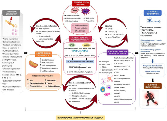
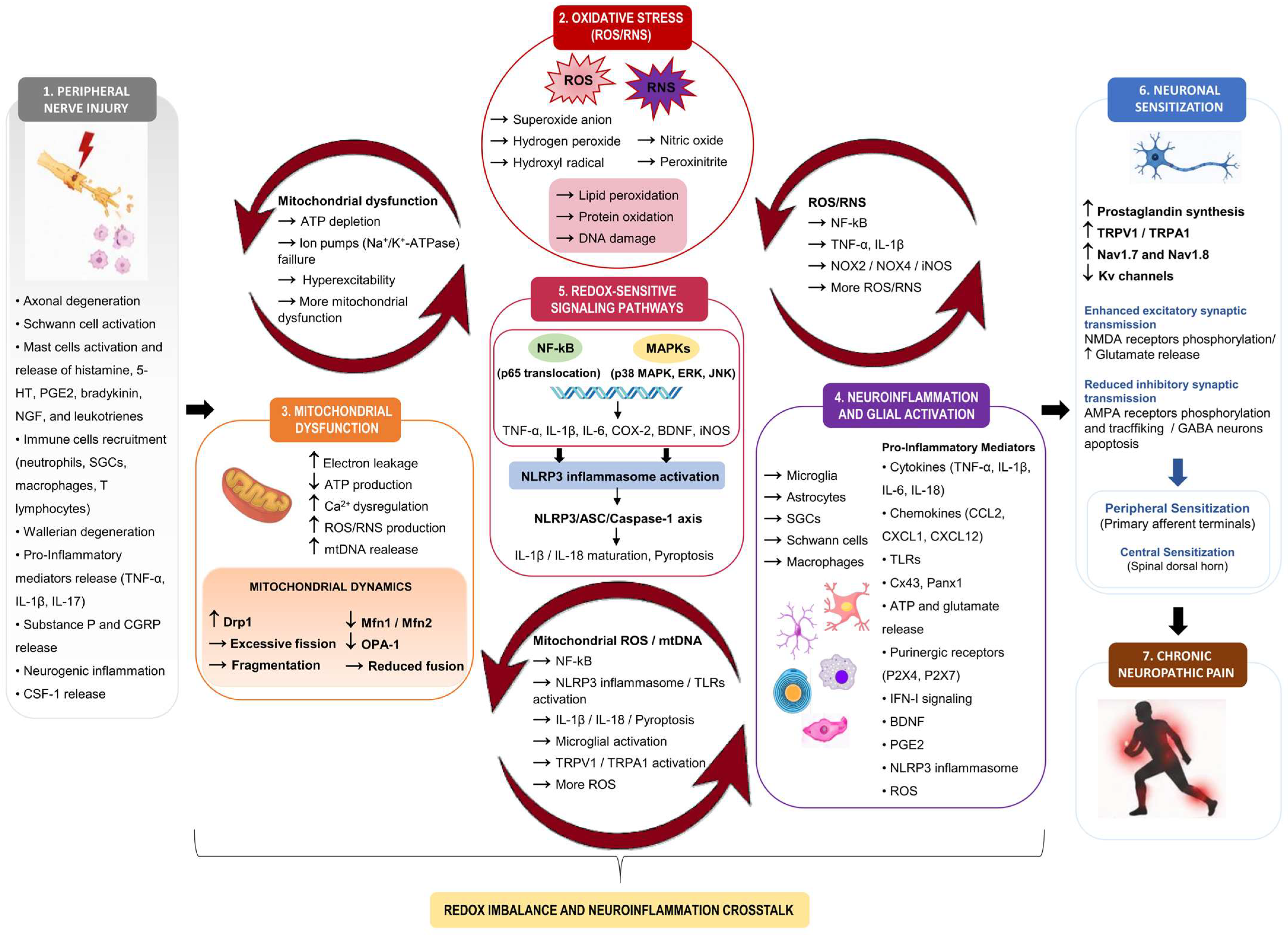

Disclaimer/Publisher’s Note: The statements, opinions and data contained in all publications are solely those of the individual author(s) and contributor(s) and not of MDPI and/or the editor(s). MDPI and/or the editor(s) disclaim responsibility for any injury to people or property resulting from any ideas, methods, instructions or products referred to in the content. |
© 2026 by the authors. Licensee MDPI, Basel, Switzerland. This article is an open access article distributed under the terms and conditions of the Creative Commons Attribution (CC BY) license.