


In the original publication [1], there was an error regarding the affiliation of the authors, Nagina Rehman and Farhat Jabeen. In addition to affiliations 1–3, the updated affiliations should include: Department of Zoology, Government College University Faisalabad, Faisalabad 38000, Pakistan.
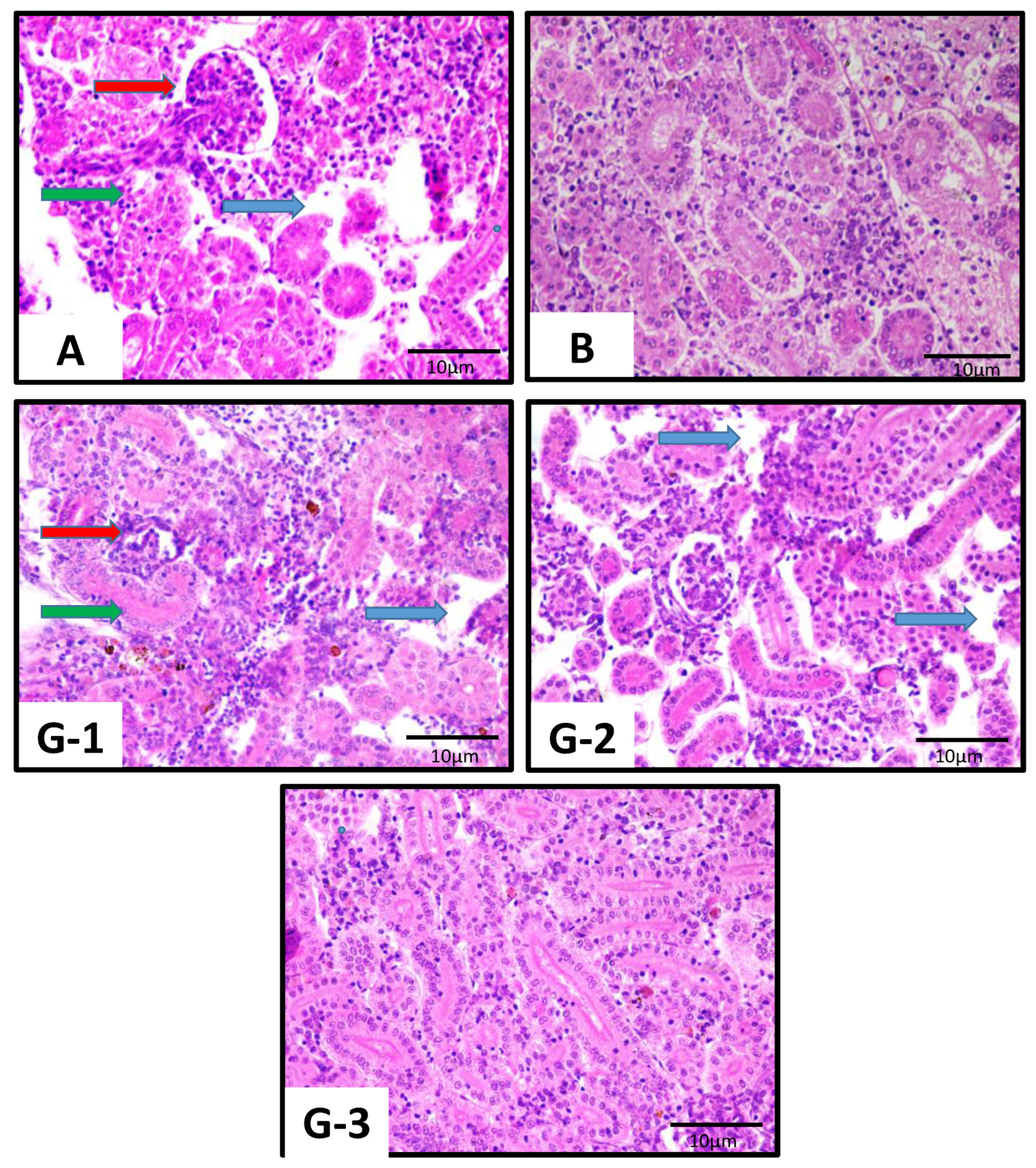
In the original publication [1], some image panels included in Figure 2 (2B, 2G-1, 2G-2), Figure 3 (3A), and Figure 4 (4G-1, 4G-2, 4G-3), showing photomicrographs (400×) of gills, liver, and kidney, respectively, of the common carp Cyprinus carpio exposed to copper oxide nanoparticles, were inadvertently duplicated during figure preparation. The corrected figures, in which all panels now represent distinct biological samples from their respective treatment groups, appear below.
The captions for Figures 2–4 have also been revised to clearly describe the pathological features and curative effects shown, with all arrow colors and annotations explained for consistency and interpretability. Figure 2B has been replaced with a photomicrograph taken at 10× magnification, matching the other panels, with the scale bar and caption updated accordingly. Moreover, the black circles previously surrounding the arrows have been removed from all images. In Figure 4B, the previously missing blue arrow has been added, and the image now accurately reflects all the histological findings, as clearly indicated by arrows whose colors have been clearly defined in the revised caption.
The authors also noted a methodological limitation regarding Figure 3, since vacuolization in the liver sections was assessed using H&E staining alone. Although the vacuolated hepatocytes showed cytoplasmic disintegration, nuclear pyknosis, and loss of normal cord arrangement, consistent with degeneration rather than physiological lipid or glycogen deposits, H&E staining alone cannot definitively distinguish between these possibilities. Accordingly, this limitation has now been addressed in the Discussion Section, immediately following the discussion of the histological findings in the carp gills, liver, and kidney. Correspondingly, the statement within the Figure 3 caption, which had previously appeared as an incomplete sentence, has also been revised to read as a complete and clear statement, so that the resulting interpretations remain appropriately cautious. The revised sentence in the Discussion Section now reads as follows:
The authors also acknowledge that vacuolization observed in liver sections (Figure 3) was interpreted based solely on hematoxylin and eosin (H&E) staining. Although the vacuolated hepatocytes exhibited cytoplasmic disintegration, nuclear pyknosis, and loss of normal cord arrangement, supporting their interpretation as degenerated cells rather than physiological lipid or glycogen deposits, it is recognized that H&E staining alone cannot definitively distinguish between these entities. This methodological limitation is noted, and all interpretations of vacuolization in this study remain cautious and evidence-based.
The authors state that the scientific conclusions are unaffected. This correction was approved by the Academic Editor. The original publication has also been updated.
Figure 2.
Photomicrographs (400×) of hematoxylin and eosin (H&E)-stained gill sections of the common carp (C. carpio) experimentally exposed for 28 days to CuO NPs (1.50 mg/L) as positive control (A; C-positive group), to no treatments as negative control (B; C-negative group), and to different concentrations of the fenugreek (T. foenum-graecum) extract at 100 mg/L (G-1), 125 mg/L (G-2), and 150 mg/L (G-3). Fish gills from the positive control showed various histological abnormalities including degenerative epithelium (green arrow), complete degeneration (red arrow), fused lamellae (blue arrow), and necrosis of primary lamella (yellow arrow). Fish from groups treated with the fenugreek showed dose-dependent curative effects on the gill epithelium. Scale bar = 10 µm. Histological observations were based on five randomly selected fields from one section per sample.
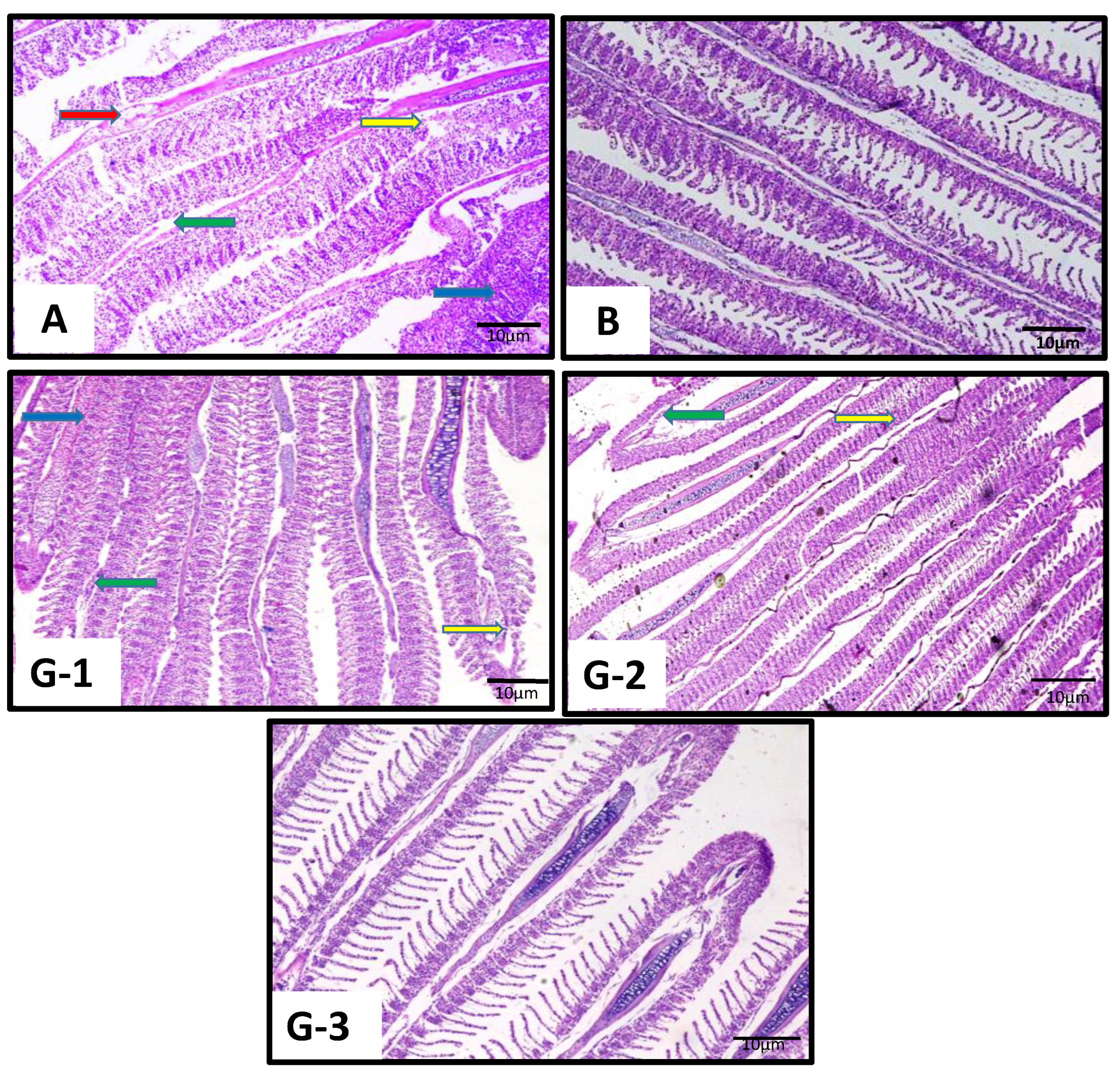
Figure 3.
Photomicrographs (400×) of hematoxylin and eosin (H&E)-stained liver sections of the common carp (C. carpio) experimentally exposed for 28 days to CuO NPs (1.50 mg/L) as positive control (A; C-positive group), to no treatments as negative control (B; C-negative group), and to different concentrations of the fenugreek (T. foenum-graecum) extract at 100 mg/L (G-1), 125 mg/L (G-2), and 150 mg/L (G-3). Fish livers from the positive control showed various histological aberrations including vacuolization (red arrow); note that vacuolization identified by H&E staining alone cannot be definitively distinguished from physiological lipid or glycogen deposits, degenerative hepatocytes (yellow arrow), dilated sinusoid (blue arrow), and complete degeneration (green arrow). Fish from groups treated with the fenugreek showed dose-dependent curative effects in their livers. Scale bar = 10 µm. Histological observations were based on five randomly selected fields from one section per sample.
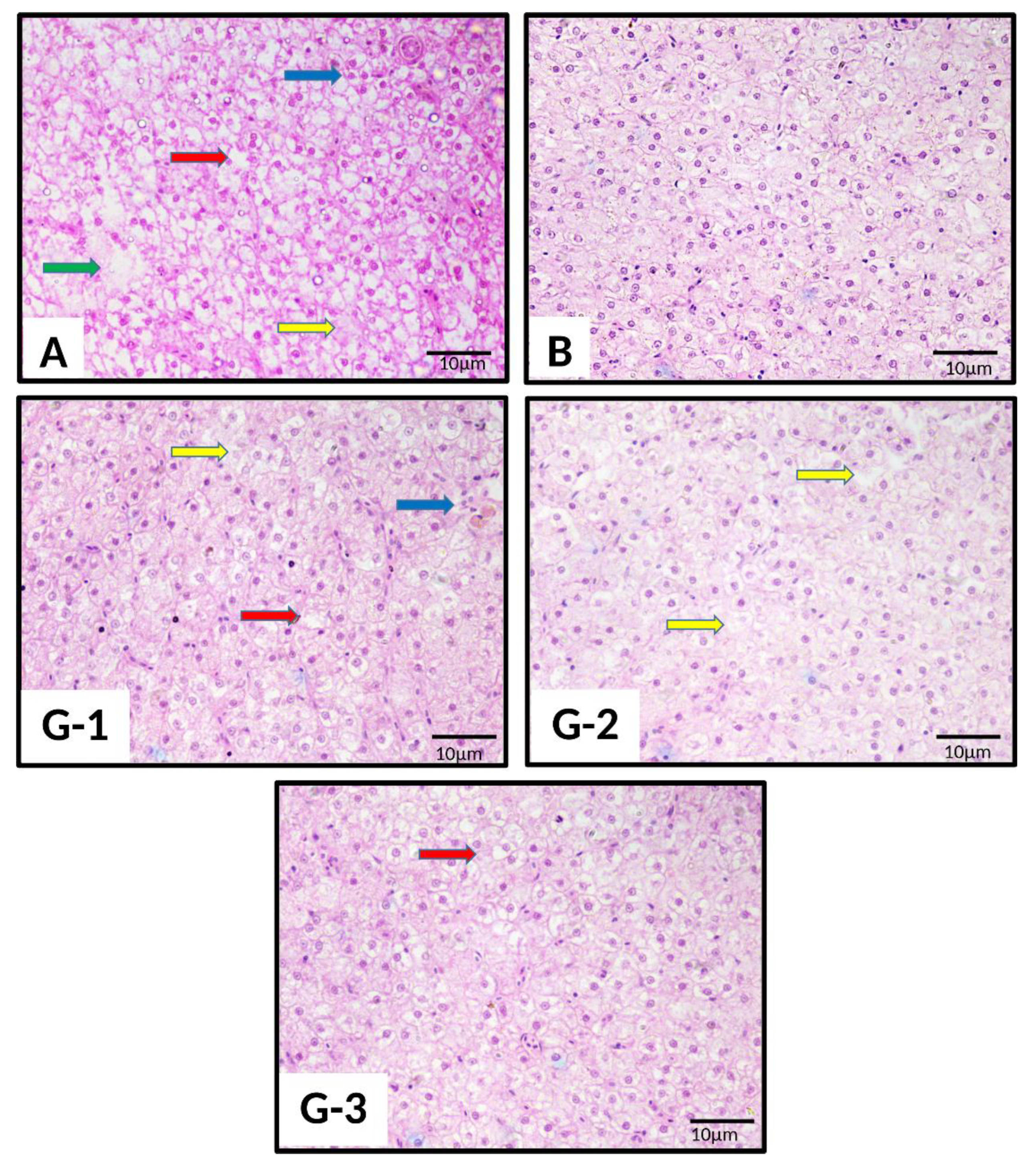
Figure 4.
Photomicrographs (400×) of hematoxylin and eosin (H&E)-stained kidney sections of the common carp (C. carpio) experimentally exposed for 28 days to CuO NPs (1.50 mg/L) as positive control (A; C-positive group), to no treatments as negative control (B; C-negative group), and to different concentrations of the fenugreek (T. foenum-graecum) extract at 100 mg/L (G-1), 125 mg/L (G-2), and 150 mg/L (G-3). Fish kidneys from the positive control showed various histological abnormalities including swollen tubules (green arrow), abnormal glomerulus (red arrow), and complete degeneration (blue arrow). Fish from groups treated with the fenugreek showed dose-dependent curative effects in their kidneys even if some tissue damages were observed. Scale bar = 10 µm. Histological observations were based on five randomly selected fields from one section per sample.
Reference
- Noureen, A.; De Marco, G.; Rehman, N.; Jabeen, F.; Cappello, T. Ameliorative Hematological and Histomorphological Effects of Dietary Trigonella foenum-graecum Seeds in Common Carp (Cyprinus carpio) Exposed to Copper Oxide Nanoparticles. Int. J. Environ. Res. Public Health 2022, 19, 13462. [Google Scholar] [CrossRef] [Scilit] [PubMed]
Disclaimer/Publisher’s Note: The statements, opinions and data contained in all publications are solely those of the individual author(s) and contributor(s) and not of MDPI and/or the editor(s). MDPI and/or the editor(s) disclaim responsibility for any injury to people or property resulting from any ideas, methods, instructions or products referred to in the content. |
© 2026 by the authors. Licensee MDPI, Basel, Switzerland. This article is an open access article distributed under the terms and conditions of the Creative Commons Attribution (CC BY) license.