


Abstract
Background and Objectives: This study aimed to compare the real-world outcomes of first-line pembrolizumab monotherapy (Mono-IO) versus pembrolizumab plus platinum-based chemotherapy (IO + ChT) in patients with metastatic non-small cell lung cancer (mNSCLC) and a PD-L1 tumor proportion score (TPS) ≥ 50%. Materials and Methods: This retrospective, multicenter, real-world study evaluated 148 patients with mNSCLC and PD-L1 expression ≥ 50% who received either first-line Mono-IO or IO + ChT. Overall survival (OS), progression-free survival (PFS), and clinical benefit rate were compared between the two treatment strategies. Additionally, clinical prognostic factors, patterns of progression, and post-progression treatments were analyzed. Survival outcomes were assessed using Kaplan–Meier estimates, multivariable Cox regression, and restricted mean survival time (RMST) analysis. Results: Patients in the Mono-IO group were significantly older (median 69 vs. 63 years; p < 0.001) and had a higher proportion of ECOG PS 2 (35.3% vs. 5.2%; p < 0.001). No significant differences were observed between Mono-IO and IO + ChT in median OS (32.0 vs. 26.0 months; p = 0.559) or PFS (19.6 vs. 19.4 months; p = 0.572). In an exploratory multivariable analysis adjusting for organ-specific metastases and antibiotic exposure, ECOG PS ≥ 1 (aHR: 1.86, 95% CI: 1.16–2.97; p = 0.009), antibiotic use (aHR: 1.85, 95% CI: 1.16–2.95; p = 0.010), and liver metastasis (aHR: 2.58, 95% CI: 1.16–5.74; p = 0.020) were independent predictors of worse OS. Conversely, PD-L1 TPS ≥ 90% (aHR: 0.56, 95% CI: 0.35–0.91; p = 0.018) was independently associated with superior OS. In exploratory subgroup analysis, a numerical survival trend favoring Mono-IO was observed in patients with PD-L1 TPS ≥ 90%, although this finding remains hypothesis-generating. Following disease progression, 64.7% of patients received no further systemic therapy. Conclusions: In this real-world cohort, no statistically significant differences in OS or PFS were detected between Mono-IO and IO + ChT; however, the wide confidence intervals do not exclude clinically meaningful differences, and equivalence cannot be inferred from these data. PD-L1 TPS ≥ 90% may potentially serve as a biomarker to identify patients who derive benefit from monotherapy, although this exploratory finding requires validation in larger cohorts. Given that the majority of patients did not transition to subsequent therapy in routine practice, likely reflecting multifactorial host frailty and rapid disease evolution, first-line therapeutic optimization warrants careful individualized consideration.
1. Introduction
Metastatic non-small cell lung cancer (mNSCLC) remains one of the leading causes of cancer-related mortality worldwide [1]. Over the past decade, the treatment landscape of advanced NSCLC has been fundamentally reshaped by immune checkpoint inhibitors (IO), particularly agents targeting the programmed death-1 (PD-1) and programmed death-ligand 1 (PD-L1) pathways [2]. In patients without actionable driver alterations and with high PD-L1 expression, defined as a tumor proportion score (TPS) ≥ 50%, pembrolizumab monotherapy (Mono-IO) has become a standard first-line option based on the survival benefit demonstrated over platinum-based chemotherapy in pivotal clinical trials such as KEYNOTE-024 and KEYNOTE-042 [3,4]. In parallel, pembrolizumab combined with platinum-based chemotherapy (IO + ChT) has also been established as an effective first-line strategy in this population [5].
Despite the availability of these two approved approaches, the optimal treatment strategy for patients with PD-L1-high mNSCLC remains uncertain in routine clinical practice [6]. A central unresolved question is whether the addition of chemotherapy to pembrolizumab provides a clinically meaningful survival advantage over monotherapy in patients already expected to benefit from IO on the basis of high PD-L1 expression. Indirect meta-analyses have suggested a possible benefit for IO + ChT in some settings [7], whereas pooled and retrospective analyses have produced less consistent results regarding differences in overall survival (OS) and progression-free survival (PFS) between the two strategies in patients with PD-L1 TPS ≥ 50% [8,9].
Real-world evidence further complicates this issue, as IO + ChT is more frequently selected for clinically aggressive patient populations, including younger individuals and those with symptomatic brain metastases. Despite these differences in treatment allocation, no substantial survival advantage has been consistently observed between the two treatment approaches in terms of OS [10]. Accordingly, direct comparison of Mono-IO and IO + ChT outside randomized settings is challenging, because outcomes may be influenced not only by treatment intensity but also by baseline clinical fitness and disease burden.
Another important issue is the heterogeneity within the broadly defined PD-L1-high population. Although TPS ≥ 50% is the accepted threshold for first-line pembrolizumab-based treatment, accumulating evidence suggests that patients with very high PD-L1 expression may represent a clinically distinct subgroup. In particular, long-term follow-up from KEYNOTE-024 confirmed the durability of benefit with pembrolizumab monotherapy [11], while chemo-immunotherapy trials have demonstrated the efficacy of combination approaches across PD-L1-defined subgroups [12]. At the same time, broader conceptual and clinical reviews have emphasized that treatment selection in metastatic NSCLC should be individualized according to multiple patient- and disease-related factors rather than PD-L1 level alone [13].
Within this PD-L1-high population, a prior real-world study explored whether very high PD-L1 expression, including TPS ≥ 90%, may identify a subgroup with distinct outcomes [6]. However, the clinical relevance of this threshold and its role in selecting between pembrolizumab monotherapy and chemo-immunotherapy remain uncertain. We therefore evaluated outcomes according to this threshold as an exploratory analysis.
In this context, real-world comparative data remain highly relevant. This multicenter study aimed to compare the clinical outcomes of first-line pembrolizumab monotherapy and pembrolizumab plus chemotherapy in patients with PD-L1 TPS ≥ 50% mNSCLC. In addition, we sought to identify clinical and biological predictors of survival, evaluate patterns of progression, and examine post-progression management in order to provide a more comprehensive perspective on treatment selection in routine practice.
2. Materials and Methods
2.1. Patient Population and Study Design
This retrospective, multicenter study was designed to evaluate real-world outcomes in patients with mNSCLC who received first-line pembrolizumab-based therapy across participating centers. Adult patients treated between January 2019 and March 2026 were included if they had a PD-L1 tumor proportion score (TPS) ≥ 50%, lacked EGFR mutations, ALK rearrangements, or other actionable driver alterations, and received either Mono-IO or IO + ChT as first-line treatment. Patients with incomplete clinical data or insufficient follow-up were excluded from the analysis.
2.2. Variables and Data Collection
Demographic and clinical characteristics, including age, sex, smoking history, ECOG PS, and histologic subtype, were systematically collected from electronic medical records. PD-L1 expression levels were recorded according to the TPS determined using validated immunohistochemical assays routinely employed in clinical practice, including 22C3 pharmDx (Agilent Technologies/Dako, Carpinteria, CA, USA), SP263 (Ventana Medical Systems/Roche Diagnostics, Tucson, AZ, USA), and SP142 (Ventana Medical Systems/Roche Diagnostics, Tucson, AZ, USA). In six patients (4.1%), the specific clone could not be identified from archival pathology reports. Although large harmonization studies, such as the Blueprint Phase 1 and 2 initiatives, have established high analytical concordance between 22C3 and SP263 at standard clinical thresholds, inter-assay variability at extreme cutoffs such as TPS ≥ 90% has not been fully characterized.
PD-L1 expression was evaluated both as a continuous variable (per 1% increment) and as a dichotomous variable using a pre-specified threshold of ≥90%. This cutoff was selected a priori based on published literature defining an ultra-high PD-L1 subset that exhibits enhanced sensitivity to immune checkpoint inhibition and was not derived through data-driven cutpoint optimization. Given the limited sample size of this subgroup, all analyses stratified by the ≥90% cutoff are strictly exploratory and hypothesis-generating.
Systemic antibiotic (ATB) exposure was defined as the administration of at least one course of oral or intravenous antibiotics within a periprocedural window from 30 days prior to the first dose of pembrolizumab through 30 days after treatment initiation, or during active first-line therapy. Topical antibiotics were excluded. In the survival models, ATB exposure was analyzed as a binary fixed covariate (yes vs. no). Where available from hospital electronic prescription systems, granular data regarding antibiotic class, clinical indication, and duration were also retrieved. In addition, the presence of organ-specific metastatic sites, including liver, bone, brain, and other metastatic locations identified at baseline or during follow-up, was recorded.
Data are presented as median (IQR) for continuous variables and n (%) for categorical variables, as appropriate. Mono-IO refers to patients who received first-line IO without chemotherapy, whereas IO + ChT refers to patients who received first-line IO in combination with chemotherapy. Comparisons between treatment groups were performed using the Mann–Whitney U test for continuous variables and the chi-square test or Fisher’s exact test, as appropriate, for categorical variables. Percentages were calculated using the number of patients with available data for each variable; therefore, denominators may vary because of missing data.
2.3. Treatment Follow-Up and Study Endpoints
The primary study endpoints were OS, defined as the time from treatment initiation to death from any cause, and PFS, defined as the time from treatment initiation to radiological progression or death, whichever occurred first.
Tumor response and disease progression were evaluated in accordance with Response Evaluation Criteria in Solid Tumors (RECIST version 1.1). Cross-sectional imaging, consisting of contrast-enhanced chest and abdominopelvic CT, brain MRI, or 18F-FDG PET/CT, was routinely obtained every 8 to 12 weeks during active treatment, or earlier if disease progression was clinically suspected. All imaging studies were interpreted by dedicated board-certified subspecialty radiologists (thoracic, abdominal, and neuroradiologists) as part of standard care. In cases of equivocal, dissociated, or atypical radiological progression, cases were reviewed and adjudicated during multidisciplinary Thoracic Oncology Tumor Board meetings.
Clinical benefit rate (CBR) was defined as the proportion of patients achieving complete response (CR), partial response (PR), or stable disease (SD) as their best overall response. All 148 patients had evaluable follow-up imaging for best response determination. Secondary endpoints also included reasons for treatment discontinuation due to toxicity or disease progression, post-progression management strategies, and patterns of disease progression categorized as either widespread metastatic progression or organ-specific progression.
2.4. Statistical Analysis
Survival outcomes were estimated using the Kaplan–Meier method, and median survival times were calculated. Overall survival (OS) was defined as the time from initiation of first-line treatment to death from any cause or last follow-up. Progression-free survival (PFS) was defined as the time from initiation of first-line treatment to the first documented disease progression or death from any cause, whichever occurred first. Differences in survival between groups were assessed using the log-rank test.
Multivariable Cox proportional hazards regression models were used to evaluate the association between treatment strategy and survival outcomes and to estimate adjusted hazard ratios (aHRs). These models were adjusted for age, sex, ECOG PS, histology, and PD-L1 expression level (TPS ≥ 90% vs. <90%). In these analyses, an aHR > 1.0 indicated an increased risk of the event in the IO + ChT group relative to the Mono-IO group. Separate univariable and multivariable Cox proportional hazards regression analyses were performed to identify factors associated with PFS, including treatment strategy, ECOG PS, histology, PD-L1 specimen site, PD-L1 TPS (per 1% increment), and ATB use. In univariable analysis, ECOG PS was evaluated as ECOG PS 2 versus 0–1; in multivariable models evaluating treatment strategy, ECOG PS 1 and 2 were combined as ECOG PS ≥ 1 to ensure model stability due to the limited number of patients with ECOG PS 2 (n = 23, 15.5%).
The proportional hazards assumption for covariates included in the multivariable Cox proportional hazards models was formally tested using Schoenfeld residual analysis against log-transformed survival time, and no statistically significant violation was detected (all p > 0.05). However, recognizing that formal goodness-of-fit tests may have limited statistical power to detect late departures from proportionality in cohorts of moderate size, and to complement the hazard ratio with an intuitively meaningful metric that does not rely on the proportional hazards assumption, Restricted Mean Survival Time (RMST) analysis was performed as a sensitivity analysis. The truncation time (τ) for the overall cohort was set at 48 months, representing the long-term follow-up horizon where patients remained actively at risk in both treatment arms without projecting into the sparse tail of the survival curve. For the exploratory PD-L1 TPS ≥ 90% subgroup, a truncation time of 36 months was chosen to ensure adequate numbers of patients at risk given the smaller sample size.
A sensitivity analysis restricted to patients evaluated with the 22C3 assay (n = 92) was additionally performed to assess the robustness of the primary findings against assay heterogeneity. To address potential residual confounding arising from baseline imbalances between treatment groups, an Inverse Probability of Treatment Weighting (IPTW) analysis was performed as a pre-specified sensitivity analysis. Propensity scores were estimated using a multivariable logistic regression model incorporating baseline covariates (age, sex, smoking status, ECOG PS, histology, PD-L1 TPS, PD-L1 TPS ≥ 90%, and brain, liver, and bone metastases). Stabilized weights trimmed at the 99th percentile were applied, and balance was evaluated using standardized mean differences (SMDs < 0.10 indicative of adequate balance).
A two-sided p-value < 0.05 was considered statistically significant. Statistical analyses were performed using SPSS version 26.0 (IBM Corp., Armonk, NY, USA).
2.5. Ethical Considerations
The study protocol was approved by the Clinical Research Ethics Committee of Mehmet Ali Aydinlar Acibadem University (Approval Date: 2023; Approval Number: 2023-07-243). The study was conducted in accordance with the principles of the Declaration of Helsinki and Good Clinical Practice guidelines. Due to the retrospective and observational nature of the study, the requirement for informed consent was waived, as all data were analyzed anonymously.
3. Results
3.1. Patient Characteristics
A total of 148 patients with mNSCLC and a PD-L1 TPS of ≥50% who received first-line pembrolizumab-based therapy were included in the study. Of these, 51 patients (34.5%) received Mono-IO and 97 patients (65.5%) received pembrolizumab in combination with IO + ChT. Baseline demographic and clinical characteristics are summarized in Table 1.
Table 1.
Baseline demographic and clinical characteristics of the study population according to first-line treatment strategy (IO + ChT vs. Mono-IO).
The overall cohort had a median age of 65 years (IQR 59–71), and the majority of patients were male (73.6%) and had a history of smoking (83.1%). Adenocarcinoma was the predominant histological subtype (74.3%). PD-L1 TPS ≥ 90% was present in 41.2% of patients. The 22C3 assay was the most frequently utilized platform (62.2%, n = 92), followed by SP263 (31.8%, n = 47) and SP142 (2.0%, n = 3), while the assay type was unspecified in six patients (4.1%) due to unavailable institutional pathology archival records. Assay distribution did not differ significantly between the Mono-IO and IO + ChT cohorts (p = 0.299; Table 1). The primary biopsy site for PD-L1 assessment was the lung (66.2%).
Comparison of baseline characteristics between treatment groups revealed significant differences in age and ECOG PS (Table 1). Patients in the Mono-IO group were significantly older than those in the IO + ChT group (median 69 vs. 63 years; p < 0.001) and had a higher proportion of ECOG PS 2 (35.3% vs. 5.2%; p < 0.001). Although squamous cell carcinoma was numerically more frequent in the Mono-IO group (33.3% vs. 15.5%), the overall histological distribution did not differ significantly between groups (p = 0.068). No significant differences were observed in sex, smoking history, PD-L1 assay type, PD-L1 specimen site, or the proportion of patients with PD-L1 TPS ≥ 90%.
3.2. Treatment Exposure and Clinical Response
First-line treatment exposure and intensity characteristics are summarized in Table 2. In the IO + ChT group, the median number of chemotherapy cycles administered was 4 (IQR 4–6). The number of IO administrations was identical between treatment groups, with a median of 12 administrations (Mono-IO: 12 [IQR 6–27] vs. IO + ChT: 12 [IQR 6–22]; p = 0.799). Overall, 29 patients (19.6%) received IO for at least 2 years, with no significant difference between the Mono-IO and IO + ChT groups (25.5% vs. 16.5%, p = 0.190).
Table 2.
Treatment Characteristics and Clinical Outcomes According to First-Line Treatment Strategy.
Disease progression was the most frequent reason for IO discontinuation, occurring in 40 patients (27.0%) overall (Mono-IO: 19.6%; IO + ChT: 30.9%). Toxicity and intolerance led to IO discontinuation in 13.5% and 2.7% of patients, respectively. At the time of analysis, 51 patients (34.5%) remained on active IO (Table 2).
3.3. Survival Outcomes
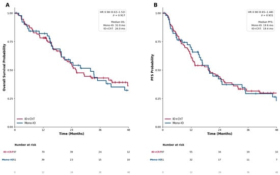
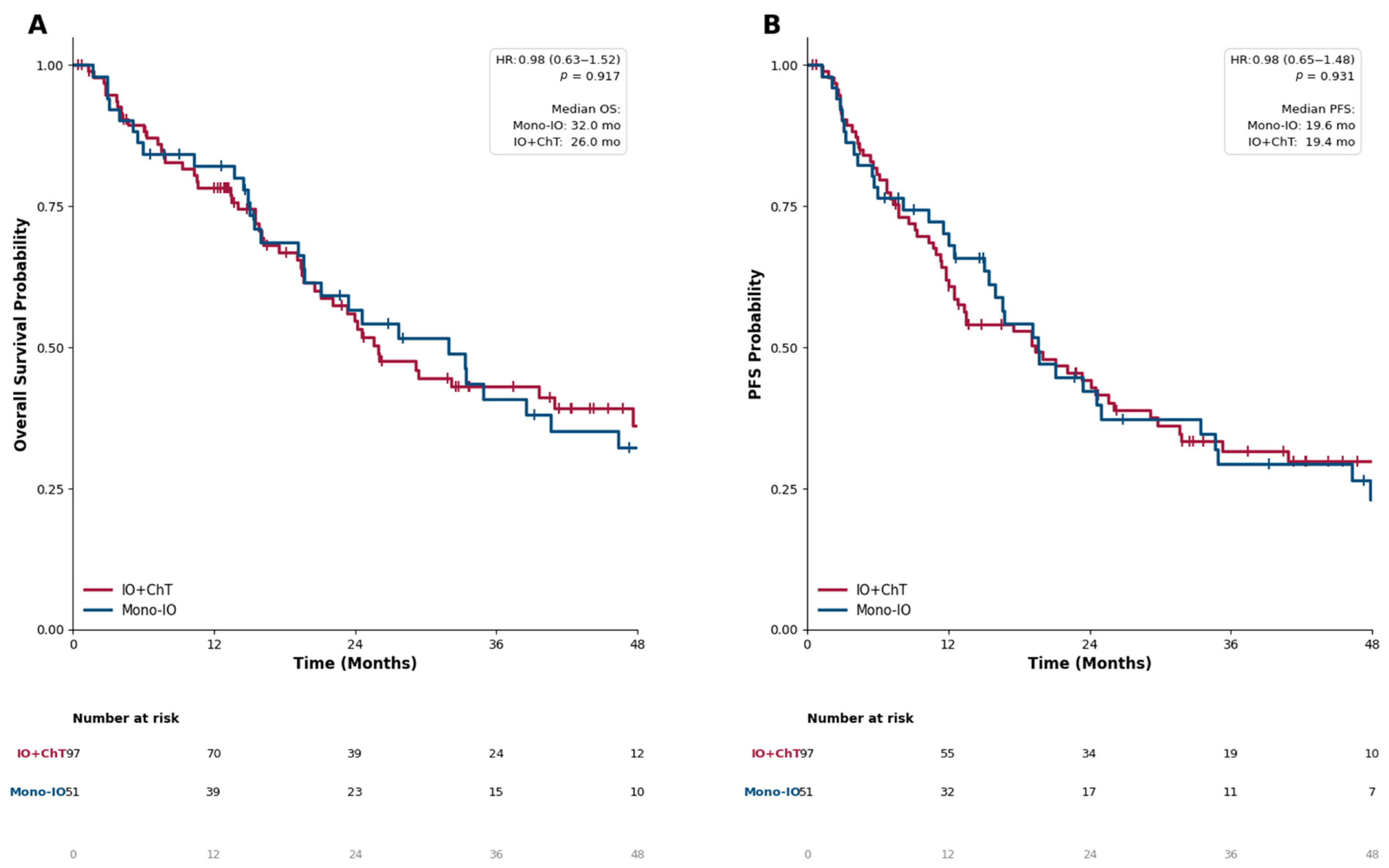
Survival outcomes are summarized in Table 3 and illustrated in Figure 1. Median OS was 32.0 months in the Mono-IO group and 26.0 months in the IO + ChT group, with no significant difference between treatment strategies (unadjusted HR 1.05, 95% CI 0.68–1.62; adjusted HR 1.16, 95% CI 0.71–1.87; p = 0.559). Median PFS was likewise similar between the two groups (19.6 vs. 19.4 months; unadjusted HR 1.04, 95% CI 0.69–1.57; adjusted HR 1.14, 95% CI 0.73–1.79; p = 0.572). Clinical benefit rates were also comparable (68.6% vs. 63.9%, p = 0.696). Of note, the confidence intervals around the adjusted hazard ratios were wide and do not exclude clinically meaningful differences between the treatment strategies.
Table 3.
Survival outcomes and Cox regression analysis (n = 148).
Figure 1.
Kaplan–Meier survival estimates according to treatment strategy. (A) Overall survival (OS) and (B) progression-free survival (PFS) in patients treated with Mono-IO (blue) or IO + ChT (red). Tick marks indicate censored observations. Median survival times, hazard ratios, and log-rank p-values are shown within the plots. At 48 months, 9 and 6 patients remained at risk for OS, and 7 and 4 for PFS, in the Mono-IO and IO + ChT groups, respectively.
Visual inspection of the Kaplan–Meier curves showed extensive overlap between treatment groups throughout follow-up, with a late visual intersection after approximately 30 months (Figure 1). Although formal Schoenfeld residual tests did not indicate a statistically significant violation of the proportional hazards assumption (p > 0.05), RMST analysis was conducted as a complementary, model-free evaluation of survival differences up to 48 months. At this time point, where patients remained actively under observation, the RMST for OS was 29.32 months in the Mono-IO group and 29.06 months in the IO + ChT group (ΔRMST: 0.26 months, 95% CI: −6.10 to 6.27; p = 0.933). Similarly, the RMST for PFS at 48 months was 24.31 months and 24.09 months, respectively (ΔRMST: 0.22 months, 95% CI: −5.82 to 6.50; p = 0.946), corroborating the Cox regression findings.
Figure 1 illustrates the survival outcomes stratified by treatment strategy. Neither OS (Figure 1A; log-rank p = 0.917) nor PFS (Figure 1B; log-rank p = 0.931) differed significantly between the Mono-IO and IO + ChT groups. The survival curves largely overlapped throughout follow-up and exhibited a late crossing pattern after approximately 30 months, suggesting a potential violation of the proportional hazards assumption. Therefore, restricted mean survival time (RMST) analysis was additionally performed. At 48 months, the RMST for OS was 29.32 months in the Mono-IO group and 29.06 months in the IO + ChT group (ΔRMST 0.26 months, 95% CI −6.10 to 6.27; p = 0.933). Similarly, the RMST for PFS was 24.31 months and 24.09 months, respectively (ΔRMST 0.22 months, 95% CI −5.82 to 6.50; p = 0.946); no statistically significant differences were observed. At the 48-month landmark, 9 patients (17.6%) in the Mono-IO group and 6 patients (6.2%) in the IO + ChT group remained at risk for OS, while 7 (13.7%) and 4 (4.1%) patients, respectively, remained at risk for PFS.
3.4. Predictors of Progression-Free Survival
In the univariate Cox regression analysis for PFS, ECOG PS 2, other/NOS histology, and ATB use during treatment were associated with significantly shorter PFS. In contrast, treatment strategy (Mono-IO vs. IO + ChT), PD-L1 specimen site, and PD-L1 TPS did not reach statistical significance in the univariate model.
In the multivariable analysis, ECOG PS 2 remained independently associated with worse PFS (aHR 2.93, 95% CI 1.72–4.96; p < 0.001), as did other/NOS histology (aHR 4.07, 95% CI 1.57–10.56; p = 0.004). Higher PD-L1 TPS was independently associated with a lower risk of progression or death (aHR 0.99 per 1% increase, 95% CI 0.97–1.00; p = 0.041). Treatment strategy was not independently associated with PFS (aHR 1.17, 95% CI 0.75–1.82; p = 0.491). Although ATB use was significantly associated with shorter PFS in the univariate analysis, its effect was attenuated after adjustment and remained of borderline significance (aHR 1.50, 95% CI 1.00–2.27; p = 0.052). Full results are presented in Table 4.
Table 4.
Univariate and Multivariable Cox Regression Analysis of Factors Associated with PFS.
3.5. Independent Predictors of Overall and Progression-Free Survival
A multivariable Cox regression model was constructed to evaluate independent prognostic factors for OS and PFS (Table 5). In this model, treatment strategy was not independently associated with either OS (aHR 1.16, 95% CI 0.71–1.87; p = 0.559) or PFS (aHR 1.14, 95% CI 0.73–1.79; p = 0.572). ECOG PS ≥ 1 was independently associated with worse OS (aHR 1.86, 95% CI 1.16–2.97; p = 0.009). Notably, a PD-L1 TPS threshold of ≥90% was identified as an independent favorable prognostic factor for both OS (aHR 0.56, 95% CI 0.35–0.91; p = 0.018) and PFS (aHR 0.60, 95% CI 0.39–0.93; p = 0.022). When analyzed as a continuous covariate in multivariable Cox regression, each 1% increase in PD-L1 TPS was independently associated with a decrease in the risk of death (aHR: 0.99, 95% CI: 0.98–1.00, p = 0.038) and disease progression or death (aHR: 0.99, 95% CI: 0.97–1.00, p = 0.041), supporting a continuous biological gradient of benefit across expression levels.
Table 5.
Multivariable Cox Regression Analysis for Overall Survival and Progression-Free Survival.
3.6. Exploratory Multivariable Analysis Including Metastatic Sites and Antibiotic Use
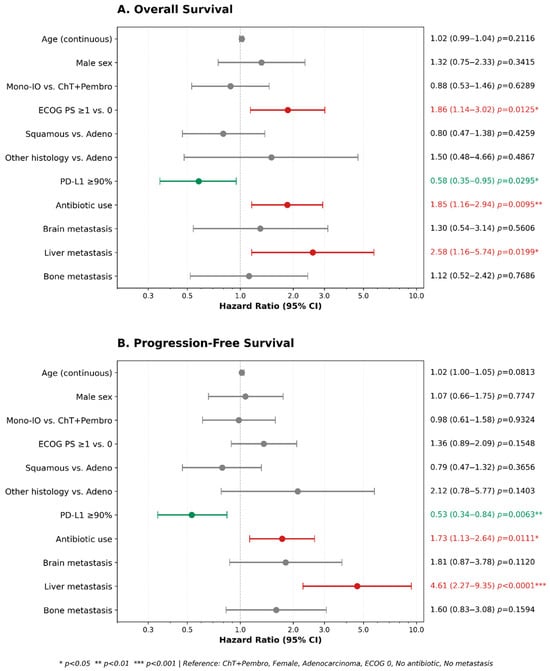
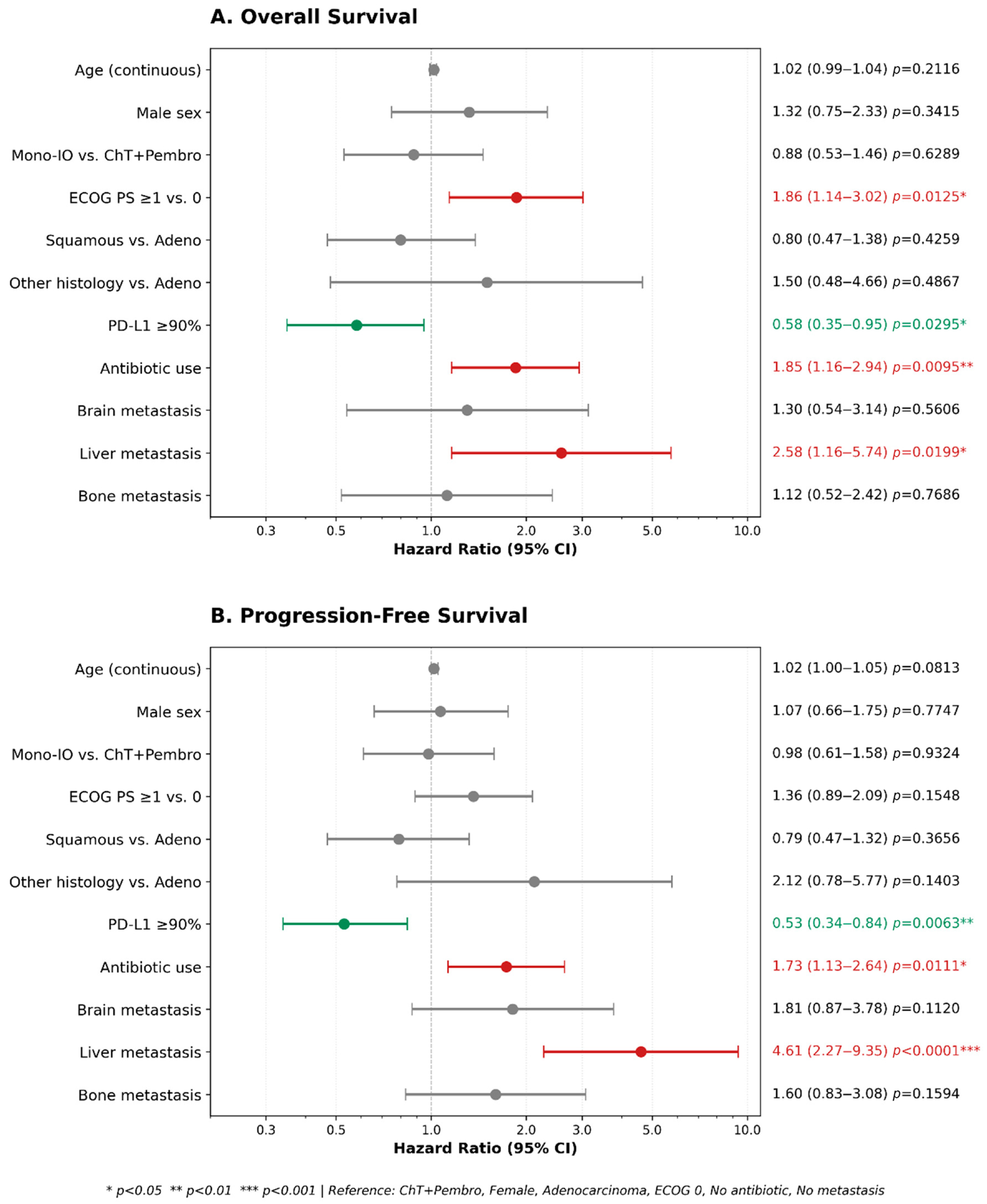
To explore the prognostic impact of disease dissemination patterns and concurrent supportive exposures, an expanded exploratory multivariable model incorporating organ-specific metastatic sites and systemic ATB exposure was evaluated (Figure 2). In this model, ECOG PS ≥ 1 (aHR: 1.86, 95% CI: 1.16–2.97; p = 0.009), ATB exposure (aHR: 1.85, 95% CI: 1.16–2.95; p = 0.010), and the presence of liver metastases (aHR: 2.58, 95% CI: 1.16–5.74; p = 0.020) were independently associated with inferior OS. PD-L1 expression ≥ 90% remained an independent favorable prognostic factor for OS (aHR: 0.56, 95% CI: 0.35–0.91; p = 0.018).
Figure 2.
Multivariable Cox proportional hazards regression analysis for Overall Survival (A) and Progression-Free Survival (B). Hazard ratios (HR) with 95% confidence intervals (CI) are presented on a logarithmic scale. Red dots indicate variables independently associated with increased risk of death or progression (p < 0.05); green dots indicate independently protective factors (p < 0.05); grey dots indicate non-significant associations (p ≥ 0.05). Reference categories: chemotherapy plus pembrolizumab (for Mono-IO), female sex, adenocarcinoma histology, ECOG performance status 0, no antibiotic use, and absence of organ-specific metastasis. Age was included as a continuous variable. Significance levels: * p < 0.05, ** p < 0.01, *** p < 0.001. Abbreviations: HR, hazard ratio; CI, confidence interval; ECOG PS, Eastern Cooperative Oncology Group performance status; PD-L1, programmed death-ligand 1; ChT, chemotherapy; Mono-IO, pembrolizumab monotherapy.
In the corresponding expanded PFS model, liver metastasis was associated with an increased risk of disease progression or death (aHR: 4.61, 95% CI: 2.14–9.92; p < 0.001). ATB use was also independently associated with shorter PFS (aHR: 1.73, 95% CI: 1.13–2.65; p = 0.011), while high PD-L1 expression (≥90%) retained its protective association (aHR: 0.53, 95% CI: 0.34–0.83; p = 0.006).
Overall, systemic ATB exposure within the defined treatment window was documented in 48 patients (32.4%). Among patients with detailed prescription records available (n = 33), beta-lactams and beta-lactamase inhibitor combinations were the most prevalent class (57.6%), followed by fluoroquinolones (24.2%) and macrolides (12.1%). The primary indication was lower respiratory tract infection (72.7%), followed by urinary tract infection (15.2%). The median duration of antibiotic administration was 10 days (IQR, 7–14 days).
3.7. Outcomes in PD-L1 TPS ≥ 90% Subgroup
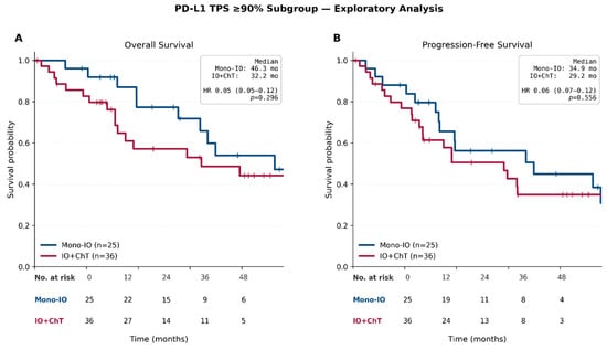
In an exploratory analysis of patients with very high PD-L1 expression (TPS ≥ 90%, n = 61), Mono-IO (n = 25) demonstrated numerically longer survival compared to IO + ChT (n = 36) for both OS and PFS (Figure 3). The median OS was 46.3 months (95% CI: 27.7-NR) in the Mono-IO group and 32.2 months (95% CI: 15.5-NR) in the IO + ChT group (HR: 0.67, 95% CI: 0.31–1.44; p = 0.293).
Figure 3.
Kaplan–Meier estimates of overall survival and progression-free survival in the PD-L1 TPS ≥ 90% subgroup. (A) Overall survival (OS) and (B) progression-free survival (PFS) in patients with a PD-L1 tumor proportion score (TPS) ≥ 90% who received first-line pembrolizumab-based therapy (n = 61). Patients were stratified by treatment strategy: Mono-immunotherapy (Mono-IO, n = 25) and Chemo-immunotherapy (IO + ChT, n = 36). PFS was defined as the time from treatment initiation to first documented disease progression or death from any cause; patients without an event were censored at the date of last follow-up. For deceased patients without a specific date of death recorded, the last follow-up date was utilized as a proxy for death. Tick marks denote censored observations. Hazard ratios (HR) were calculated using Cox proportional hazards models, and p-values were derived from log-rank tests. Shaded areas represent 95% confidence intervals. At the 36-month landmark, 7 patients (28.0%) in the Mono-IO group and 9 patients (25.0%) in the IO + ChT group remained at risk for OS; the corresponding numbers for PFS were 6 (24.0%) and 6 (16.7%), respectively.
Regarding PFS in this subgroup, the median was 34.9 months (95% CI: 16.8-NR) with Mono-IO versus 29.2 months (95% CI: 14.1-NR) with IO + ChT (HR: 0.82, 95% CI: 0.43–1.57; p = 0.546). RMST analysis at 36 months showed an absolute survival difference of 5.35 months for OS (95% CI: −3.81 to 14.51; p = 0.252) and 3.18 months for PFS (95% CI: −4.92 to 11.28; p = 0.441) in favor of the Mono-IO group.
3.8. Patterns of Progression and Post-Progression Management
Among the 65 patients who experienced disease progression, widespread metastasis was the most frequent pattern in both treatment groups (28.6% for Mono-IO vs. 38.6% for IO + ChT) (Table 6). Lung progression occurred in 47.6% and 25.0% of patients, respectively. Brain-only progression was identified exclusively in the IO + ChT group (9.1%). Detailed progression patterns according to treatment strategy are presented in Table 6. Exploratory subgroup analyses did not reveal significant interactions between treatment modality and clinical variables for either survival endpoint (all interaction p > 0.05).
Table 6.
Pattern of progression according to treatment group.
Among the 148 patients treated with first-line pembrolizumab, disease progression or death occurred in 102 patients (69.0%). Following progression, the majority of patients (64.7%) did not receive any subsequent systemic therapy. Among those who received further treatment, chemotherapy was administered more frequently than immunotherapy (31.4% vs. 12.7%, respectively).
The most common reason for discontinuation of first-line pembrolizumab was disease progression (39.2%). Notably, patients who remained on pembrolizumab despite progression accounted for 22.5% of the progression subgroup (15.5% of the overall cohort). Treatment discontinuation due to adverse events occurred in 14.7% of patients (10.1% of the overall cohort), whereas discontinuation for financial reasons was observed in 9.8% (6.8% of the overall cohort) (Table 7).
Table 7.
Treatment Discontinuation Patterns and Post-Progression Management in the Pembrolizumab Cohort.
4. Discussion
In this multicenter real-world study, no significant differences in OS or PFS were observed between first-line Mono-IO and IO + ChT in patients with PD-L1 TPS ≥ 50% mNSCLC. The absence of statistically significant differences in OS and PFS, supported by RMST analyses, suggests that treatment intensification alone may not necessarily translate into superior outcomes in this population. However, given the wide confidence intervals and the non-randomized design, these findings should not be interpreted as evidence of equivalence between the two strategies. Conversely, poor ECOG PS, liver metastasis, and ATB exposure were identified as adverse prognostic factors, whereas PD-L1 TPS ≥ 90% emerged as a favorable prognostic marker. Collectively, these findings suggest that clinical and biological patient selection may be more important determinants of outcome than treatment intensity alone.
4.1. Consistency with Previous Literature
Randomized trials established pembrolizumab monotherapy as a standard first-line option for patients with PD-L1 TPS ≥ 50%, with KEYNOTE-024 and KEYNOTE-042 demonstrating survival benefits over chemotherapy [3,4]. Long-term follow-up of KEYNOTE-024 confirmed the durability of this benefit despite substantial crossover from the chemotherapy arm [11]. Although indirect comparisons and retrospective studies have suggested that IO + ChT may provide an advantage in selected patients, real-world studies consistently show that combination regimens are more frequently used in younger patients and those with clinically aggressive disease, which complicates direct comparisons between treatment strategies [5,10,12]. Our findings are consistent with these observations, as Mono-IO achieved comparable outcomes despite being preferentially administered to older patients and those with poorer ECOG PS.
4.2. The Significance of the PD-L1 TPS ≥ 90% Subgroup
While PD-L1 TPS ≥ 50% is the established threshold for first-line pembrolizumab-based treatment in mNSCLC, this broad population remains biologically and clinically heterogeneous. Recent studies suggest that patients with very high PD-L1 expression (TPS ≥ 90%) may represent a distinct subgroup with a more favorable prognostic profile than those with TPS 50–89% [13]. Consistent with this concept, Mildanoglu et al. reported significantly superior OS and PFS among patients with TPS ≥ 90% and identified this threshold as an independent prognostic factor for OS [6]. In our study, PD-L1 TPS ≥ 90% was independently associated with improved OS and PFS. Furthermore, within the TPS ≥ 90% subgroup, Mono-IO was associated with numerically longer OS and PFS than IO + ChT.
While our continuous analyses confirm that PD-L1 operates as a quantitative biological spectrum where each incremental increase in TPS confers favorable survival, the dichotomous ≥ 90% cutoff was pre-specified to align with previous landmark studies investigating “ultra-high” expressors. However, whether clinical benefit exhibits an abrupt threshold or a gradual continuum remains a subject of investigation. Because our sample size in this ultra-high stratum was modest (n = 61), these findings should be regarded purely as hypothesis-generating, and caution is warranted against over-interpreting this threshold until prospective trials or pooled individual patient data analyses provide definitive confirmation.
An important methodological aspect regarding PD-L1 evaluation in our real-world cohort is the use of different diagnostic clones. Notably, 94.0% of our patients were evaluated using either 22C3 or SP263, assays known to exhibit high analytical concordance and interchangeable scoring at the standard TPS ≥ 50% threshold based on the Blueprint Phase 2 trial. However, clinical concordance at ultra-high levels (TPS ≥ 90%) has not been definitively demonstrated across platforms, and minor staining intensity variations might influence categorization around this high cutoff.
4.3. Antibiotics and the Microbiome
The relationship between IO efficacy and the gut microbiota has received increasing attention in mNSCLC. Our finding that ATB exposure was associated with inferior OS and PFS aligns with extensive literature and recent meta-analyses demonstrating that antibiotic-induced gut dysbiosis impairs anti-tumor immune priming [14,15].
However, attributing causality from real-world observational data requires extreme caution. In oncology, antibiotic use often correlates with underlying clinical deterioration, severe comorbidities, and infections, making it difficult to separate the drug’s biological effect from confounding related to patient frailty. Furthermore, because antibiotics administered after treatment initiation were modeled as a fixed rather than a time-dependent covariate, the possibility of immortal time bias cannot be entirely excluded, even though the majority of exposures occurred peri-treatment initiation. Consequently, our findings should not be interpreted as definitive mechanistic proof, but rather as supporting the practice of prudent antibiotic stewardship during immune checkpoint inhibitor therapy. Future prospective studies incorporating detailed information on ATB indication, class, timing, duration, and microbiome composition are needed to clarify the biological and clinical relevance of this association.
4.4. Performance Status and Metastatic Sites
Poor ECOG PS was an important adverse prognostic factor in our cohort, consistent with previous reports of substantially shorter survival among patients with ECOG PS ≥ 2 receiving IO-based treatment [16]. Together with liver metastasis, poor ECOG PS may identify a clinically high-risk phenotype characterized by limited clinical fitness and potential resistance to IO-based therapies.
Lung and intrathoracic lymph node metastases were not independently associated with worse OS or PFS. This finding is compatible with the hypothesis that thoracic sites may be relatively immune-responsive because of higher PD-L1 expression and tumor-infiltrating lymphocyte density [17]. Similarly, bone, brain, pleural, and adrenal metastases were not independently associated with survival, in line with previous findings that brain and bone metastases may not retain independent prognostic relevance after adjustment, whereas abdominal organ involvement, particularly liver metastasis, is associated with worse outcomes [18]. In our cohort, liver metastasis was independently associated with inferior OS and PFS, consistent with evidence that liver involvement may promote systemic immunosuppression and attenuate IO efficacy [19].
4.5. Histology and Progression Patterns
Although differential IO responsiveness between squamous cell carcinoma and adenocarcinoma has been suggested [20], we observed no significant differences in OS or PFS between these subtypes. This is consistent with meta-analytic evidence and subgroup data from KEYNOTE-024 and KEYNOTE-042 indicating broadly comparable outcomes across major histologic categories in PD-L1-high disease [4,21]. Thus, PD-L1 expression and clinical risk factors may be more prognostically relevant than histology alone in this population.
Other/NOS histology was independently associated with worse PFS (aHR 4.07, 95% CI 1.57–10.56; p = 0.004). This result may reflect poorly differentiated or biologically heterogeneous tumors, but should be interpreted cautiously because of the small subgroup size.
Lung-limited progression was numerically more frequent with Mono-IO, whereas brain-only progression occurred exclusively with IO + ChT. Although these differences were not statistically significant, they are compatible with reported organ-specific and oligoprogressive patterns during PD-(L)1 inhibitor treatment [22,23], possibly reflecting site-specific mechanisms of immune resistance.
4.6. Post-Progression Management
Rheinheimer et al. reported that oligoprogression is more prevalent under Mono-IO compared to IO + ChT (20% vs. 10%), with the lymph nodes (42%) and lungs (24%) being the most frequently involved sites [22]. Due to its status as an immune-privileged site, the brain parenchyma remains a critical focus where progression can occur despite systemic control under Mono-IO; similarly, progression may be observed in IO + ChT combinations even when deep systemic responses are achieved [23].
In our study, an analysis of progression patterns revealed a higher rate of lung-limited progression in the Mono-IO group, while brain-only progression was exclusively observed in the IO + ChT group. Although this finding did not reach statistical significance, the development of oligoprogression in specific organs while maintaining long-term systemic control under IO supports the hypothesis that immune resistance mechanisms may exhibit organ-specific characteristics.
Regarding the reasons for treatment discontinuation, disease progression was the most common cause in both cohorts. Real-world literature confirms that progression remains the primary reason for IO discontinuation, accounting for rates as high as 62% in clinical practice [24]. This underscores that while IO can deliver durable responses, acquired resistance eventually develops in a substantial proportion of patients.
Notably, the rate of treatment discontinuation due to toxicity was numerically higher in the Mono-IO arm (21.6% vs. 9.3%). This finding is likely attributable to the higher prevalence of elderly patients and those with poorer ECOG PS in the Mono-IO cohort. Literature suggests that an ECOG PS ≥ 2 increases vulnerability to severe immune-related adverse events, such as cardiac toxicities, playing a decisive role in treatment termination among fragile patients [25].
In our cohort, 64.7% of patients who experienced disease progression did not receive subsequent systemic therapy, reflecting the substantial attrition commonly observed in real-world cohorts compared to trial populations [26]. However, the inability to transition to second-line therapy cannot be solely ascribed to the initial therapeutic regimen. In routine clinical practice, receipt of subsequent therapy is heavily dictated by host fitness (declining performance status, advancing age, and cumulative organ dysfunction), severe disease burden or fulminant symptomatic progression, cumulative treatment toxicities, financial barriers, and patient preference [27]. Although these multifactorial barriers demonstrate that overall survival reflects a complex continuum of care rather than first-line efficacy alone, they also highlight the high clinical stakes of frontline regimen selection, as the initial treatment may realistically represent the only systemic intervention a patient is fit enough to receive.
4.7. Limitations
This study has several limitations inherent to its real-world, multicenter, and observational design.
First, treatment selection was non-randomized, introducing baseline imbalances and potential residual confounding. Patients receiving Mono-IO were significantly older and had poorer performance status than those treated with IO + ChT. Although multivariable models adjusted for major prognostic variables (including age, sex, ECOG PS, histology, and PD-L1 expression), unmeasured confounding and channeling bias cannot be fully eliminated in the absence of propensity-score matching. Furthermore, the lack of statistically significant differences between treatment strategies does not establish therapeutic equivalence; the wide confidence intervals around the adjusted hazard ratios (OS: 0.71–1.87; PFS: 0.73–1.79) do not exclude clinically meaningful differences in either direction, and this study was not powered for non-inferiority testing.
Second, our sample size was modest, particularly for exploratory subgroup evaluations. While continuous multivariable models supported a dose-dependent benefit per 1% increment in PD-L1 TPS, dichotomizing expression at the pre-specified ≥ 90% threshold inevitably entails some loss of information and potential categorization bias. Consequently, survival trends observed in the ultra-high PD-L1 subgroup should be interpreted as strictly hypothesis-generating until validated in larger prospective cohorts.
Third, PD-L1 expression was evaluated using different immunohistochemical platforms across participating institutions (predominantly 22C3 [62.2%] and SP263 [31.8%], with SP142 in 2.0% and unspecified clones in 4.1%). Although 22C3 and SP263 demonstrate high analytical concordance at the standard 50% cutoff, potential inter-assay discordance at the extreme ≥ 90% boundary cannot be excluded. In addition, the lack of central pathology review is a recognized limitation of pragmatic multicenter cohorts.
Fourth, routine restaging scans were assessed locally by treating oncologists and multidisciplinary tumor boards according to RECIST 1.1 without blinded independent central review (BICR) or protocol-mandated imaging intervals. For deceased patients without a recorded exact date of death, the last clinical encounter was utilized as a proxy; nevertheless, sensitivity analyses using alternative proxy dates confirmed the stability of survival estimates.
Finally, systemic antibiotic exposure was analyzed as a fixed binary covariate rather than a time-varying exposure due to the lack of exact daily administration timelines across all centers, which introduces the potential for immortal time bias. Although beta-lactams and fluoroquinolones for respiratory infections predominated among patients with granular records, granular data regarding cumulative dosage, broad-spectrum coverage, and gut microbiome profiling were not available.
Despite these recognized limitations, this study provides valuable real-world evidence from a homogeneous cohort of patients with PD-L1-high advanced NSCLC, shedding light on frontline regimen selection, organ-specific prognostic factors, progression patterns, and the sobering clinical reality of post-progression therapeutic attrition.
5. Conclusions
In this multicenter retrospective cohort, no statistically significant differences in OS or PFS were observed between patients receiving pembrolizumab monotherapy and those receiving pembrolizumab plus chemotherapy. These findings do not establish equivalence between the two treatment approaches; given the wide confidence intervals and the potential for residual confounding, the results should be interpreted cautiously. The achievement of comparable outcomes despite the older age and poorer ECOG PS of patients in the Mono-IO cohort supports the use of Mono-IO as an effective first-line treatment option in appropriately selected patients.
Poor ECOG PS, liver metastasis, and ATB use were identified as adverse prognostic factors, whereas PD-L1 TPS ≥ 90% emerged as an independent favorable prognostic factor for both OS and PFS. Furthermore, the numerical survival advantage observed with Mono-IO in this subgroup suggests that very high PD-L1 expression may represent a clinically meaningful biomarker for treatment selection, although these exploratory findings remain hypothesis-generating and require validation in larger cohorts.
The high proportion of patients unable to receive subsequent systemic therapy highlights the vulnerability of real-world populations. While post-progression attrition is multifactorial and heavily driven by clinical deterioration, age, and comorbidities, it reinforces the necessity of carefully weighing risks, toxicities, and patient fitness when tailoring the initial systemic regimen. Although these findings require validation in prospective studies, our results suggest that treatment intensity alone may not determine clinical outcomes in PD-L1-high mNSCLC and that appropriate patient selection remains a key determinant of therapeutic success.
Author Contributions
Conceptualization, Ö.M.C. and B.O.; Methodology, Ö.M.C. and B.O.; Formal analysis, Ö.M.C. and B.O.; Investigation, Ö.M.C., B.K., H.M., M.M.M., Ç.K., M.U., H.İ.E., S.B., M.M.K., S.T., S.S., A.Y. and A.A.Ç.; Resources, Ö.E., F.S., A.B., N.K., M.A., S.K. (Serdar Karakaya), A.K., İ.Y., G.K., M.Ö., T.Ş., B.D., S.K. (Saadettin Kılıçkap), A.S., F.K., İ.D., D.K.S. and B.O.; Data curation, Ö.M.C., B.K., H.M., M.M.M., Ç.K., M.U., H.İ.E., S.B., M.M.K., S.T., S.S., A.Y. and A.A.Ç.; Validation, Ö.E., F.S., A.B., N.K., M.A., S.K. (Serdar Karakaya), A.K., İ.Y., G.K., M.Ö., T.Ş., B.D., S.K. (Saadettin Kılıçkap), A.S., F.K., İ.D., D.K.S. and B.O.; Writing—original draft, Ö.M.C. and B.O.; Supervision, B.O.; Project administration, Ö.M.C. and B.O. All authors have read and agreed to the published version of the manuscript.
Funding
This research received no external funding.
Institutional Review Board Statement
The study was conducted in accordance with the Declaration of Helsinki, and approved by the Clinical Research Ethics Committee of Mehmet Ali Aydinlar Acibadem University (Approval Date: 28 April 2023; Approval Number: 2023-07-243).
Informed Consent Statement
Due to the retrospective and observational nature of the study, the requirement for in-formed consent was waived, as all data were analyzed anonymously.
Data Availability Statement
The data presented in this study are available on request from the corresponding author.
Conflicts of Interest
The authors declare no conflicts of interest.
References
- Somasundaram, A.; Burns, T.F. The next generation of immunotherapy: Keeping lung cancer in check. J. Hematol. Oncol. 2017, 10, 87. [Google Scholar] [CrossRef] [Scilit] [PubMed]
- Punekar, S.R.; Shum, E.; Grello, C.M.; Lau, S.C.; Velcheti, V. Immunotherapy in non-small cell lung cancer: Past, present, and future directions. Front. Oncol. 2022, 12, 877594. [Google Scholar] [CrossRef] [Scilit] [PubMed]
- Reck, M.; Rodríguez-Abreu, D.; Robinson, A.G.; Hui, R.; Csőszi, T.; Fülöp, A.; Gottfried, M.; Peled, N.; Tafreshi, A.; Cuffe, S.; et al. Pembrolizumab versus Chemotherapy for PD-L1-Positive Non-Small-Cell Lung Cancer. N. Engl. J. Med. 2016, 375, 1823–1833. [Google Scholar] [CrossRef] [Scilit] [PubMed]
- Mok, T.S.K.; Wu, Y.-L.; Kudaba, I.; Kowalski, D.M.; Cho, B.C.; Turna, H.Z.; Castro, G.; Srimuninnimit, V.; Laktionov, K.K.; Bondarenko, I.; et al. Pembrolizumab versus chemotherapy for previously untreated, PD-L1-expressing, locally advanced or metastatic non-small-cell lung cancer (KEYNOTE-042): A randomised, open-label, controlled, phase 3 trial. Lancet 2019, 393, 1819–1830. [Google Scholar] [CrossRef] [Scilit] [PubMed]
- Dudnik, E.; Moskovitz, M.; Rottenberg, Y.; Lobachov, A.; Mandelboim, R.; Shochat, T.; Urban, D.; Wollner, M.; Nechushtan, H.; Rotem, O.; et al. Pembrolizumab as a monotherapy or in combination with platinum-based chemotherapy in advanced non-small cell lung cancer with PD-L1 tumor proportion score ≥ 50%: Real-world data. Oncoimmunology 2021, 10, 1865651. [Google Scholar] [CrossRef] [Scilit] [PubMed]
- Mildanoglu, M.M.; Yucel, M.H.; Delipoyraz, E.E.; Sunger, E.; Ozcelik, H.; Fidan, S.; Terzioglu, C.; Muglu, H.; Hamdard, J.; Demirel, B.C.; et al. Immunotherapy versus chemo-immunotherapy as first-line treatment in metastatic non-small cell lung cancer patients with PD-L1 TPS ≥ 50%: A real-world retrospective study. J. Clin. Med. 2026, 15, 2406. [Google Scholar] [CrossRef] [Scilit] [PubMed]
- Zhou, Y.; Lin, Z.; Zhang, X.; Chen, C.; Zhao, H.; Hong, S.; Zhang, L. First-line treatment for patients with advanced non-small cell lung carcinoma and high PD-L1 expression: Pembrolizumab or pembrolizumab plus chemotherapy. J. Immunother. Cancer 2019, 7, 120. [Google Scholar] [CrossRef] [Scilit] [PubMed]
- Akinboro, O.; Vallejo, J.J.; Nakajima, E.C. Outcomes of anti–PD-(L)1 therapy with or without chemotherapy (chemo) for first-line (1L) treatment of advanced non–small cell lung cancer (NSCLC) with PD-L1 score ≥ 50%: FDA pooled analysis. J. Clin. Oncol. 2022, 40, 9000. [Google Scholar] [CrossRef] [Scilit]
- Svaton, M.; Knetki-Wroblewska, M.; Hosek, P.; Chowaniecova, G.; Spacek, J.; Fischer, O.; Bilek, O.; Hrnciarik, M.; Kauffmann-Guerrero, D. Pembrolizumab versus pembrolizumab plus chemotherapy in non-small cell lung cancer with high PD-L1 expression: Multicenter real-world evidence study. J. Cancer 2025, 16, 3015–3023. [Google Scholar] [CrossRef] [Scilit] [PubMed]
- Pons-Tostivint, E.; Hulo, P.; Guardiolle, V.; Bodot, L.; Rabeau, A.; Porte, M.; Hiret, S.; Demontrond, P.; Curcio, H.; Boudoussier, A.; et al. Real-world multicentre cohort of first-line pembrolizumab alone or in combination with platinum-based chemotherapy in non-small cell lung cancer PD-L1 ≥ 50. Cancer Immunol. Immunother. 2023, 72, 1881–1890. [Google Scholar] [CrossRef] [Scilit] [PubMed]
- Reck, M.; Rodríguez-Abreu, D.; Robinson, A.G.; Hui, R.; Csőszi, T.; Fülöp, A.; Gottfried, M.; Peled, N.; Tafreshi, A.; Cuffe, S.; et al. Updated Analysis of KEYNOTE-024: Pembrolizumab Versus Platinum-Based Chemotherapy for Advanced Non-Small-Cell Lung Cancer with PD-L1 Tumor Proportion Score of 50% or Greater. J. Clin. Oncol. 2019, 37, 537–546. [Google Scholar] [CrossRef] [Scilit] [PubMed]
- Chen, Y.; Wang, Y.; Yang, Z.; Hu, M.; Zhang, Y.; Qian, F.; Zhang, W.; Zhang, B.; Han, B. Pembrolizumab Alone or Combined with Chemotherapy in Advanced NSCLC with PD-L1 ≥ 50%: Results of a Retrospective Study. Front. Oncol. 2021, 11, 691519. [Google Scholar] [CrossRef] [Scilit] [PubMed]
- Pacheco, J.M.; Camidge, D.R.; Doebele, R.C.; Schenk, E. A Changing of the Guard: Immune Checkpoint Inhibitors with and Without Chemotherapy as First Line Treatment for Metastatic Non-small Cell Lung Cancer. Front. Oncol. 2019, 9, 195. [Google Scholar] [CrossRef] [Scilit] [PubMed]
- Elkrief, A.; Méndez-Salazar, E.O.; Maillou, J.; Vanderbilt, C.M.; Gogia, P.; Desilets, A.; Messaoudene, M.; Kelly, D.; Ladanyi, M.; Hellmann, M.D.; et al. Antibiotics are associated with worse outcomes in lung cancer patients treated with chemotherapy and immunotherapy. npj Precis. Oncol. 2024, 8, 143. [Google Scholar] [CrossRef] [Scilit] [PubMed]
- Nyein, A.F.; Bari, S.; Hogue, S.; Zhao, Y.; Maller, B.; Sha, S.; Gomez, M.F.; Rollison, D.E.; Robinson, L.A. Effect of prior antibiotic or chemotherapy treatment on immunotherapy response in non-small cell lung cancer. BMC Cancer 2022, 22, 101. [Google Scholar] [CrossRef] [Scilit] [PubMed]
- Sehgal, K.; Gill, R.R.; Widick, P.; Bindal, P.; McDonald, D.C.; Shea, M.; Rangachari, D.; Costa, D.B. Association of performance status with survival in patients with advanced non-small cell lung cancer treated with pembrolizumab monotherapy. JAMA Netw. Open 2021, 4, e2037120. [Google Scholar] [CrossRef] [Scilit] [PubMed]
- Botticelli, A.; Cirillo, A.; Scagnoli, S.; Cerbelli, B.; Strigari, L.; Cortellini, A.; Pizzuti, L.; Vici, P.; De Galitiis, F.; Di Pietro, F.R.; et al. The Agnostic Role of Site of Metastasis in Predicting Outcomes in Cancer Patients Treated with Immunotherapy. Vaccines 2020, 8, 203. [Google Scholar] [CrossRef] [Scilit] [PubMed]
- Tamura, T.; Kurishima, K.; Nakazawa, K.; Kagohashi, K.; Ishikawa, H.; Satoh, H.; Hizawa, N. Specific organ metastases and survival in metastatic non-small-cell lung cancer. Mol. Clin. Oncol. 2015, 3, 217–221. [Google Scholar] [CrossRef] [Scilit] [PubMed]
- Yang, K.; Li, J.; Bai, C.; Sun, Z.; Zhao, L. Efficacy of Immune Checkpoint Inhibitors in Non-small-cell Lung Cancer Patients with Different Metastatic Sites: A Systematic Review and Meta-Analysis. Front. Oncol. 2020, 10, 1098. [Google Scholar] [CrossRef] [Scilit] [PubMed]
- Li, F.; Zhai, S.; Lv, Z.; Yuan, L.; Wang, S.; Jin, D.; Yi, H.; Fu, L.; Mao, Y. Effect of histology on the efficacy of immune checkpoint inhibitors in advanced non-small cell lung cancer: A systematic review and meta-analysis. Front. Oncol. 2022, 12, 968517. [Google Scholar] [CrossRef] [Scilit] [PubMed]
- Choucair, K.; Jang, H.; Jalal, S.I.; Hadid, T.; Uprety, D.; Kim, S.; Mamdani, H. 529 Impact of histology and PD-L1 status on immune checkpoint inhibitors in the treatment of non-small cell lung cancer. J. Immunother. Cancer 2023, 11, A613. [Google Scholar] [CrossRef] [Scilit]
- Rheinheimer, S.; Heussel, C.-P.; Mayer, P.; Gaissmaier, L.; Bozorgmehr, F.; Winter, H.; Herth, F.J.; Muley, T.; Liersch, S.; Bischoff, H.; et al. Oligoprogressive non-small-cell lung cancer under treatment with PD-(L)1 inhibitors. Cancers 2020, 12, 1046. [Google Scholar] [CrossRef] [Scilit] [PubMed]
- Wei, Y.; Li, R.; Liu, X.; Shi, Y.; Xu, Y.; Zhao, J.; Chen, M.; Wang, M. Clinical outcomes and predictive factors of immunotherapy efficacy in non-small-cell lung cancer brain metastases: A comparative study. BMC Cancer 2026, 26, 198. [Google Scholar] [CrossRef] [Scilit] [PubMed]
- Virtanen, S.; Pihlman, H.; Silvoniemi, M.; Vihinen, P.; Jaakkola, P.; Mattila, K.E. Reasons for Treatment Discontinuation and Their Effect on Outcomes of Immunotherapy in Southwest Finland: A Retrospective, Real-World Cohort Study. Cancers 2024, 16, 709. [Google Scholar] [CrossRef] [Scilit] [PubMed]
- Moskalenko, Y.; Yazykov, O.; Vasylieva, O.; Smiian, K.; Ivakhniuk, T.; Budko, H.; Moskalenko, R. The Impact of Age on the Effectiveness of Immune Checkpoint Inhibitors Therapy in Patients with Metastatic Non-Small-Cell Lung Cancer. Geriatrics 2025, 10, 85. [Google Scholar] [CrossRef] [Scilit] [PubMed]
- Davies, J.; Patel, M.; Gridelli, C.; de Marinis, F.; Waterkamp, D.; McCusker, M.E. Real-world treatment patterns for patients receiving second-line and third-line treatment for advanced non-small cell lung cancer: A systematic review of recently published studies. PLoS ONE 2017, 12, e0175679. [Google Scholar] [CrossRef] [Scilit] [PubMed]
- Velcheti, V.; Moore, J.; Wang, H.; Zhu, X.; Samyshkin, Y.; Ward, T.; Christensen, A.; Solem, C. Real-world treatment patterns and outcomes in US patients with advanced NSCLC after platinum-based chemo-therapy and anti–PD-(L)1 treatment. J. Clin. Oncol. 2024, 42, 8627. [Google Scholar]

Disclaimer/Publisher’s Note: The statements, opinions and data contained in all publications are solely those of the individual author(s) and contributor(s) and not of MDPI and/or the editor(s). MDPI and/or the editor(s) disclaim responsibility for any injury to people or property resulting from any ideas, methods, instructions or products referred to in the content. |
© 2026 by the authors. Published by MDPI on behalf of the Lithuanian University of Health Sciences. Licensee MDPI, Basel, Switzerland. This article is an open access article distributed under the terms and conditions of the Creative Commons Attribution (CC BY) license.