


Simple Summary
Glioblastoma is the most aggressive brain tumor in adults, and current treatments offer only limited survival. Tumor cells depend on a protein called RRM2 to supply the building blocks needed for DNA replication, making RRM2 an attractive therapeutic target. The natural plant compound pectolinarigenin (PECT) has shown anti-glioblastoma activity, but how it switches off RRM2 remained unknown. In this study, we found that PECT first increases oxidative stress (reactive oxygen species) in glioblastoma cells, which suppresses a master cell-cycle regulator called E2F1. Reduced E2F1 then shuts down RRM2 gene transcription through a specific DNA element in its promoter, confirmed by mutation experiments; restoring E2F1 only partially protected tumor cells from PECT, and our earlier work showed that restoring RRM2 likewise provides only partial protection, indicating that additional mechanisms contribute. These findings are therefore consistent with a druggable ROS–E2F1–RRM2 pathway and support further preclinical investigation of PECT-based strategies for glioblastoma.
Abstract
Background: Glioblastoma (GBM) exhibits marked dependence on ribonucleotide reductase regulatory subunit M2 (RRM2) to cope with endogenous replication stress. Pectolinarigenin (PECT) suppresses GBM partly by inhibiting RRM2, yet the upstream mechanism conveying PECT’s effect to the RRM2 promoter remains undefined. Methods: Bioinformatic prediction (PROMO, SwissRegulon, JASPAR, TFsearch) and TCGA/GTEx analyses (GEPIA2) were used to identify and characterize candidate regulators of RRM2. U251 and U87 GBM cells were analyzed by DCFH-DA staining, MTT assay, qRT-PCR, and Western blotting with siRNA knockdown, overexpression, NAC rescue, and dual-luciferase reporter assays using wild-type and binding-site-mutant RRM2 promoter reporters. Results: PECT-induced ROS accumulation and ROS-dependent cytotoxicity in GBM cells. E2F1 was upregulated in GBM, correlated with RRM2 expression (R = 0.71), and predicted poor overall survival (HR = 1.5, log-rank p = 0.028); E2F1 knockdown reduced RRM2 at both mRNA and protein levels. PECT dose-dependently suppressed E2F1 and RRM2, with E2F1 mRNA declining prior to RRM2, and NAC reversed PECT-mediated E2F1 suppression, demonstrating ROS dependence. A predicted GC-rich E2F1-response element (5′-CGGGCGCGAGA−3′) in the RRM2 promoter was required for E2F1-mediated transactivation and for PECT-induced repression, and ectopic E2F1 partially rescued GBM cells from PECT-induced cytotoxicity. Conclusions: Taken together, our data support a model in which PECT suppresses RRM2 transcription through ROS-dependent downregulation of its upstream activator E2F1, identifying the ROS–E2F1–RRM2 axis as a candidate for therapeutic targeting in GBM.
1. Introduction
Glioblastoma (GBM) is the most common and aggressive primary malignant tumor of the adult central nervous system, with a median overall survival of approximately 15 months despite maximal surgical resection, radiotherapy, and temozolomide-based chemotherapy [1]. Its dismal prognosis stems from pronounced intratumoral heterogeneity, diffuse infiltration, and marked therapeutic resistance [2]. A hallmark of GBM biology is constitutive replication stress—an imbalance between DNA replication demand and the capacity to sustain it—driven by oncogenic signaling and accelerated proliferation [3]. To tolerate supraphysiological levels of replication stress, GBM cells become dependent on nucleotide supply pathways, rendering the enzymes that govern deoxynucleotide triphosphate (dNTP) production an Achilles’ heel of these tumors [4].
Ribonucleotide reductase (RNR), the rate-limiting enzyme of dNTP synthesis, consists of a large catalytic subunit (RRM1) and a small regulatory subunit (RRM2), the latter controlling RNR activity during S phase [5]. Elevated RRM2 expression has been documented across multiple malignancies, where supraphysiological dNTP pools fuel genomic instability and tumor progression [6]. In GBM specifically, RRM2 has been shown to protect tumor cells from endogenous replication stress and DNA damage; accordingly, RRM2 positivity increases with glioma grade, RRM2-high tumors associate with poor patient survival, and pharmacological inhibition of RRM2 with the iron chelator triapine suppresses GBM growth in preclinical models [4,7]. These findings position RRM2 as an attractive therapeutic target in GBM, but clinically tractable strategies to suppress RRM2 at the transcriptional level remain scarce.
Transcriptional control of RRM2 is governed principally by the E2F family of cell-cycle transcription factors. E2F1 was identified early as a direct transactivator of the RRM2 promoter [8], and subsequent studies confirmed that E2F-family switching dynamically controls RRM2 expression [9]. In GBM, E2F1 cooperates with BRCA1 to sustain RRM2 transcription and thereby buffers tumor cells against replication stress [4]. Moreover, E2F1 is upregulated in high-grade gliomas and has been linked to adverse clinical features and poor survival in glioma patients [10]. Nevertheless, whether the E2F1–RRM2 axis can be selectively disabled by small molecules—and through which upstream signaling event—has not been systematically addressed.
Reactive oxygen species (ROS) have emerged as critical regulators of cancer cell fate and as actionable vulnerabilities in redox-adapted tumors [11]. GBM cells exhibit elevated baseline oxidative stress and DNA damage signaling, and further ROS accumulation can exceed the tolerable threshold and trigger cell death [12]. Notably, the ROS–E2F1 relationship is context-dependent: acute genotoxic stress can stabilize and activate E2F1 through ATM/ATR-dependent phosphorylation [13], and redox signaling intersects with cell-cycle control in a phase-specific manner [14]; whether oxidative stress stabilizes or suppresses E2F1 therefore depends on the intensity and duration of the insult. Natural products that perturb cellular redox homeostasis therefore represent a promising source of anti-GBM leads. Pectolinarigenin (PECT), a flavonoid originally isolated from Cirsium species and later identified in multiple medicinal plants, exerts antitumor activity in several cancer models, including gastric, lung, hepatocellular, and triple-negative breast cancers [15,16,17]. Our previous work demonstrated that PECT suppresses GBM progression by inhibiting RRM2 transcription and promoting RRM2 protein degradation via the autophagy–lysosome pathway [18]; however, the upstream transcriptional machinery that conveys PECT’s inhibitory effect to the RRM2 promoter—and the initiating signal that sets this cascade in motion—remained unknown. Addressing this question is conceptually distinct from re-establishing the anti-GBM efficacy of PECT, which we have previously validated in vitro and in vivo; the present study instead asks how the PECT signal is initiated and relayed to the RRM2 promoter.
In the present study, we sought to define the mechanism by which PECT silences RRM2 transcription in GBM. Through promoter-based bioinformatic prediction and public database analysis, we identified E2F1 as the candidate regulator of RRM2, confirmed its prognostic significance in GBM, and demonstrated that PECT triggers ROS-dependent suppression of E2F1, which precedes and drives RRM2 downregulation. Using site-directed mutagenesis of a predicted GC-rich E2F1-response element in the RRM2 promoter together with loss- and gain-of-function experiments, we established that the E2F1–RRM2 axis is a functional determinant of PECT sensitivity. Our findings delineate a druggable ROS–E2F1–RRM2 signaling cascade in GBM and provide a mechanistic rationale for targeting this axis therapeutically. To our knowledge, this study provides the first definition of a ROS-initiated, E2F1-executed transcriptional checkpoint governing RRM2 expression in GBM, and maps PECT sensitivity to a single promoter element at nucleotide resolution.
2. Materials and Methods
2.1. Cell Culture
Human GBM cell lines U251 (resource ID: 3101HUMTCHu58) and U87 (resource ID: 1101HUM-PUMC000208) were obtained from the China Infrastructure of Cell Line Resource (National Science and Technology Infrastructure). Cells were cultured in DMEM supplemented with 10% fetal bovine serum (AllBio Science, Taichung, Taiwan) and 1% penicillin–streptomycin at 37 °C in a 5% CO2 humidified incubator. All experiments were performed with mycoplasma-free cells within 20 passages.
2.2. Reagents
Pectolinarigenin (PECT, purity ≥ 99.98%) was purchased from MCE and dissolved in DMSO (final concentration ≤ 0.1%). N-acetyl-L-cysteine (NAC; Cat. No. HY-B0215; MedChemExpress, Shanghai, China) was dissolved in PBS. Antibodies: anti-E2F1 (ab4070, 1:500, Abcam, Cambridge, UK), anti-RRM2 (BS7520, 1:1000, BioWorld Technology, Nanjing, China), anti-β-actin (66009–1-Ig, 1:20000, Proteintech, Rosemont, IL, USA).
2.3. Bioinformatic Analysis
Promoter sequences of RRM2 were retrieved from UCSC Genome Browser (GRCh38/hg38). Transcription factor binding sites were predicted using PROMO (release TF_8.3) and SwissRegulon, with the E2F1 position weight matrix from JASPAR (MA0024.1; relative profile score threshold ≥80%) and TFsearch (threshold ≥ 85%). Gene expression, correlation, and survival analyses were performed using the GEPIA2 database (http://gepia2.cancer-pku.cn/; accessed 7 October 2026), based on the TCGA and GTEx datasets (GBM, n = 163; LGG, n = 518; normal, n = 207). Overall survival was analyzed using the Kaplan–Meier method with log-rank tests; the cutoff values were set at 60% (high) and 60% (low). All analyses were performed in accordance with the TCGA and GTEx data usage policies. All data used are publicly available and deidentified; therefore, no additional ethical approval was required for this study.
2.4. Cell Viability Assay
Cells were seeded at a density of 5000 cells per well in 96-well plates and treated with PECT for 48 h. For NAC rescue experiments, cells were pretreated with NAC (5 mM) for 3 h, followed by co-treatment with PECT (20 µM) for 48 h. After treatment, cells were incubated with MTT (0.5 mg/mL; Sigma, Sigma, St. Louis, MO, USA, M5655) at 37 °C for 4 h, the medium was removed, and the formazan crystals were dissolved in DMSO. Absorbance was measured at 570 nm using a microplate reader, and cell viability was normalized to the vehicle control. PECT concentrations (10 and 20 μM) were selected based on the dose range established in our previous study [18], in which PECT inhibited GBM cell viability in the low-micromolar range; 20 μM approximates the half-maximal inhibitory concentration under the 48 h conditions used here, and 10 μM serves as a sub-maximal dose to assess dose dependence.
2.5. ROS Measurement
Intracellular ROS levels were detected using the Reactive Oxygen Species Assay Kit (S0033, Beyotime, Shanghai, China) according to the manufacturer’s instructions. Briefly, cells were incubated with 10 µM DCFH-DA at 37 °C for 30 min, washed three times with serum-free medium to remove excess probe, and imaged immediately by fluorescence microscopy (excitation/emission: 488/525 nm). DCFH-DA-positive cells were counted in ≥5 random fields per group using ImageJ (version 1.52a; National Institutes of Health, Bethesda, MD, USA).
2.6. siRNA Knockdown and Plasmid Transfection
siRNAs targeting E2F1 (siE2F1 #1/#2/#3; sequences listed in Supplementary Table S1) and negative control (siNC) were transfected at 50 nM using Lipofectamine 3000 (Invitrogen, Carlsbad, CA, USA) according to the manufacturer’s protocol; knockdown was assessed 48 h post-transfection. For overexpression, the human E2F1 coding sequence (NM_005225.3) cloned into pcDNA3.1(+) (2 μg/well in 6-well plates) was transfected using Lipofectamine 3000; empty vector (EV) served as a control. Three siRNAs targeting E2F1 (NM_005225.3) were selected from previously validated sequences [19,20,21,22]; siE2F1#1 and siE2F1#2 target an overlapping high-efficacy region, whereas siE2F1#3 targets a non-overlapping region. All three achieved efficient knockdown (Figure 2E), and the concordant phenotypes across siRNAs minimize concerns of off-target effects.
2.7. Dual-Luciferase Reporter Assay
The human RRM2 promoter (−2465/+23) was cloned into the pGL3-Basic vector (WT); the mutant (Mut) reporter was generated by site-directed mutagenesis of the core sequence of the predicted E2F1-response element (5′-CGGGCGCGAGA−3′→5′-CGGATATGAGA−3′) and verified by Sanger sequencing. U251 cells were co-transfected with the WT or Mut reporter and pRL-TK (Firefly∶Renilla = 10:1), with or without the E2F1 expression plasmid, using Lipofectamine 3000. Next, 48 h post-transfection, cells were treated with PECT (20 μM) for another 48 h, and luciferase activities were measured using the Dual-Luciferase Reporter Assay System (Promega, E1910, Madison, WI, USA). Firefly activity was normalized to Renilla activity.
2.8. RNA Extraction and qRT-PCR
Total RNA was extracted from treated cells using TRIzol reagent (15596026; Thermo Fisher Scientific/Invitrogen, Waltham, MA, USA). according to the manufacturer’s protocol, and RNA concentration and purity were assessed with a NanoDrop spectrophotometer (NanoDrop Technologies, Wilmington, DE, USA). One microgram of total RNA was reverse-transcribed into cDNA using the ReverTra Ace qPCR RT Kit (Toyobo, Osaka, Japan). Quantitative real-time PCR was performed in triplicate on an Applied Biosystems 7500 Fast Real-Time PCR System (Thermo Fisher Scientific) using SYBR Green master mix (Roche, Basel, Switzerland), with primers for E2F1, RRM2, and GAPDH listed in Supplementary Table S1. The amplification program consisted of an initial denaturation at 95 °C for 10 min, followed by 40 cycles of 95 °C for 15 s and 60 °C for 1 min. Specificity of amplification was confirmed by melting curve analysis. GAPDH served as the internal control, and relative mRNA expression was calculated using the 2−ΔΔCt method.
2.9. Western Blotting
Cells were lysed in RIPA buffer with protease/phosphatase inhibitors; protein (20 μg) was resolved by SDS-PAGE and transferred to PVDF membranes. After blocking, membranes were incubated with primary antibodies overnight at 4 °C, followed by HRP-conjugated secondary antibodies, and detected by ECL. β-actin served as the loading control; band intensities were quantified using ImageJ.
2.10. Statistical Analysis
Data are presented as mean ± SD from at least three independent experiments. Comparisons between two groups were performed using a two-tailed Student’s t test, comparisons among multiple groups were performed using one-way ANOVA followed by Tukey’s multiple comparisons test, and experiments involving two factors (e.g., promoter construct × treatment) were analyzed by two-way ANOVA including the interaction term, followed by Šídák’s multiple comparisons test. Survival curves were compared using the log-rank test. A p value < 0.05 was considered statistically significant (* p < 0.05, ** p < 0.01, *** p < 0.001, **** p < 0.0001; ns, not significant). All analyses were performed using GraphPad Prism 10 (GraphPad Software, San Diego, CA, USA). Unless otherwise indicated, n denotes the number of independent biological experiments, and each plotted data point represents one independent biological replicate, measured as the mean of technical replicates.
3. Results
3.1. PECT Triggers ROS Accumulation to Mediate Its Cytotoxic Effects in GBM Cells
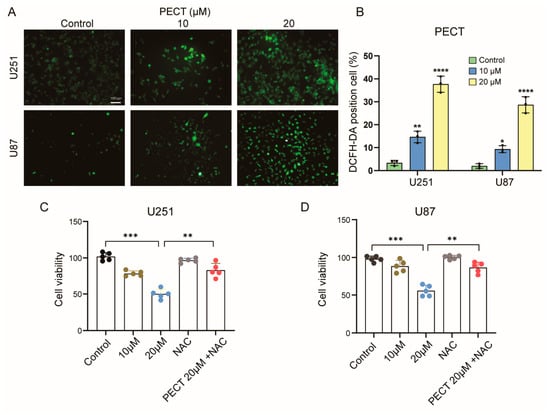
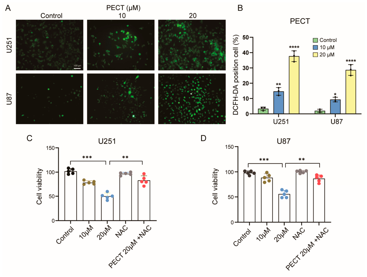
To investigate whether PECT induces oxidative stress in GBM cells, we measured intracellular ROS levels using the DCFH-DA probe. Fluorescence imaging revealed a dose-dependent accumulation of ROS in both U251 and U87 cells following PECT treatment, with the most pronounced signal observed at 20 µM (Figure 1A). Quantitative analysis confirmed a marked increase in the percentage of DCFH-DA-positive cells in PECT-treated groups compared with controls (Figure 1B). Consistently, MTT assays showed that PECT significantly reduced cell viability in a dose-dependent manner, whereas co-treatment with the ROS scavenger N-acetyl-L-cysteine (NAC) substantially rescued cell viability in both cell lines (Figure 1C,D). These results indicate that ROS accumulation is a critical upstream event mediating PECT-induced cytotoxicity in GBM cells.
Figure 1.
PECT Triggers ROS Accumulation to Mediate its Cytotoxic Effects in GBM Cells. (A) Representative fluorescence images of DCFH-DA-stained U251 and U87 cells treated with PECT (0, 10, or 20 μM) for 48 h (representative of three independent experiments). Scale bar, 100 µm. (B) Quantification of DCFH-DA-positive cells in (A); bars represent mean ± SD from n = 3 independent biological experiments. (C,D) Cell viability of U251 (C) and U87 (D) cells pretreated with NAC (5 mM) for 3 h and co-treated with PECT (10 or 20 µM) for 48 h, as determined by MTT assay. Data are presented as mean ± SD from n = 5 independent biological experiments; each data point represents one independent biological replicate, measured as the mean of technical replicates. * p < 0.05, ** p < 0.01, *** p < 0.001, **** p < 0.0001.
3.2. E2F1 Is Upregulated in GBM, Predicts Poor Prognosis, and Transcriptionally Activates RRM2
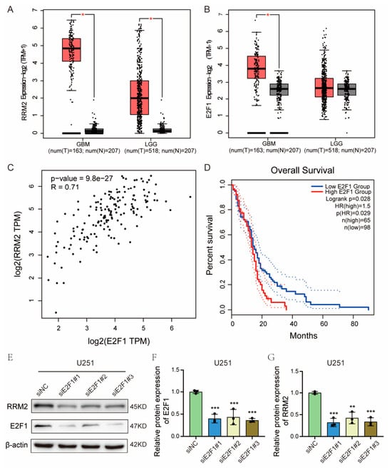
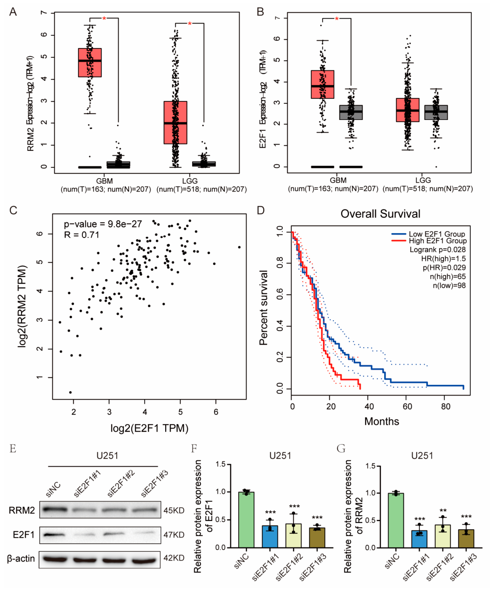
Our previous work demonstrated that PECT exerts anti-GBM effects by suppressing RRM2 transcription [18]; however, the upstream transcriptional regulator of RRM2 in GBM remained undefined. To identify candidate transcription factors, we performed promoter-based bioinformatic predictions using PROMO and SwissRegulon, which nominated E2F1 as a potential regulator of RRM2 transcription. Consistent with this prediction, GEPIA2-based analysis of the TCGA and GTEx datasets revealed marked upregulation of both RRM2 and E2F1 in GBM (n = 163) and LGG (n = 518) compared with normal brain tissues (n = 207) (Figure 2A,B). E2F1 and RRM2 mRNA levels were strongly and positively correlated in GBM specimens (R = 0.71, p = 9.8 × 10⁻²⁷; Figure 2C), and high E2F1 expression was associated with significantly shorter overall survival (HR = 1.5, p (HR) = 0.029, log-rank p = 0.028; cutoff-high 60%, cutoff-low 60%; n(high) = 65, n(low)=98) (Figure 2D). To functionally validate this regulatory relationship, U251 cells were transfected with three E2F1-targeting siRNAs. E2F1 knockdown potently reduced RRM2 expression at both the mRNA (Figure S1A,B) and protein levels (Figure 2E–G), confirming that E2F1 positively regulates RRM2 transcription in GBM cells.
Figure 2.
E2F1 is Upregulated in GBM, Predicts Poor Prognosis, and Transcriptionally Activates RRM2 (A,B) Box plots showing RRM2 (A) and E2F1. (B) mRNA expression in glioblastoma (GBM, n = 163), lower-grade glioma (LGG, n = 518), and normal brain tissues (n = 207) from the TCGA and GTEx databases (GEPIA2). TPM, transcripts per million. (C) Pearson correlation analysis of E2F1 and RRM2 mRNA levels in GBM (R = 0.71, p = 9.8 × 10⁻²⁷). (D) Kaplan–Meier overall survival analysis of GBM patients stratified by E2F1 expression (cutoff-high = 60%, n = 65; cutoff-low = 60%, n = 98). Log-rank p = 0.028; HR = 1.5, p = 0.029. (E) Western blot analysis of E2F1 and RRM2 protein levels in U251 cells transfected with siE2F1 (#1, #2, #3) or siNC; β-actin served as the loading control. (F,G) Quantification of E2F1 (F) and RRM2 (G) protein expression in (E). Data are presented as mean ± SD (n = 3 independent experiments). * p < 0.05, ** p < 0.01, *** p < 0.001.
3.3. PECT Suppresses the E2F1–RRM2 Axis in a ROS-Dependent Manner in GBM Cells
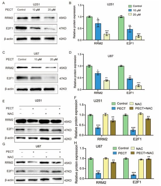
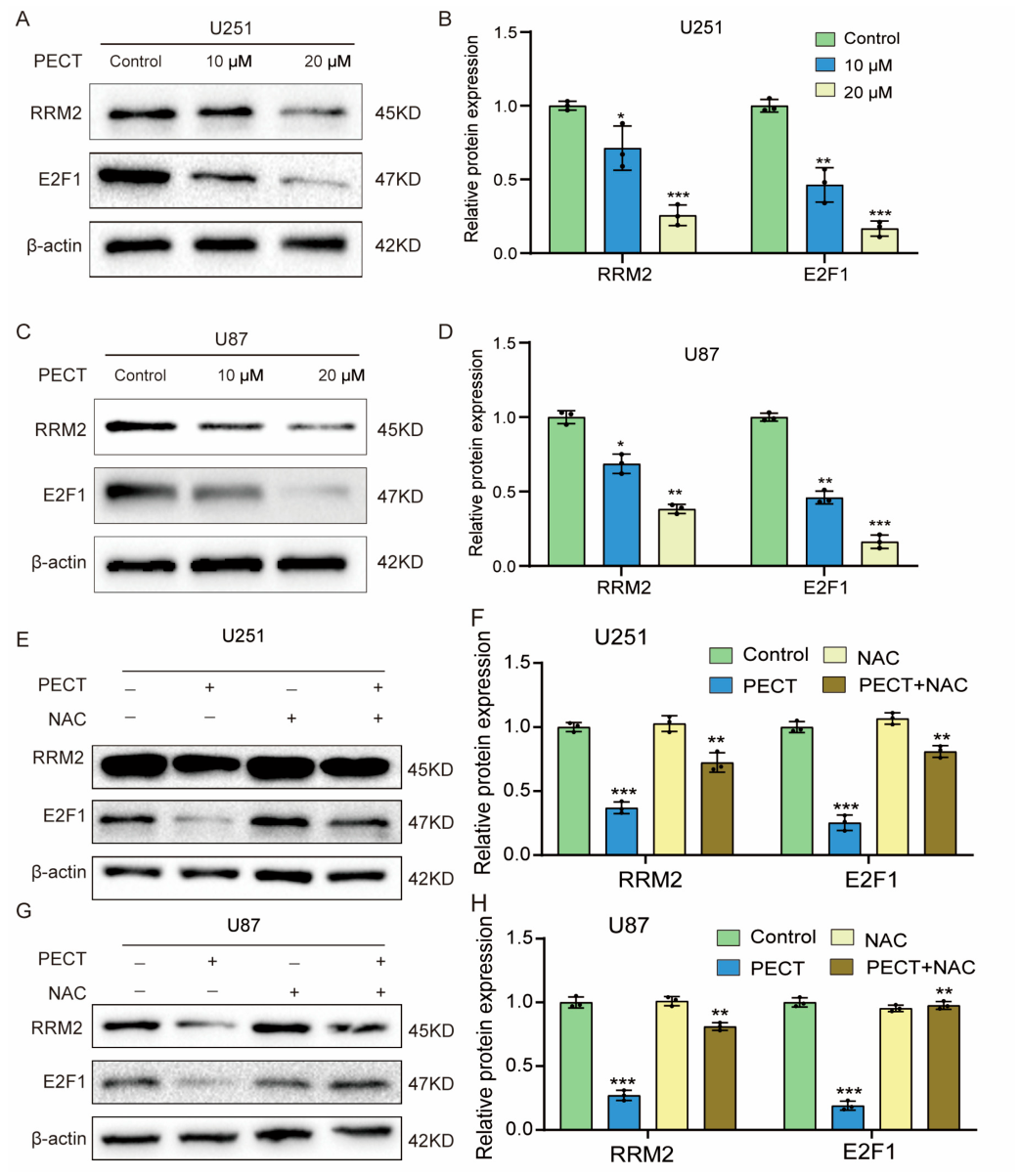
Having established that ROS mediates PECT-induced cytotoxicity (Figure 1) and that E2F1 drives RRM2 transcription (Figure 2), we next asked whether PECT targets this newly identified axis. Western blot analysis revealed that PECT dose-dependently reduced both E2F1 and RRM2 protein levels in U251 and U87 cells, with maximal inhibition observed at 20 μM (Figure 3A–D). At the transcriptional level, PECT similarly suppressed E2F1 and RRM2 mRNA in a dose-dependent manner (Figure S2A,B). Time-course analysis further demonstrated that E2F1 mRNA declined as early as 12 h after PECT exposure, whereas RRM2 mRNA reduction became significant only at 24 h (Figure S2C,D), indicating that E2F1 suppression precedes RRM2 downregulation and supporting the upstream position of E2F1 in this cascade. To determine whether ROS mediates PECT-dependent E2F1 suppression, cells were pretreated with NAC (5 mM) for 3 h prior to PECT exposure. Notably, NAC substantially restored E2F1 protein expression in both cell lines, whereas NAC alone had no detectable effect; importantly, NAC co-treatment also restored RRM2 protein levels in both cell lines (Figure 3E–H), demonstrating that PECT suppresses the E2F1–RRM2 axis in a ROS-dependent manner. Collectively, these findings integrate our prior observations into a coherent signaling cascade: PECT induces intracellular ROS accumulation, which suppresses E2F1 at both the mRNA and protein levels, leading to reduced RRM2 transcription.
Figure 3.
PECT Suppresses the E2F1–RRM2 Axis in a ROS-Dependent Manner in GBM cells. (A,C) Western blot analysis of RRM2 and E2F1 protein levels in U251 (A) and U87 (C) cells treated with PECT (0, 10, or 20 μM) for 48 h. β-actin served as the loading control. (B,D) Quantification of RRM2 and E2F1 protein expression in (A,C), respectively. (E,G) Western blot analysis of RRM2 and E2F1 protein levels in U251 (E) and U87 (G) cells pretreated with NAC (5 mM) for 3 h, followed by co-treatment with PECT (20 μM) for 48 h. (F,H) Quantification of RRM2 and E2F1 protein expression in (E) and (G), respectively. Data are presented as mean ± SD (n = 3 independent experiments). * p < 0.05, ** p < 0.01, *** p < 0.001.
3.4. A Predicted E2F1-Binding Motif in the RRM2 Promoter Mediates E2F1-Dependent Transcription and Its Suppression by PECT
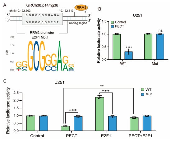
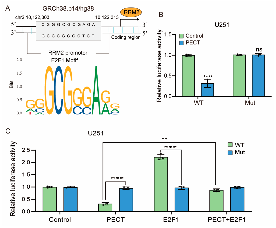
PROMO, SwissRegulon, JASPAR and TFsearch predicted a GC-rich E2F1-response element (5′-CGGGCGCGAGA−3′, chr2:10,122,303–10,122,313, GRCh38/hg38) within the RRM2 promoter (−2465/+23) (Figure 4A). We therefore generated wild-type (WT) and mutant (Mut; core GCGC → ATAT) RRM2 promoter reporters. The Mut construct retained basal activity comparable to WT (Figure 4B), indicating that the mutation did not compromise basal promoter function. PECT markedly suppressed WT reporter activity, whereas the Mut reporter was refractory to PECT (Figure 4B), demonstrating that PECT inhibits RRM2 transcription through this element. Furthermore, ectopic E2F1 robustly activated the WT reporter, and this activation was (i) significantly attenuated by PECT co-treatment and (ii) largely abolished by mutation of the element (Figure 4C), establishing that E2F1-dependent transactivation of the RRM2 promoter operates through this predicted site. This functional assignment is consistent with prior chromatin immunoprecipitation evidence that E2F1 directly occupies the RRM2 promoter in GBM cells [4]. We next asked whether this axis is functionally required for PECT-mediated cytotoxicity. E2F1 was efficiently overexpressed at both the mRNA (Figure S3A) and protein (Figure S3B,C) levels in U251 and U87 cells. E2F1 overexpression had no effect on basal viability but significantly attenuated PECT-induced loss of viability in both cell lines (Figure S3D,E). Similarly, in our previous work, RRM2 overexpression partially rescued GBM cells from PECT-induced proliferation inhibition and G2/M cell-cycle arrest in the same cell models [18], indicating that restoring the E2F1–RRM2 axis partially protects GBM cells from PECT.
Figure 4.
A predicted E2F1-Binding Motif in the RRM2 Promoter Mediates E2F1-Dependent Transcription and its Suppression By PECT (A) Schematic of the predicted GC-rich E2F1-response element (5′-CGGGCGCGAGA−3′; chr2:10,122,303–10,122,313, GRCh38/hg38; −313 to −303 bp relative to the transcription start site of RRM2 transcript variant 1, NM_001165931) within the RRM2 promoter region (−2465/+23); the E2F1 position weight matrix (JASPAR MA0024.1) is shown below. (B) Dual-luciferase reporter assay in U251 cells co-transfected with the wild-type (WT) or mutant (Mut) RRM2 promoter reporter together with the internal control reporter pRL-TK, followed by treatment with PECT (20 μM) for 48 h. (C) Dual-luciferase reporter assay in U251 cells co-transfected with the indicated RRM2 promoter reporters together with an E2F1 expression plasmid, treated with PECT (20 μM) for 48 h as indicated. Firefly luciferase activity was normalized to Renilla luciferase activity. Data are presented as mean ± SD (n = 3 independent experiments). ns, not significant; ** p < 0.01, *** p < 0.001, **** p < 0.0001. For panels (B,C), data were analyzed by two-way ANOVA (promoter construct × treatment, including the interaction term) followed by Šídák’s multiple comparisons test.
4. Discussion
Glioblastoma remains one of the most lethal human malignancies, and the identification of druggable nodes within its core survival circuitry is urgently needed. In this study, we delineate a previously unrecognized upstream mechanism governing RRM2 expression in GBM and show that the natural compound PECT therapeutically exploits this vulnerability. Our principal findings can be summarized as follows: PECT induces ROS accumulation that is required for its cytotoxicity; ROS-dependent suppression of E2F1 precedes and drives RRM2 downregulation; and a single GC-rich E2F1-response element in the RRM2 promoter is required for both E2F1-mediated transactivation and PECT-induced repression. Together, these findings define a linear pathway: PECT→ROS accumulation→E2F1 suppression→loss of RRM2 transcription→impaired GBM cell survival, and identify the ROS–E2F1–RRM2 axis as a candidate therapeutic target in GBM.
The oncogenic relevance of the E2F1–RRM2 axis in GBM is well precedented. E2F1 was identified as a direct transcriptional activator of RRM2 more than two decades ago [8], and its binding to the RRM2 promoter has since been confirmed by chromatin immunoprecipitation-based approaches [4]. In GBM, Rasmussen et al. demonstrated that the BRCA1–E2F1–RRM2 module protects GBM cells from endogenous replication stress, that RRM2 is a negative prognostic factor in glioma, and that the RRM2 inhibitor triapine suppresses GBM growth and extends survival in orthotopic models [4,23]. Pharmacological disruption of the E2F1–RRM2 axis by natural compounds has precedent in pancreatic cancer [24]. Our findings are concordant with and extend this paradigm in two directions: First, TCGA analysis links E2F1 itself—not merely its partners or downstream effectors—to poor GBM outcome. Second, by combining site-directed mutagenesis with reporter assays, we map the transcriptional response to a single GC-rich element at nucleotide resolution. In short, earlier work explained why GBM cells depend on RRM2; our work shows how this dependence can be switched off pharmacologically at the transcriptional level.
Our previous work demonstrated that PECT suppresses GBM progression by inhibiting RRM2 transcription and promoting RRM2 protein degradation via the autophagy–lysosome pathway [18]. That study, however, left a central mechanistic question unanswered: which factor conveys PECT’s inhibitory signal to the RRM2 promoter, and what initiates the cascade? The present study complements and extends that report in three respects. First, we identify E2F1 as the upstream transcriptional regulator that executes PECT-mediated RRM2 repression, thereby filling this mechanistic gap. Second, we establish ROS accumulation as the proximal initiating event, as ROS scavenging with NAC substantially reversed PECT-dependent E2F1 suppression. Third, through complementary loss- and gain-of-function experiments (E2F1 siRNA knockdown and overexpression rescue), we demonstrate that the E2F1–RRM2 axis is not merely a correlate but a functional determinant of PECT sensitivity. Notably, ectopic E2F1 only partially rescued PECT-induced viability loss. Because E2F1 governs a broad transcriptional program spanning cell-cycle progression, DNA replication, and apoptosis, this rescue cannot be ascribed to RRM2 restoration alone. Importantly, however, RRM2 overexpression has previously been shown to partially rescue GBM cells from PECT-induced proliferation inhibition, G2/M cell-cycle arrest, and autophagic-flux alterations in the same U251 and U87 models, providing published evidence that RRM2 downregulation contributes to the anticancer effects of PECT. The partial nature of both rescues indicates that PECT simultaneously engages additional mechanisms, most plausibly the reported autophagy–lysosome-mediated degradation of RRM2 protein [18]; accordingly, we propose that effective RRM2 targeting may therefore require simultaneous disruption of both its transcriptional and post-transcriptional regulation. The placement of ROS upstream of E2F1 suppression warrants discussion. Although ROS have classically been viewed as genotoxic by-products, growing evidence implicates redox signaling in the control of cell-cycle transcriptional machinery [11]. E2F1 activity is tightly coupled to proliferative and stress-adaptive signaling, and sustained oxidative stress can remodel the transcriptional landscape that sustains S-phase gene expression [12]. Our observation that PECT rapidly induces ROS and that E2F1 mRNA and protein decline within hours—preceding the RRM2 decrease—supports a model in which PECT-triggered oxidative stress dismantles the E2F1-driven transcriptional program upstream of RRM2. The molecular step linking ROS to E2F1 downregulation (for example, redox-sensitive modulation of E2F1-stabilizing interactions or of upstream signaling) remains to be defined and represents an important direction for future work. Notably, the convergence of PECT-induced ROS with RRM2 suppression is reminiscent of the mode of action of triapine, an iron-chelating RRM2 inhibitor that likewise elevates ROS and oxidative DNA lesions in GBM [4,23]; this convergence suggests that redox perturbation and RRM2 targeting may constitute a therapeutically exploitable synthetic relationship in GBM.
From a translational standpoint, our findings carry several implications. E2F1 overexpression defines a GBM subgroup with dismal prognosis and high RRM2 dependence, nominating E2F1–RRM2 axis activity as a candidate stratification biomarker. PECT may therefore represent a candidate modulator of this axis; however, our current evidence is limited to two GBM cell lines without normal-cell controls, and any therapeutic extrapolation would be premature. Its ROS-dependent mechanism nevertheless offers a rationale for future combination strategies—for instance, with PARP inhibitors, given the established sensitivity of replication-stressed GBM cells to PARP blockade [25], or RRM2-directed agents like triapine. Pharmacokinetic profiling, blood–brain barrier penetration, normal-glial toxicity, and orthotopic efficacy studies will be essential before PECT, alone or in such combinations, can be evaluated in these settings.
Several limitations should be acknowledged. First, direct occupancy of the predicted promoter element by endogenous E2F1 was inferred from bioinformatic prediction, loss- and gain-of-function data, and site-directed mutagenesis; formal confirmation by chromatin immunoprecipitation is pending, and the contribution of other GC-box-binding factors to this element cannot be formally excluded; ChIP-qPCR verification of endogenous E2F1 occupancy at this element is planned in our ongoing follow-up study. We further note that direct RRM2 overexpression rescue of PECT-treated GBM cells has been reported in our previous study [18]; nevertheless, whether ectopic E2F1 restores endogenous RRM2 protein expression under PECT treatment remains to be directly measured. Second, our mechanistic analyses were performed in U251 and U87 cell lines without normal glial controls, and validation in patient-derived GBM models, organoids, and in vivo systems is required. Third, ROS was measured at a single 48 h endpoint matched to the viability assay; early-kinetic ROS measurements and direct ROS quantification in NAC-rescued cells would further strengthen the causal chain. We note that RRM2 immunoblotting has now been incorporated into the NAC rescue experiment (Figure 3E–H), showing that NAC co-restores RRM2 together with E2F1 and further supporting the ROS dependence of the entire axis. Fourth, although NAC reversal establishes ROS dependence, the precise redox-sensitive step that silences E2F1 remains unidentified. Finally, the partial nature of the E2F1 rescue indicates that additional effectors contribute to PECT cytotoxicity, which we consider a fertile ground for further dissection rather than a shortcoming of the model.
5. Conclusions
In conclusion, we identify a ROS–E2F1–RRM2 signaling cascade in GBM and show that PECT disables this axis by oxidatively suppressing E2F1 and thereby silencing RRM2 transcription through a defined promoter element. Given the established addiction of GBM to the replication-stress-protective functions of RRM2, pharmacological disruption of the E2F1–RRM2 transcriptional module—whether by PECT or by rationally designed combinations—merits further preclinical exploration, including validation in normal glial cells and in vivo models.
Supplementary Materials
The following supporting information can be downloaded at https://www.mdpi.com/article/10.3390/cimb48111255/s1.
Author Contributions
Conceptualization, methodology, software, resources, writing—original draft preparation, writing—review and editing and visualization, H.J.; validation, formal analysis, investigation and data curation, F.C. and W.L.; supervision and project administration, S.Z. and J.X.; funding acquisition, H.J. and J.X. All authors have read and agreed to the published version of the manuscript.
Funding
This work was supported by the Scientific Research Project of Guangdong Provincial Administration of Traditional Chinese Medicine (grant no. 20251204), Collaborative Traditional Chinese and Western Medicine Specialty Cultivation Project (1039K20250018) and the high-level talents scientific research start-up funds of the Affliated Hospital of Guangdong Medical University: GCC2022050 and GCC2023007.
Institutional Review Board Statement
The study was conducted in accordance with the Declaration of Helsinki. As the data were obtained from publicly accessible databases (TCGA, GTEx, and GEPIA2) and were fully deidentified data, in accordance with the U.S. Department of Health and Human Services regulations for the protection of human subjects (45 CFR §46.104(d)(4)), the research was exempted from ethics committee review.
Informed Consent Statement
Informed consent was exempted, as the study used only deidentified, publicly available data, in accordance with the U.S. Department of Health and Human Services regulations for the protection of human subjects (45 CFR §46.104(d)(4)).
Data Availability Statement
The original contributions presented in the study are included in the article/Supplementary Material. Publicly available datasets analyzed in this study include The Cancer Genome Atlas (TCGA, https://www.cancer.gov/tcga, accessed on 15 November 2026), the Genotype-Tissue Expression project (GTEx, https://gtexportal.org/home/), and the GEPIA2 platform (http://gepia2.cancer-pku.cn/). The human reference genome (GRCh38/hg38) was obtained from the UCSC Genome Browser (https://genome.ucsc.edu). Further inquiries can be directed to the corresponding authors.
Acknowledgments
We would like to thank Editage (www.editage.cn) for English language editing. The authors confirm that all individuals included in this section have provided consent to be acknowledged.
Conflicts of Interest
The authors declare no conflicts of interest.
Abbreviations
The following abbreviations are used in this manuscript:
| RRM2 | Ribonucleotide reductase regulatory subunit M2 |
| GBM | Glioblastoma |
| ROS | Reactive oxygen species |
| PECT | Pectolinarigenin |
| dNTP | deoxynucleotide triphosphate |
References
- Cloughesy, T.F.; Cavenee, W.K.; Mischel, P.S. Glioblastoma: From molecular pathology to targeted treatment. Annu Rev. Pathol. 2014, 9, 1–25. [Google Scholar] [CrossRef] [Scilit]
- Patel, A.P.; Tirosh, I.; Trombetta, J.J.; Shalek, A.K.; Gillespie, S.M.; Wakimoto, H.; Cahill, D.P.; Nahed, B.V.; Curry, W.T.; Martuza, R.L.; et al. Single-cell RNA-seq highlights intratumoral heterogeneity in primary glioblastoma. Science 2014, 344, 1396–1401. [Google Scholar] [CrossRef] [Scilit]
- Bartkova, J.; Hamerlik, P.; Stockhausen, M.T.; Ehrmann, J.; Hlobilkova, A.; Laursen, H.; Kalita, O.; Kolar, Z.; Poulsen, H.S.; Broholm, H.; et al. Replication stress and oxidative damage contribute to aberrant constitutive activation of DNA damage signalling in human gliomas. Oncogene 2010, 29, 5095–5102. [Google Scholar] [CrossRef] [Scilit]
- Rasmussen, R.D.; Gajjar, M.K.; Tuckova, L.; Jensen, K.E.; Maya-Mendoza, A.; Holst, C.B.; Mollgaard, K.; Rasmussen, J.S.; Brennum, J.; Bartek, J., Jr.; et al. BRCA1-regulated RRM2 expression protects glioblastoma cells from endogenous replication stress and promotes tumorigenicity. Nat. Commun. 2016, 7, 13398, Correction in Nat. Commun. 2018, 9, 5396. https://doi.org/10.1038/s41467-018-07892-6. [Google Scholar] [CrossRef] [Scilit]
- Greene, B.L.; Kang, G.; Cui, C.; Bennati, M.; Nocera, D.G.; Drennan, C.L.; Stubbe, J. Ribonucleotide Reductases: Structure, Chemistry, and Metabolism Suggest New Therapeutic Targets. Annu Rev. Biochem. 1989, 89, 45–75. [Google Scholar] [CrossRef] [Scilit]
- Wang, Y.; Chen, R.; Zhang, J.; Zeng, P. A comprehensive analysis of ribonucleotide reductase subunit M2 for carcinogenesis in pan-cancer. PLoS ONE 2024, 19, e0299949. [Google Scholar] [CrossRef] [Scilit]
- Perrault, E.N.; Shireman, J.M.; Ali, E.S.; Lin, P.; Preddy, I.; Park, C.; Budhiraja, S.; Baisiwala, S.; Dixit, K.; James, C.D.; et al. Ribonucleotide reductase regulatory subunit M2 drives glioblastoma TMZ resistance through modulation of dNTP production. Sci. Adv. 2023, 9, eade7236. [Google Scholar] [CrossRef] [Scilit]
- DeGregori, J.; Kowalik, T.; Nevins, J.R. Cellular targets for activation by the E2F1 transcription factor include DNA synthesis- and G1/S-regulatory genes. Mol. Cell. Biol. 1995, 15, 4215–4224, Erratum in Mol. Cell. Biol. 1995, 15, 5846–5847. [Google Scholar] [CrossRef] [Scilit]
- Aird, K.M.; Zhang, G.; Li, H.; Tu, Z.; Bitler, B.G.; Garipov, A.; Wu, H.; Wei, Z.; Wagner, S.N.; Herlyn, M.; et al. Suppression of nucleotide metabolism underlies the establishment and maintenance of oncogene-induced senescence. Cell Rep. 2003, 3, 1252–1265. [Google Scholar] [CrossRef] [Scilit]
- Yu, H.; Li, Z.; Wang, M. Expression and prognostic role of E2F transcription factors in high-grade glioma. CNS Neurosci. Ther. 2020, 26, 741–753. [Google Scholar] [CrossRef] [Scilit]
- Glorieux, C.; Liu, S.; Trachootham, D.; Huang, P. Targeting ROS in cancer: Rationale and strategies. Nat. Rev. Drug Discov. 2024, 23, 583–606. [Google Scholar] [CrossRef] [Scilit]
- Nowacka, A.; Sniegocki, M.; Ziolkowska, E. Oxidative Stress and Antioxidants in Glioblastoma: Mechanisms of Action, Therapeutic Effects and Future Directions. Antioxidants 2025, 14, 1121. [Google Scholar] [CrossRef] [Scilit]
- Lin, W.C.; Lin, F.T.; Nevins, J.R. Selective induction of E2F1 in response to DNA damage, mediated by ATM-dependent phosphorylation. Genes Dev. 2001, 15, 1833–1844. [Google Scholar]
- Burhans, W.C.; Heintz, N.H. The cell cycle is a redox cycle: Linking phase-specific targets to cell fate. Free Radic. Biol. Med. 2009, 47, 1282–1293. [Google Scholar] [CrossRef] [Scilit]
- Xu, F.; Gao, X.; Pan, H. Pectolinarigenin inhibits non-small cell lung cancer progression by regulating the PTEN/PI3K/AKT signaling pathway. Oncol. Rep. 2018, 40, 3458–3468. [Google Scholar] [CrossRef] [Scilit]
- Wu, T.; Dong, X.; Yu, D.; Shen, Z.; Yu, J.; Yan, S. Natural product pectolinarigenin inhibits proliferation, induces apoptosis, and causes G2/M phase arrest of HCC via PI3K/AKT/mTOR/ERK signaling pathway. OncoTargets Ther. 2018, 11, 8633–8642. [Google Scholar] [CrossRef] [Scilit]
- Arjsri, P.; Semmarath, W.; Srisawad, K.; Intanil, I.; Thippraphan, P.; Dejkriengkraikul, P. Pectolinarigenin from Tiliacora triandra Exhibits Potent Anticancer Activity in Triple-Negative Breast Cancer Cells Through Cell Cycle Arrest, Apoptosis, and MAPK Signaling Inhibition. Pharmaceuticals 2026, 19, 384. [Google Scholar] [CrossRef] [Scilit]
- Jiang, H.; Zhang, D.; Aleksandrovich, K.D.; Ye, J.; Wang, L.; Chen, X.; Gao, M.; Wang, X.; Yan, T.; Yang, H.; et al. RRM2 Mediates the Anti-Tumor Effect of the Natural Product Pectolinarigenin on Glioblastoma Through Promoting CDK1 Protein Degradation by Increasing Autophagic Flux. Front. Oncol. 2022, 12, 887294. [Google Scholar] [CrossRef] [Scilit]
- Subtil-Rodríguez, A.; Vázquez-Chávez, E.; Ceballos-Chávez, M.; Rodríguez-Paredes, M.; Martín-Subero, J.I.; Esteller, M.; Reyes, J.C. The chromatin remodeller CHD8 is required for E2F-dependent transcription activation of S-phase genes. Nucleic Acids Res. 2014, 42, 2185–2196. [Google Scholar] [CrossRef] [Scilit]
- Moniz, S.; Bandarra, D.; Biddlestone, J.; Campbell, K.J.; Komander, D.; Bremm, A.; Rocha, S. Cezanne regulates E2F1-dependent HIF2α expression. J. Cell Sci. 2015, 128, 3082–3093. [Google Scholar] [CrossRef] [Scilit]
- Sang, X.B.; Zong, Z.H.; Wang, L.L.; Wu, D.D.; Chen, S.; Liu, B.L.; Zhao, Y. E2F-1 targets miR-519d to regulate the expression of the ras homolog gene family member C. Oncotarget 2017, 8, 14777–14793. [Google Scholar] [CrossRef] [Scilit]
- Jesenko, T.; Kranjc Brezar, S.; Pisljar, Z.; Bozic, T.; Markelc, B.; Cazzato, M.; Grassi, G.; Cemazar, M. Effective targeting of E2F1 transcription factor via siRNA gene electrotransfer in HT-29 colorectal carcinoma xenografts. Bioelectrochemistry 2025, 165, 108994. [Google Scholar] [CrossRef] [Scilit]
- Corrales-Guerrero, S.; Cui, T.; Castro-Aceituno, V.; Yang, L.; Nair, S.; Feng, H.; Venere, M.; Yoon, S.; DeWees, T.; Shen, C.; et al. Inhibition of RRM2 radiosensitizes glioblastoma and uncovers synthetic lethality in combination with targeting CHK1. Cancer Lett. 2023, 570, 216308. [Google Scholar] [CrossRef] [Scilit]
- Xia, G.; Wang, H.; Song, Z.; Meng, Q.; Huang, X.; Huang, X. Gambogic acid sensitizes gemcitabine efficacy in pancreatic cancer by reducing the expression of ribonucleotide reductase subunit-M2 (RRM2). J. Exp. Clin. Cancer Res. 2017, 36, 107. [Google Scholar] [CrossRef] [Scilit]
- Venere, M.; Hamerlik, P.; Wu, Q.; Rasmussen, R.D.; Song, L.A.; Vasanji, A.; Tenley, N.; Flavahan, W.A.; Hjelmeland, A.B.; Bartek, J.; et al. Therapeutic targeting of constitutive PARP activation compromises stem cell phenotype and survival of glioblastoma-initiating cells. Cell Death Differ. 2014, 21, 258–269. [Google Scholar] [CrossRef] [Scilit]
Disclaimer/Publisher’s Note: The statements, opinions and data contained in all publications are solely those of the individual author(s) and contributor(s) and not of MDPI and/or the editor(s). MDPI and/or the editor(s) disclaim responsibility for any injury to people or property resulting from any ideas, methods, instructions or products referred to in the content. |
© 2026 by the authors. Licensee MDPI, Basel, Switzerland. This article is an open access article distributed under the terms and conditions of the Creative Commons Attribution (CC BY) license.