


Abstract
Background: Energy drinks are widely consumed stimulant beverages among young adults. Its long-term effects on male reproductive function remain poorly defined. Objective: This study investigates the histopathological, endocrine, and inflammatory consequences of chronic energy drink consumption on testicular tissue and evaluates the protective role of quercetin in adult male rats. Methods: Eighteen adult male rats were randomly assigned into three groups, control, energy drink, and energy drink + quercetin-treated groups (n = 6/group), and orally treated for 12 weeks. Serum testosterone was measured to assess steroidogenic function. Testicular histopathology was evaluated by H&E staining. The immunoreactivity of IL-6, TNF-α, and NF-κB was assessed in spermatogenic and interstitial compartments. Gene expression levels of IL-6 and TNF-α were quantified by qRT-PCR. Results: Chronic energy drink consumption significantly decreased serum testosterone, induced marked testicular damage, upregulated TNF-α and IL-6 mRNA expression, and enhanced immunoreactivity of TNF-α, IL-6, and NF-κB. Quercetin co-administration significantly ameliorated these alterations, restoring testosterone toward control levels, preserving testicular architecture, and suppressing inflammatory markers. Conclusion: Chronic energy drink consumption impairs testicular steroidogenesis and structural integrity, accompanied by NF-κB-mediated inflammatory activation. Quercetin exerts substantial protection by preserving testicular structure and attenuating pro-inflammatory responses, suggesting a mechanistic role for antioxidant-mediated suppression of testicular inflammation.
Keywords:
energy drinks; testosterone; testis; NF-κB; IL-6; TNF-α; quercetin; oxidative stress; spermatogenesis 1. Introduction
Over the last decade, the global consumption of energy drinks has significantly increased, raising many concerns of potential negative health impacts [1]. Energy drinks usually contain a mixture of large amounts of caffeine, sugar, taurine, and other vitamins and herbs to improve taste and effect [2]. Several studies have investigated the deleterious effects of chronic energy drink consumption on the nervous and cardiovascular systems, gastrointestinal tract, and renal system [3,4,5]. Several studies have linked the detrimental effects caused by ED consumption to the ability of these drinks to promote tissue injury primarily by altering the redox balance via depleting endogenous antioxidants and inducing an inflammatory response. Both oxidative stress and inflammatory response initiate a group of metabolic disturbances and hence endothelial dysfunction in various tissues, resulting in many studies testing the ability of known antioxidants such as blueberries, omega-3 fatty acids and vitamin E to protect against ED-induced damage [6,7]. Recently, it has been shown that regular intake of energy drinks can negatively influence male fertility and reproductive health. Chronic consumption reduced sperm quality and reproductive organ weight, and disrupted hormonal balance [8,9]. Specifically, low serum testosterone levels, alongside disrupted follicle-stimulating (FSH) and luteinizing hormone (LH) levels, have been observed in test animals, indicating disruption of the hypothalamus–pituitary–gonadal pathway [8,9,10,11]. Reduced testosterone levels as reported in these studies were accompanied by decreased seminiferous tubule diameter and degeneration of different cell types. Whereas some studies reported no significant change in relative testicular weight at low-to-moderate doses of ED [12], others reported decrease in relative testicular weight upon prolonged exposure to large doses of caffeine [13]. Furthermore, metabolic disruptions such as hyperglycemia and hyperlipidemia as well as renal dysfunction were reported in subjects chronically consuming energy drinks [14,15,16].
Mechanistically, oxidative stress and activation of inflammatory signaling pathways have become increasingly recognized as main players in testicular pathogenesis in response to different environmental and nutritional factors. Testicular tissue is particularly vulnerable to oxidative damage due to the rapid cell turnover in the germ cells and the high lipid content in sperm cell membranes. One of the most important molecular pathways aiding inflammatory response is the activation of the nuclear factor-kappa B (NF-κB) signaling pathway which regulates the complex, multifactorial inflammatory processes [17]. A previous study reported an upregulation of NF-κB/p65 and COX2 in animals exposed to energy drinks for 3 months. The effects of energy drinks were extended to histopathological changes in the testicular tissue that included decreased tubule diameter and degenerating germinal cells [18]. Moreover, NF-κB regulates the transcription and activation of various downstream pro-inflammatory cytokines, such as tumor necrosis factor-alpha (TNF-α) and interleukin-6 (IL-6). Both TNF-α and IL-6 were reported to negatively affect the blood–testis barrier and impair spermatogenesis [19].
Quercetin is a bioactive flavonoid that occurs naturally in various plants, with proven safety and significant cytoprotective effects against oxidative stress, inflammation, and other harmful stimuli [20]. It was reported to exert antioxidant, anti-inflammatory, anti-apoptotic, anti-viral, anti-aging and antiallergic effects [21]. The protective effect of quercetin was shown to be mediated via balancing the redox state, inhibiting the NF-κB signaling pathway, and downregulating the levels of various inflammatory cytokines, e.g., IL-1β and IL-6 [22,23,24]. Quercetin was reported to prevent oxidative damage of testicular tissue, retrieve tissue architecture and restore steroid production after exposure to various toxicants [21,25].
Although exposure to caffeine and sugar as isolated compounds has been clearly associated with oxidative stress and inflammation in different organs [18], the testicular damage in response to chronic consumption of multi-component energy drinks and the potential protective effect of quercetin against this damage remain to be further elucidated. To our knowledge, this is the first study to address this gap in vivo. Accordingly, this study aimed at investigating the structural, steroidogenic, and inflammatory alterations in testicular tissue induced by chronic energy drink consumption in adult male rats, and exploring the protective effects of quercetin against these alterations by assessing the levels of key inflammatory markers NF-κB, Il-6 and TNF-α in the different test groups, along with evaluation of the histopathological and hormonal changes regarding testosterone levels.
2. Materials and Methods
2.1. Materials
Quercetin was purchased from Merk KGaA [CAS Number 117-39-5, Darmstadt, Germany]. The commercially available energy drink Power Horse® (S. Spitz GmbH, Attnang-Puchheim, Austria) was obtained from local retailers in Al-Madinah Al-Munawara, Saudi Arabia. The detailed composition of Power Horse® energy drink per 100 mL is summarized in Table 1. RNAlater® solution and RNA extraction reagents were obtained from Thermo Fisher Scientific (Waltham, MA, USA). All other chemicals and reagents used in this study were of analytical grade and were purchased from Sigma-Aldrich (St. Louis, MO, USA) or equivalent commercial suppliers.
Table 1.
Composition of Power Horse® energy drink (S. Spitz GmbH, Attnang-Puchheim, Austria) contained in each 100 mL of energy drink.
2.2. Experimental Animals and Study Design
Eighteen adult male Albino rats weighing 200 ± 20 g (8 weeks old) were obtained from the Experimental Animal Facility, College of Pharmacy, Taibah University, Al-Madinah Al-Munawara, Saudi Arabia. Animals were acclimatized for one week before initiation of the experiment under standard laboratory conditions, including room temperature of 25 ± 2 °C, relative humidity of 45–55%, and a 12 h light/12 h dark cycle. Rats had free access to standard laboratory chow and water throughout the experimental period.
All experimental procedures were approved by the Research Ethics Committee of the College of Pharmacy, Taibah University (Approval No. COPTU-REC-118-20241222), and were conducted in accordance with the Guide for the Care and Use of Laboratory Animals and applicable national regulations governing the use of laboratory animals.
Following acclimatization, animals were randomly assigned to three experimental groups (n = 6 per group) as follows:
Group I (control) received distilled water (12 mL/kg/day) by oral gavage for 12 consecutive weeks.
Group II (energy drink, ED) received Power Horse® energy drink (12 mL/kg/day, equivalent to 3.84 mg/kg/day caffeine) by oral gavage [16] for the same duration [18].
Group III (ED + quercetin) received Power Horse® energy drink (12 mL/kg/day) together with quercetin (50 mg/kg/day) [26] by oral gavage for 12 consecutive weeks.
Animals were observed twice daily and those showing any of the following signs, lethargy, unresponsiveness, tremors, dehydration, reduced food and water intake, were excluded from the study and humanely euthanized.
At the end of the experimental period, body weights were recorded, and animals were deeply anesthetized using pentobarbital sodium (50 mg/kg, intraperitoneally). Blood samples were collected by cardiac puncture, and serum was separated for evaluation of testosterone levels. Following confirmed euthanasia, both testes were excised, cleared of adherent tissues, and rinsed with cold physiological saline. One testis from each animal was fixed in 10% neutral-buffered formalin for histopathological and immunohistochemical analyses, while the contralateral testis was immediately preserved in RNAlater® solution and stored at −80 °C until molecular analysis.
2.3. Assessment of Serum Testosterone
Serum testosterone concentrations were determined using a commercially available enzyme-linked immunosorbent assay (ELISA) kit according to the manufacturer’s instructions (ab108666, Abcam, Cambridge, UK). Absorbance was measured using a microplate reader, and testosterone concentrations were calculated based on the corresponding standard curve. The results were expressed as ng/mL.
2.4. Quantitative Real-Time PCR Analysis
Total RNA was extracted from testicular tissues preserved in RNAlater® using the miRNeasy® Mini Kit (Qiagen, Helden, Germany) according to the manufacturer’s instructions. RNA concentration and purity were assessed spectrophotometrically before reverse transcription into complementary DNA (cDNA).
Quantitative real-time polymerase chain reaction (qRT-PCR) was performed using SYBR Green Master Mix (Bio-Rad, Hercules, CA, USA) and gene-specific primers for TNF-α and IL-6. Hypoxanthine phosphoribosyltransferase (HPRT) was used as the housekeeping gene for normalization. The sequences of the primers used in this study are presented in Table 2.
Table 2.
Primer sequences used for quantitative real-time PCR (qRT-PCR) analysis.
Amplification reactions were carried out using a Bio-Rad CFX Real-Time PCR System. Relative gene expression levels were calculated using the comparative Ct (2−ΔΔCt) method according to the following equations:
ΔCt = Ct (target gene) − Ct (housekeeping gene)
ΔΔCt = ΔCt (treated group) − ΔCt (control group)
Gene expression data were expressed as fold change relative to the control group. All samples were analyzed in triplicate.
2.5. Histopathological Evaluation
Testicular tissues fixed in 10% neutral-buffered formalin for 24 h were dehydrated through ascending concentrations of ethanol, cleared in xylene, and embedded in paraffin wax. Serial sections (6 μm thick) prepared using a rotary microtome were deparaffinized and rehydrated before staining.
For routine histological examination, tissue sections were stained with hematoxylin and eosin (H&E) according to the manufacturer’s instructions and examined under a Zeiss Axiovert A1 microscope (Carl Zeiss Microscopy GmbH, Jena, Germany). Histological evaluation was performed by an experienced histopathologist blinded to experimental groups.
Testicular injury was assessed using a semi-quantitative histopathological scoring system. Briefly, tissue alterations were graded on a scale from 0 to 4, where 0 indicated no detectable lesion, 1 indicated ≤25% tissue involvement, 2 indicated 26–50% tissue involvement, 3 indicated 51–75% tissue involvement, and 4 indicated 76–100% tissue involvement. The evaluated histopathological parameters included congestion of testicular blood vessels, interstitial edema, thickening of the seminiferous tubular basement membrane, degeneration of spermatogenic cells, depletion of spermatozoa, and Leydig cell activity [27].
2.6. Morphometric Analysis
Morphometric measurements were performed using calibrated digital micrographs obtained from H&E-stained sections. For each experimental group, fifteen, non-overlapping microscopic fields from different animals per group were utilized to obtain morphometric data. The diameter of seminiferous tubules, thickness of spermatogenic cell layers, number of Leydig cells, number of primary spermatocytes, and number of spermatids were quantified in three randomly selected non-overlapping microscopic fields per tissue section. Measurements obtained from each animal were averaged and used for subsequent statistical analysis.
2.7. Immunohistochemical Evaluation
Immunohistochemical staining was performed to evaluate the expression of inflammatory markers within testicular tissue according to Xi et al.’s protocol with slight modifications [28]. Paraffin-embedded sections were deparaffinized, rehydrated and non-specific binding sites were masked by incubation with blocking solution (goat serum). The sections were then incubated with primary antibodies against IL-6 (Abcam, Cat. No. ab259341), TNF-α (Abcam, Cat. No. ab220210), and NF-κB (Abcam, Cat. No. ab32536) followed by incubation with the corresponding secondary antibodies according to the manufacturers’ protocols. Immunoreactive signals were visualized using 3,3′-diaminobenzidine (DAB) as a substrate according to the manufacturer’s instructions, and examined using the Zeiss Axiovert A1 microscope (Carl Zeiss, Jena, Germany). Representative images were captured using a digital imaging system attached to the microscope.
2.8. Statistical Analysis
Data were analyzed using GraphPad Prism software version 6.0 (GraphPad Software Inc., La Jolla, CA, USA). Results are presented as mean ± standard deviation (SD). Statistical comparisons among experimental groups were performed using one-way analysis of variance (ANOVA) followed by Tukey–Kramer post hoc test for group comparison. Differences were considered statistically significant at p < 0.05.
3. Results
3.1. Effect of Quercetin on Serum Testosterone Levels
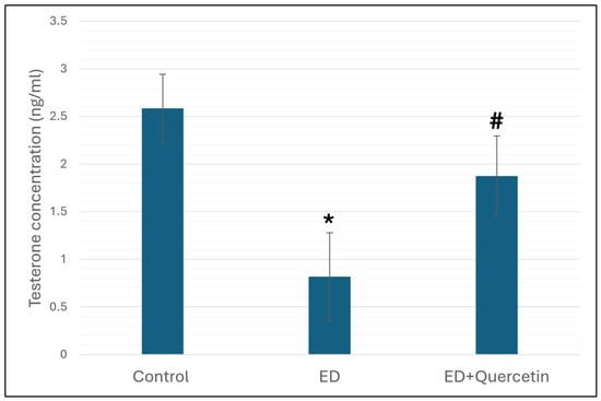
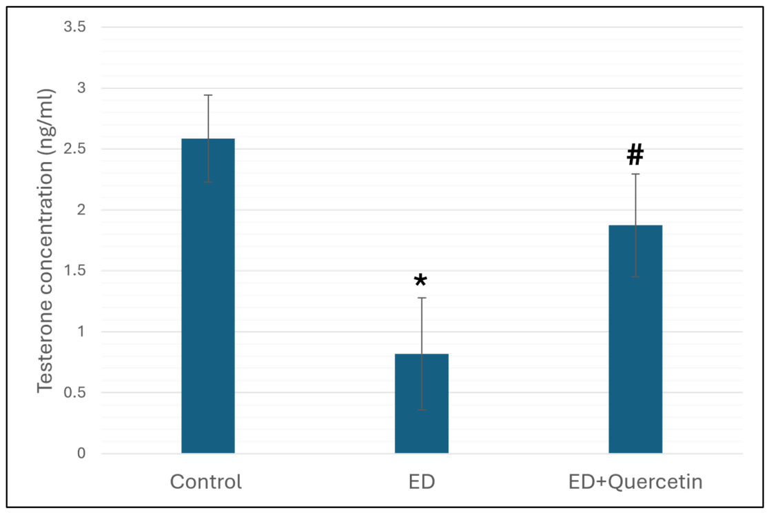
Evaluation of testosterone levels in blood is considered as a marker for testicular function. For this, the serum levels of testosterone were evaluated in samples collected from the different test groups. Animals of the ED group showed a significant reduction in serum testosterone compared to the healthy control (p < 0.05). In contrast, co-administration of quercetin along with the energy drink protected against the reduction in testosterone levels, resulting in levels that were significantly higher than those of the ED group (p < 0.05) and comparable to healthy control values (p > 0.05) (Figure 1).
Figure 1.
Effect of administration of energy drink and/or quercetin on serum levels of testosterone. Values are presented as mean ± SD. * indicates significant difference compared to healthy control, # indicates significant difference compared to energy drink-treated group at p value <0.05, n = 6 animals/group.
3.2. Effect of Quercetin on Gene Expression Levels of Inflammatory Markers
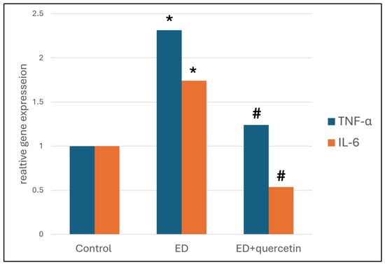
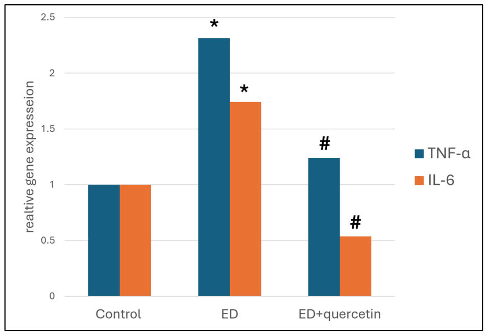
Relative gene expression of the inflammatory markers TNF-α and IL-6 were assessed to investigate the effects of the different treatments on the inflammatory response, where the expression level in healthy control was set to 1. Treating animals with energy drinks for 12 weeks provoked an inflammatory response in the form of a significant upregulation in the gene expression of TNF-α and IL-6 compared to the control (p < 0.05). Notably, co-administration of quercetin and energy drinks significantly ameliorated the inflammatory response and alleviated the gene expression of both cytokines compared to animals receiving energy drinks alone (p < 0.05). It is to be mentioned that TNF-α and IL-6 gene expression values in animals treated with energy drinks and quercetin were comparable to those of healthy controls (p > 0.05, Figure 2).
Figure 2.
Relative expression levels of TNF-α and IL-6 genes across the experimental groups. Gene expression was quantified using qRT-PCR and normalized to the healthy control group, which was assigned a value of 1. Data are presented as the mean ± SD. * indicates significant difference versus the healthy control group; # indicates significant difference versus the energy drink-treated group at p value < 0.05, n = 3.
3.3. Histological Assessment of Testicular Tissue
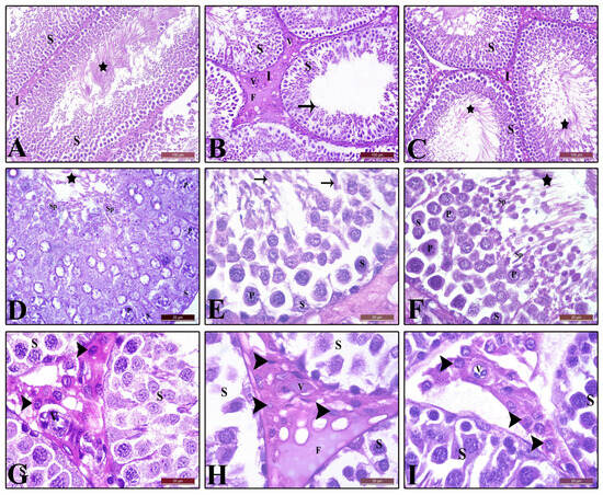
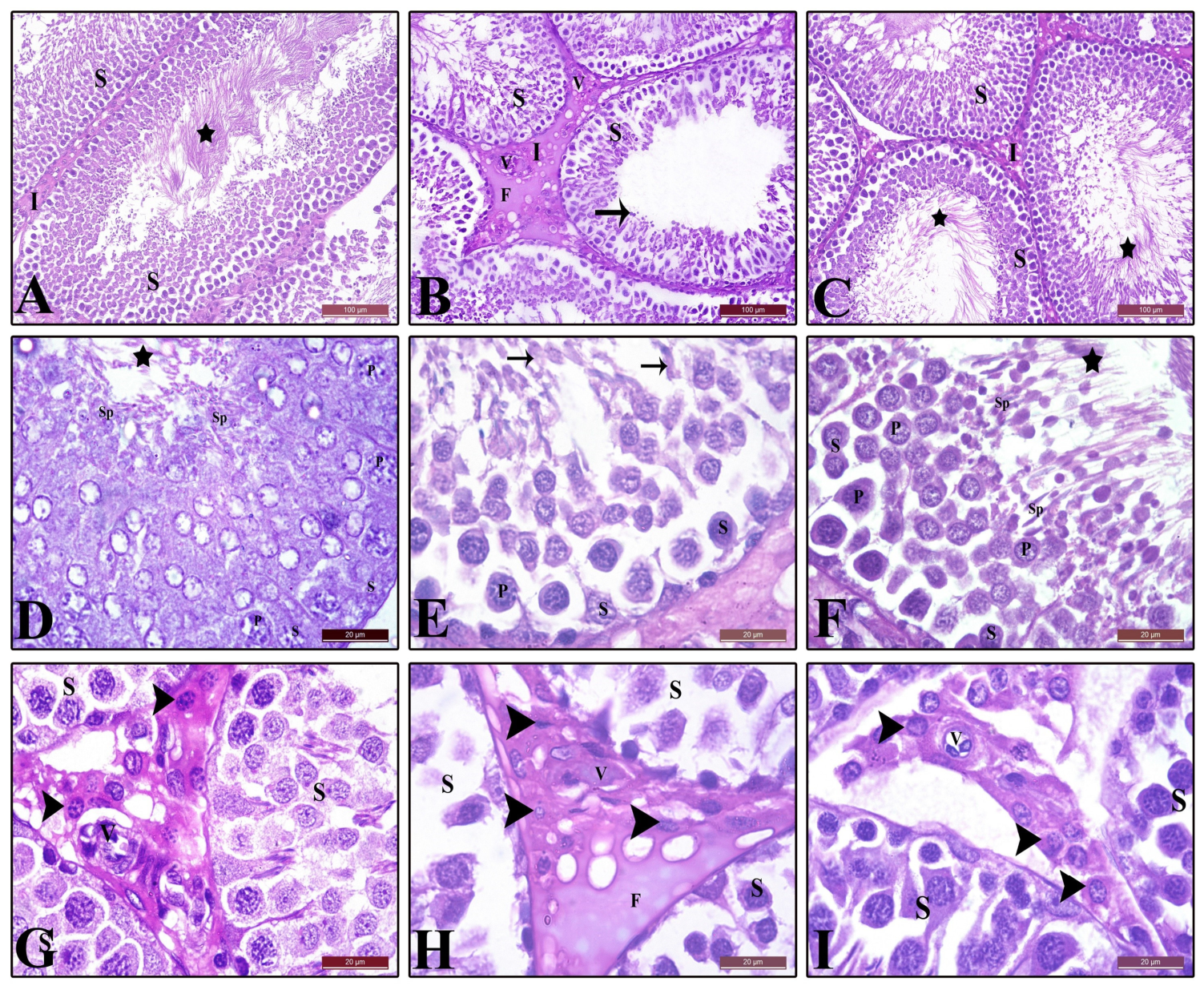
Chronic administration of energy drink effectively induced histological changes in the testicular tissue of the corresponding animals, where it induced interstitial edema and congestion of testicular blood vessels (Figure 3B,E). In addition, this treatment resulted in the degeneration of spermatogenic cells, and a diminishing number of active Leydig cells and spermatids compared to healthy control rats (Figure 3A,D). In contrast, rats administered energy drinks along with quercetin revealed improved histological features compared to animals receiving energy drinks alone. Testicular tissues from such group showed almost no signs of edema or blood vessel congestion (V, Figure 3C,F). Also, retrieval of the number of active Leydig cells and spermatids could be achieved in quercetin-treated animals, which showed almost normal histological features. It is to be noted that the changes in all histomorphometric parameters of the ED group were significant compared to the control group (p < 0.05) except for the number of Leydig cells, which was decreased compared to the control; however, the effect was non-significant (p > 0.05). Interestingly, quercetin administration prevented the damaging effect of ED on the different morphometric parameters (Table 3 and Table 4).
Figure 3.
Histopathological micrographs of testicular tissues from the different experimental groups (H&E staining). (A,D) Healthy control group showing normal seminiferous tubules with highly proliferative spermatogenic cells, including spermatogonia (S), primary spermatocytes (P), spermatids (Sp), and abundant spermatozoa (stars), together with normal interstitial tissue (I). (B,E) Energy drink (ED)-treated group showing degenerating and apoptotic spermatogenic cells, including degenerating spermatogonia (S) and primary spermatocytes (P), and accumulation of highly acidophilic fluid (F). Desquamated and degenerating cells are visible within the tubular lumen (arrows), along with a thickened interstitial membrane (I) and congested blood vessels (V). (C,F) ED + quercetin-treated group showing normal, highly proliferative spermatogenic cells, including spermatogonia (S), primary spermatocytes (P), spermatids (Sp), and abundant spermatozoa (stars). Normal interstitial tissue (I) is also evident. (G) Healthy control group showing active Leydig cells (arrowhead), characterized by prominent vesicular nuclei and highly acidophilic granular cytoplasm, surrounding normal blood vessels (V). (H) ED-treated group showing few, inactive Leydig cells (arrowhead), characterized by condensed nuclei and vacuolated cytoplasm, surrounding congested blood vessels (V). (I) ED + quercetin-treated group showing active Leydig cells (arrowhead), with vesicular nuclei and acidophilic granular cytoplasm, surrounding normal blood vessels (V). Scale bars: (A–C) = 100 µm; (D–I) = 20 µm.
Table 3.
Lesion scores of testicular tissues across the experimental groups.
Table 4.
Histomorphometric analysis of testicular tissues across the experimental groups.
3.4. Immunohistochemical Assessment of Inflammatory Markers in Testicular Tissues
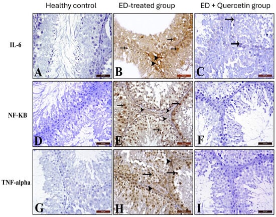
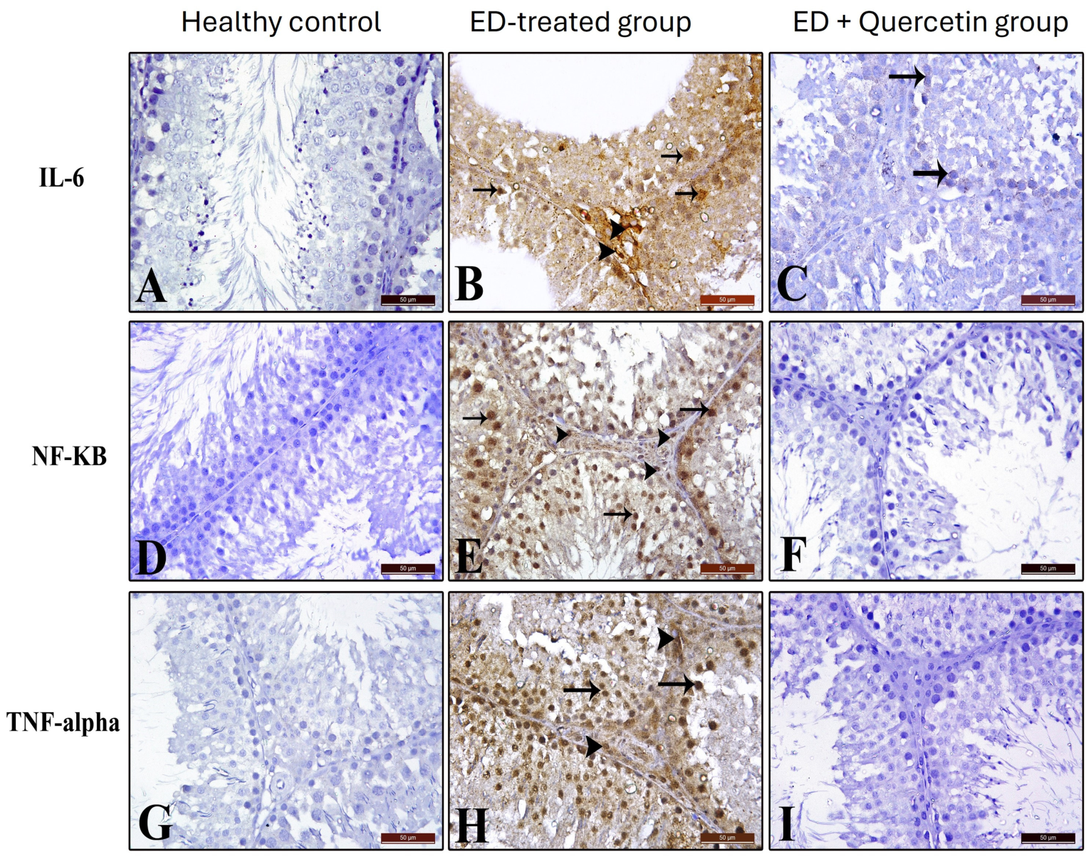
Testicular sections from the energy drink-treated group showed a significant expression of the inflammatory markers IL-6, NF-κB, and TNF-α in spermatogenic cells (arrows) and interstitial tissue (arrowhead, Figure 4B,E,H) compared to healthy control animals, which showed almost no immune reactions (Figure 4A,D,G). Interestingly, co-administration of energy drinks with quercetin virtually prevented the energy drink-induced inflammatory response, as can be observed in the form of almost complete absence of expression signals for all three markers except for IL-6, which showed a weak signal in spermatogenic cells (arrows) (Figure 4C,F,I). Quantitative analysis of immunoreactive signal intensity further confirmed these findings, revealing significantly increased IL-6, NF-κB, and TNF-α immunoreactivity in the energy drink-treated group compared with the control group (p < 0.05). Quercetin co-administration significantly reduced the immunoreactivity of all three inflammatory markers compared with the energy drink-treated group (p < 0.05), as presented in Table 5.
Figure 4.
Expression of the inflammatory markers IL-6, NF-κB, and TNF-α in the testicular tissues of the experimental animals. (A,D,G) Testicular sections from the healthy control group showing negative expression of IL-6, NF-κB, and TNF-α throughout the tissue. (B,E,H) Sections from the energy drink (ED)-treated group showing strong expression of all three markers in the spermatogenic cells (arrows) and interstitial tissue (arrowheads). (C,F,I) Sections from the ED + quercetin-treated group showing no expression of NF-κB or TNF-α and very weak IL-6 expression in the spermatogenic cells (arrows in panel C) with no expression in the interstitial tissue. Scale bars = 50 µm.
Table 5.
Quantitative assessment of immunoreactive signal intensity for inflammatory cytokines in testicular tissue of test groups.
4. Discussion
The current study reveals the development of severe testicular dysfunction upon chronic ED intake in adult male rats, involving alteration in testosterone serum levels, provoking inflammation as shown by elevated Il-6, TNF-α and NF-κB expression levels, and inducing widespread histopathological changes in the testicular tissue as well. Co-administration of quercetin with energy drinks for the same period alleviated these effects, where testosterone serum levels and inflammatory cytokine expression were comparable to those of healthy control animals. Quercetin effectively protected testicular tissue from the damaging effects of ED, revealing an almost normal architecture and retrieving spermatid count and Leydig cell activity. The observed effects of ED strongly correlate the chronic ED administration with the deterioration of testis structure and function as previously reported [7,9,18].
A key finding of this study is the significant reduction in serum testosterone levels following chronic ED administration, accompanied by degeneration and functional impairment of Leydig cells. Testosterone synthesis is primarily regulated by the hypothalamic–pituitary–gonadal axis and is highly dependent on intact Leydig cell steroidogenic activity [29,30]. The observed reduction in testosterones may be attributed, at least in part, to the pharmacological effects of caffeine as a major component of energy drinks. Caffeine acts as a non-selective adenosine receptor antagonist and may interfere with intracellular cAMP signaling pathways, which are essential for steroidogenesis in Leydig cells [31]. Disruption of this signaling cascade could therefore contribute to reduced testosterone production and impaired reproductive function [32].
It is to be mentioned, however, that the connection between caffeine intake and its effect on testosterone has been contradictory in the literature. Numerous studies have demonstrated positive effects for caffeine intake on performance by regulating serum levels of certain hormones such as testosterone and cortisol. However, these effects seemed to be dose-dependent, as low doses of caffeine exerted a positive effect on testosterone levels but high doses of caffeine were inversely linked with testosterone levels [31]. In addition, caffeine effect on testosterone levels could vary greatly depending on genetics and polymorphism, health conditions, and lifestyles [33]. It is to be noted that early and prepubertal exposure to caffeine negatively impacted the development and maturation of testicular tissue [32].
The chronic exposure to ED also induced an inflammatory response within the testicular tissue where expression levels of the inflammatory cytokines NF-κB, TNF-α, and IL-6 were upregulated both at the mRNA and protein levels. NF-κB is a transcription factor that plays a crucial role in aiding various physiological effects in addition to its role in initiating the cellular inflammatory response and mediating transcriptional activation of multiple, downstream pro-inflammatory cytokines like TNF-α and IL-6 [34]. NF-κB can also activate some enzymes, such as inducible nitric oxide synthase (iNOS), resulting in excessive production of nitric oxide (NO), which induces vasodilatation. Consequently, tissue perfusion declines allowing for edema and tissue/organ damage. Another dark side for NO is its ability to interact with ROS, producing the cytotoxic peroxynitrite radicals that can induce further tissue damage [18]. A study from Ahmed AM reported a significant upregulation in NF-κB and COX-2 upon intake of ED when he tried to investigate the effects on murine testis after chronic intake [18].
High levels of NF-κB, TNF-α and IL-6 were shown to disrupt the blood–testis barrier (BTB) by affecting Sertoli cells and tight junction proteins, interfering with spermatogenesis. However, other factors such as oxidative stress and the presence of different environmental toxins could be involved in damaging BTB [35]. In contrast, a recent study reported that NF-κB activation may help promote cell regeneration after damage and inflammation [36] and, thus, might stimulate spermatogenesis rather than hampering it.
Chronic ED administration in the current study resulted in many histopathological alterations in testicular tissue including interstitial edema, vascular congestion, and degeneration of germinal cells. The observed tissue damage can be attributed to high levels of oxidative stress that provoked inflammation, in agreement with previously published data [3,12].
Testes from ED-exposed rats showed a significant decrease in seminiferous tubular diameter, germinal epithelial thickness, spermatogenic cell count, and Leydig cell count. These findings indicate injury to testicular architecture, which is fundamental for proper spermatogenesis. The current results are consistent with previously published data showing dose-dependent testicular damage upon energy drink intake [32]. Long-term consumption of energy rinks, e.g., Code Red®, was reported to induce lipid peroxidation, diminish the endogenous antioxidants SOD and GSH and provoke apoptosis in addition to histological damage of seminiferous tubules [7]. In contrast to these findings, some researchers reported that regular use of stimulants and caffeine-containing beverages can possess insignificant effects on testicle condition and function [37,38]. It is to be taken into consideration that the variability of individual responses to these chemicals could be attributed to differences in genetics, lifestyles, and the general health of the participants.
In the current study, the concomitant administration of ED with quercetin protected against the pathological changes brought about by ED. This was observed in the form of preserved histologic architecture of testicular tissue accompanied by the significant restoration of serum testosterone levels as well as the alleviation of the inflammatory status marked by normalized inflammatory cytokine expression.
Quercetin is emerging as an important multifunctional flavonoid compound that can modulate essential molecular pathways associated with inflammatory and steroidogenic disorders [39]. Interestingly, quercetin was reported to protect bovine sperms against ferrous-ascorbate-induced oxidative damage [24]. Similar findings were observed when quercetin was used in testicular damage models induced by gentamicin and lead acetate, where it alleviated oxidative damage and apoptosis, and retrieved normal tissue architecture and functions [23,26]. The cytoprotective effects of quercetin can be in part attributed to its well-known antioxidant and anti-inflammatory activities [23,24,40], where its anti-inflammatory capacity is a result of a multi-mechanism approach. On the one hand, it induces the synthesis of reduced glutathione (GSH) via activating the corresponding enzymes and directly scavenges reactive oxygen species (ROS), thus preventing oxidative stress, the main factor driving the inflammatory response in different tissues. Interestingly, quercetin was reported to protect bovine sperms against ferrous-ascorbate-induced oxidative damage via these two effects [24]. In addition to the direct ROS scavenging capacity of quercetin, it can indirectly mitigate oxidative stress by enhancing the levels of antioxidant enzymes like superoxide dismutase (SOD), catalase (CAT), and glutathione peroxidase (GPx). In fact, the antioxidant effect of quercetin provides efficient protection against lipid peroxidation and DNA damage, maintaining cell membrane integrity in lipid-rich tissues such as the testicular tissue [41]. On the other hand, quercetin inhibits the inflammatory responses via its ability to interfere with NF-κB signaling, which is a major driver of the inflammatory process [42]. Through suppression of inflammatory responses and preservation of the integrity of steroidogenic cells, quercetin appears to counteract functional as well as structural damage caused by ED exposure.
NF-κB signaling pathway is a main checkpoint controlling expression and activation of downstream cytokines. Under oxidative stress conditions, activation of IκB kinase (IKK) results in the phosphorylation and breakdown of IκBα, leading to activation and nuclear translocation of NF-κB (p65/p50) with the subsequent transcriptional activation of the pro-inflammatory cytokines, e.g., TNF-α, IL-6, and IL-1β. The anti-inflammatory effects of quercetin were linked to its ability to prevent phosphorylation and breakage of IκBα, interfering with nuclear translocation of NF-κB protein (p65/p50) and suppressing cytokine production [7,43,44].
In addition to its anti-inflammatory actions, quercetin was reported to play an important role in preserving the steroidogenic ability of Leydig cells [39]. Biosynthesis of testosterone is facilitated by the presence of the steroidogenic acute regulatory protein (StAR) together with other enzymes, namely the cholesterol side-chain cleavage enzyme (CYP11A1), 3β-hydroxysteroid dehydrogenase (3β-HSD), and 17β-hydroxysteroid dehydrogenase (17β-HSD). According to experimental evidence, oxidative stress and inflammatory cytokines adversely affect the expression of StARs and impair the transfer of cholesterol to mitochondria, thereby limiting the amount of testosterone produced by the affected Leydig cells [45,46]. Quercetin has been found to restore the expression of StARs and mitochondrial functionality, thus maintaining the steroidogenic potential of Leydig cells exposed to any toxin-related insults [39,45,47]. Additionally, by inhibiting TNF-α signaling, quercetin counteracts the cytokine-mediated inhibition of luteinizing hormone receptor (LHR)-mediated signaling, which is crucial for the activation of steroidogenesis via the increase in cAMP concentration. In view of this dual action on inflammatory responses and steroidogenic enzymes, quercetin can account for the preservation of testosterone levels and cellular structure of Leydig cells observed in experimental reproductive toxicity studies.
The findings of this study also correspond with other findings showing that excessive energy drink intake is associated with systemic metabolic dysfunction and multi-organ toxicity, including disorders in lipid metabolism and kidney damage [15]. Since the testes are highly metabolically sensitive organs, these systemic problems can result in enhanced oxidative and inflammatory damage to the testis, ultimately leading to reproductive impairment [48].
5. Conclusions
In conclusion, the prolonged intake of energy drinks can result in considerable testicular injury in the form of hormonal inhibition and histological damage in response to the oxidative stress and inflammatory status. These deleterious effects can be effectively ameliorated by the co-administration of quercetin via its antioxidant and anti-inflammatory properties, where it scavenges free radicals generated after energy drink consumption and prevents the inflammatory cascade.
6. Limitations and Future Directions
To our knowledge, this is the first study to assess the protective effect of quercetin against chronic energy drink-induced testicular damage. The study, however, has some limitations that must be acknowledged, such as evaluating the changes in endogenous antioxidant capacity (GSH, catalase, SOD and lipid peroxidation) in addition to fresh semen analysis, sperm DNA integrity and fertility indices. These assessments were out of the scope for the current study, which focused on molecular and histological findings. Future studies should focus on investigating the andrological and fertility parameters and assessing the endogenous antioxidant capacity, in addition to replicating the study in human subjects to clarify the relevance of such transfer.
Author Contributions
Conceptualization, A.A.B. and A.J.A.; methodology, M.S.A., A.J.A. and M.A.; software, W.A.M.-S. and H.E.; validation, S.S.A.T. and H.E.; formal analysis, M.S.A., E.A.A., W.A.M.-S. and M.A.; resources, E.A.A., S.M.E. and M.A.; data curation, W.A.M.-S.; writing—original draft preparation, M.A. and H.E.; writing—review and editing, M.A.A., M.S.A., E.A.A., Z.E., F.R.H. and M.A.; visualization, S.M.E.; supervision, A.A.B., S.M.E. and M.A.; funding acquisition, M.A.A., Z.E., F.R.H., S.S.A.T. and A.J.A. All authors have read and agreed to the published version of the manuscript.
Funding
This research received no external funding.
Institutional Review Board Statement
The animal study protocol was approved by the Institutional Review Board of the College of Pharmacy, Taibah University (Approval No. COPTU-REC-118-20241222), on 22 December 2024.
Informed Consent Statement
Not applicable.
Data Availability Statement
The original contributions presented in this study are included in the article. Further inquiries can be directed to the corresponding authors.
Conflicts of Interest
The authors declare no conflicts of interest.
Abbreviations
The following abbreviations are used in this manuscript:
| ED | Energy drink |
| NF-κB | Nuclear factor-kappa B |
| TNF-α | Tumor necrosis factor-α |
References
- Visram, S.; Cheetham, M.; Riby, D.M.; Crossley, S.J.; Lake, A.A. Consumption of energy drinks by children and young people: A rapid review examining evidence of physical effects and consumer attitudes. BMJ Open 2016, 6, e010380. [Google Scholar] [CrossRef] [Scilit] [PubMed]
- Peacock, A.; Martin, F.H.; Carr, A. Energy drink ingredients. Contribution of caffeine and taurine to performance outcomes. Appetite 2013, 64, 1–4. [Google Scholar] [CrossRef] [Scilit] [PubMed]
- Costantino, A.; Maiese, A.; Lazzari, J.; Casula, C.; Turillazzi, E.; Frati, P.; Fineschi, V. The Dark Side of Energy Drinks: A Comprehensive Review of Their Impact on the Human Body. Nutrients 2023, 15, 3922. [Google Scholar] [CrossRef] [Scilit] [PubMed]
- Burrows, T.; Pursey, K.; Neve, M.; Stanwell, P. What are the health implications associated with the consumption of energy drinks? A systematic review. Nutr. Rev. 2013, 71, 135–148. [Google Scholar] [CrossRef] [Scilit] [PubMed]
- Ali, F.; Rehman, H.; Babayan, Z.; Stapleton, D.; Joshi, D.D. Energy drinks and their adverse health effects: A systematic review of the current evidence. Postgrad. Med. 2015, 127, 308–322. [Google Scholar] [CrossRef] [Scilit] [PubMed]
- Rajeh, N.A.; Al-Shaikh, T.M. Ameliorating effect of blueberry consumption on energy drink-induced testicular damage in rats: Histological and immunohistochemical study. J. Basic Appl. Zool. 2023, 84, 9. [Google Scholar] [CrossRef] [Scilit]
- Naqvi, Q.A.; Fatima, S.A.; Haider, A. Protective role of omega-3 fatty acids against energy drink–induced structural changes in the testes of adult albino Wistar rats. J. Shalamar Med. Dent. Coll. 2025, 6, 90–96. [Google Scholar] [CrossRef] [Scilit]
- Schuchowsky, E.; Schaefer, D.; Salvador, R.A.; do Nascimento, A.E.; Til, D.; Senn, A.P.; Amaral, V.L.L. Effects of energy drinks on biochemical and sperm parameters in Wistar rats. Nutrire 2017, 42, 22. [Google Scholar] [CrossRef] [Scilit]
- Murad, W.A.; Abou-Elghait, A.T.; Abdelzaher, L.A.; Abbass, T.; Ali, S.S. Comparative Histological and Immunohistochemical Study of the Effects of Energy Drinks and Cessation on The Testicular Structure of Adult and Young Male Albino Rats. Int. J. Drug Deliv. Technol. 2026, 16, 1596–1619. [Google Scholar] [CrossRef] [Scilit]
- Al-Hayder, M.N.; Al-Mayyahi, R.S.; Almahdi, Z.A. Pathological Effects on the Thyroid and Testes Induced by Prolonged Consumption of Caffeinated Energy Drinks in Adolescent Wistar Rats. Curr. Nutr. Food Sci. 2026, 22, 377–384. [Google Scholar] [CrossRef] [Scilit]
- Jouda, J. Incomplete spermatogenesis, leukocytosis and thrombocytosis appeared with energy drink consumption in mice at differences consuming periods and concentrations. Adv. Anim. Vet. Sci. 2019, 7, 356–360. [Google Scholar] [CrossRef] [Scilit]
- Amanjide, F.O.; Nwakanma, A.A.; Idaguko, C.A.; Okafor, S.R. Energy Drink (Fearless) Effect on Sperm Parameters and Testicular Histology of Wistar rats. J. Exp. Clin. Anat. 2023, 20, 1–7. [Google Scholar]
- Nwobodo, E.; Orjiako, R.; Dike, H.; Okechukwu, E.; Chukwuanukwu, T.; Nzube Ngini, P.; Eze Okafor, E. Effects of excessive administration of caffeine on the size and histology of the testesof adult male Wistar rats. Magna Sci. Adv. Res. Rev. 2026, 17, 120–127. [Google Scholar] [CrossRef] [Scilit]
- Costa-Valle, M.T.; Tonieto, B.D.; Altknecht, L.; Cunha, C.D.; Fao, N.; Cestonaro, L.V.; Goethel, G.; Garcia, S.C.; Leal, M.B.; Dallegrave, E.; et al. Energy drink and alcohol combination leads to kidney and liver alterations in rats. Toxicol. Appl. Pharmacol. 2018, 355, 138–146. [Google Scholar] [CrossRef] [Scilit] [PubMed]
- Alhajri, A.S. Chronic consumption of energy drinks and the risk of developing metabolic syndrome in rats. PeerJ 2026, 14, e20926. [Google Scholar] [CrossRef] [Scilit] [PubMed]
- Eltahir, H.M.; Alamri, G.; Alamri, A.; Aloufi, A.; Nazmy, M.; Elbadawy, H.; Bahashwan, S.; Elbadawy, H.M.; Alahmadi, Y.M.; Abouzied, M. The metabolic disorders associated with chronic consumption of soft and energy drinks in rats. Acta Biochim. Pol. 2020, 67, 79–84. [Google Scholar] [CrossRef] [Scilit] [PubMed]
- Xu, S.; Li, X.; Shen, X.; Guo, X.; Peng, Y.; Liu, Y.; Zhao, Q.; Feng, S.; Wang, F. Activation of NF-κ B signalling pathway by inflammatory regulators and associated drug discovering. Internet J. Med. Update 2010, 5, 38–44. [Google Scholar] [CrossRef] [Scilit]
- Mohamed, A.A. Expression of transcription factor NF-KAPPA B/P65 and cyclooxygenase-2(COX-2) in testicular damage induced by Red Bull energy drink in rat. Int. J. Adv. Appl. Sci. 2016, 3, 49–56. [Google Scholar] [CrossRef] [Scilit]
- Lampiao, F.; du Plessis, S.S. TNF-alpha and IL-6 affect human sperm function by elevating nitric oxide production. Reprod. Biomed. Online 2008, 17, 628–631. [Google Scholar] [CrossRef] [Scilit] [PubMed]
- Çelikezen, F.Ç. Quercetin: From Molecular Structure to Therapeutic Horizons; Academician Publishing: Singapore, 2026. [Google Scholar]
- Zhang, X.; Tang, Y.; Lu, G.; Gu, J. Pharmacological Activity of Flavonoid Quercetin and Its Therapeutic Potential in Testicular Injury. Nutrients 2023, 15, 2231. [Google Scholar] [CrossRef] [Scilit] [PubMed]
- Bastin, A.; Teimouri, M.; Faramarz, S.; Shabani, M.; Doustimotlagh, A.H.; Sadeghi, A. In vitro and Molecular Docking Analysis of Quercetin as an Anti-inflammatory and Antioxidant. Curr. Pharm. Des. 2023, 29, 883–891. [Google Scholar] [CrossRef] [Scilit] [PubMed]
- Behairy, A.; Hashem, M.M.; Abo-El-Sooud, K.; El-Metwally, A.E.; Hassan, B.A.; Abd-Elhakim, Y.M. Quercetin Abates Aluminum Trioxide Nanoparticles and Lead Acetate Induced Altered Sperm Quality, Testicular Oxidative Damage, and Sexual Hormones Disruption in Male Rats. Antioxidants 2022, 11, 2133. [Google Scholar] [CrossRef] [Scilit] [PubMed]
- Tvrda, E.; Tusimova, E.; Kovacik, A.; Paal, D.; Libova, L.; Lukac, N. Protective Effects of Quercetin on Selected Oxidative Biomarkers in Bovine Spermatozoa Subjected to Ferrous Ascorbate. Reprod. Domest. Anim. 2016, 51, 524–537. [Google Scholar] [CrossRef] [Scilit] [PubMed]
- Nna, V.U.; Ujah, G.A.; Mohamed, M.; Etim, K.B.; Igba, B.O.; Augustine, E.R.; Osim, E.E. Cadmium chloride-induced testicular toxicity in male wistar rats; prophylactic effect of quercetin, and assessment of testicular recovery following cadmium chloride withdrawal. Biomed. Pharmacother. 2017, 94, 109–123. [Google Scholar] [CrossRef] [Scilit] [PubMed]
- Biyik, A.F.; Yulug, E.; Yenilmez, E.; Kutlu, A.; Alver, A.; Erdem, S. Effects of quercetin on gentamicin-induced experimental testicular injury in rats. Rom. J. Morphol. Embryol. 2024, 65, 69–80. [Google Scholar] [CrossRef] [Scilit] [PubMed]
- Gibson-Corley, K.N.; Olivier, A.K.; Meyerholz, D.K. Principles for valid histopathologic scoring in research. Vet. Pathol. 2013, 50, 1007–1015. [Google Scholar] [CrossRef] [Scilit] [PubMed]
- Xi, L.; Wang, C.; Chen, P.; Yang, Q.; Hu, R.; Zhang, H.; Weng, Q.; Xu, M. Expressions of IL-6, TNF-alpha and NF-kappaB in the skin of Chinese brown frog (Rana dybowskii). Eur. J. Histochem. 2017, 61, 2834. [Google Scholar] [CrossRef] [Scilit] [PubMed]
- Huhtaniemi, I.; Toppari, J. Endocrine, paracrine and autocrine regulation of testicular steroidogenesis. Adv. Exp. Med. Biol. 1995, 377, 33–54. [Google Scholar] [CrossRef] [Scilit] [PubMed]
- Yang, Z.W.; Kong, L.S.; Guo, Y.; Yin, J.Q.; Mills, N. Histological changes of the testis and epididymis in adult rats as a result of Leydig cell destruction after ethane dimethane sulfonate treatment: A morphometric study. Asian J. Androl. 2006, 8, 289–299. [Google Scholar] [CrossRef] [Scilit] [PubMed]
- Glover, F.E.; Caudle, W.M.; Del Giudice, F.; Belladelli, F.; Mulloy, E.; Lawal, E.; Eisenberg, M.L. The association between caffeine intake and testosterone: NHANES 2013-2014. Nutr. J. 2022, 21, 33. [Google Scholar] [CrossRef] [Scilit] [PubMed]
- Bae, J.; Choi, H.; Choi, Y.; Roh, J. Dose- and time-related effects of caffeine on the testis in immature male rats. Exp. Anim. 2017, 66, 29–39. [Google Scholar] [CrossRef] [Scilit] [PubMed]
- Advani, A. Introduction: Defining the role of testosterone in the male. J. Pharm. Pract. 2011, 24, 365. [Google Scholar] [PubMed]
- Mitchell, J.P.; Carmody, R.J. NF-kappaB and the Transcriptional Control of Inflammation. Int. Rev. Cell Mol. Biol. 2018, 335, 41–84. [Google Scholar] [CrossRef] [Scilit] [PubMed]
- Feng, R.; Adeniran, S.O.; Huang, F.; Li, Y.; Ma, M.; Zheng, P.; Zhang, G. The ameliorative effect of melatonin on LPS-induced Sertoli cells inflammatory and tight junctions damage via suppression of the TLR4/MyD88/NF-kappaB signaling pathway in newborn calf. Theriogenology 2022, 179, 103–116. [Google Scholar] [CrossRef] [Scilit] [PubMed]
- Liu, D.; Zhong, Z.; Karin, M. NF-kappaB: A Double-Edged Sword Controlling Inflammation. Biomedicines 2022, 10, 1250. [Google Scholar] [CrossRef] [Scilit] [PubMed]
- Tóthová, Ľ.; Hodosy, J.; Mettenburg, K.; Fábryová, H.; Wagnerová, A.; Bábíčková, J.; Okuliarová, M.; Zeman, M.; Celec, P. No harmful effect of different Coca-cola beverages after 6 months of intake on rat testes. Food Chem. Toxicol. 2013, 62, 343–348. [Google Scholar] [CrossRef] [Scilit] [PubMed]
- Smith, A.P.; Richards, G. Energy drinks, caffeine, junk food, breakfast, depression and academic attainment of secondary school students. J. Psychopharmacol. 2018, 32, 893–899. [Google Scholar] [CrossRef] [Scilit] [PubMed]
- Wu, X.; Zhao, H.; Huang, X.; Lu, P.; Zhang, R.; Guan, Q.; Yu, C. The role and mechanism of quercetin in improving late-onset hypogonadism through network analysis and experimental validation. Naunyn Schmiedebergs Arch. Pharmacol. 2025, 398, 12031–12046. [Google Scholar] [CrossRef] [Scilit] [PubMed]
- Qi, W.; Qi, W.; Xiong, D.; Long, M. Quercetin: Its Antioxidant Mechanism, Antibacterial Properties and Potential Application in Prevention and Control of Toxipathy. Molecules 2022, 27, 6545. [Google Scholar] [CrossRef] [Scilit] [PubMed]
- Boots, A.W.; Haenen, G.R.; Bast, A. Health effects of quercetin: From antioxidant to nutraceutical. Eur. J. Pharmacol. 2008, 585, 325–337. [Google Scholar] [CrossRef] [Scilit] [PubMed]
- Martinez-Florez, S.; Gutierrez-Fernandez, B.; Sanchez-Campos, S.; Gonzalez-Gallego, J.; Tunon, M.J. Quercetin attenuates nuclear factor-kappaB activation and nitric oxide production in interleukin-1beta-activated rat hepatocytes. J. Nutr. 2005, 135, 1359–1365. [Google Scholar] [CrossRef] [Scilit] [PubMed]
- David, A.V.A.; Arulmoli, R.; Parasuraman, S. Overviews of biological importance of quercetin: A bioactive flavonoid. Pharmacogn. Rev. 2016, 10, 84. [Google Scholar] [CrossRef] [Scilit] [PubMed]
- Li, Y.; Yao, J.; Han, C.; Yang, J.; Chaudhry, M.T.; Wang, S.; Liu, H.; Yin, Y. Quercetin, inflammation and immunity. Nutrients 2016, 8, 167. [Google Scholar] [CrossRef] [Scilit] [PubMed]
- Manna, P.R.; Dyson, M.T.; Stocco, D.M. Regulation of the steroidogenic acute regulatory protein gene expression: Present and future perspectives. Mol. Hum. Reprod. 2009, 15, 321–333. [Google Scholar] [CrossRef] [Scilit] [PubMed]
- Diemer, T.; Allen, J.A.; Hales, K.H.; Hales, D.B. Reactive oxygen disrupts mitochondria in MA-10 tumor Leydig cells and inhibits steroidogenic acute regulatory (StAR) protein and steroidogenesis. Endocrinology 2003, 144, 2882–2891. [Google Scholar] [CrossRef] [Scilit] [PubMed]
- Cormier, M.; Ghouili, F.; Roumaud, P.; Martin, L.J.; Touaibia, M. Influence of flavonols and quercetin derivative compounds on MA-10 Leydig cells steroidogenic genes expressions. Toxicol. Vitr. 2017, 44, 111–121. [Google Scholar] [CrossRef] [Scilit] [PubMed]
- Moustafa, A. Effect of Omega-3 or Omega-6 Dietary Supplementation on Testicular Steroidogenesis, Adipokine Network, Cytokines, and Oxidative Stress in Adult Male Rats. Oxid. Med. Cell. Longev. 2021, 2021, 5570331. [Google Scholar] [CrossRef] [Scilit] [PubMed]

Disclaimer/Publisher’s Note: The statements, opinions and data contained in all publications are solely those of the individual author(s) and contributor(s) and not of MDPI and/or the editor(s). MDPI and/or the editor(s) disclaim responsibility for any injury to people or property resulting from any ideas, methods, instructions or products referred to in the content. |
© 2026 by the authors. Licensee MDPI, Basel, Switzerland. This article is an open access article distributed under the terms and conditions of the Creative Commons Attribution (CC BY) license.