


Abstract
Sotos syndrome, an autosomal dominant neurodevelopmental disorder caused by pathogenic variants in the NSD1 gene, exhibits a distinct genome-wide DNA methylation episignature characterized by widespread hypomethylation. While genome-wide methylation signatures provide robust diagnostic accuracy, their implementation in routine clinical practice is limited by cost and technical complexity. This study aimed to reduce the Sotos syndrome episignature to a confined set of CpG sites intended for Sanger bisulfite sequencing. We selected four loci containing CpG dinucleotides with the most significant methylation changes based on published data. Bisulfite Sanger sequencing was performed on patients with the molecularly confirmed Sotos syndrome and healthy controls. The obtained methylation levels of the analyzed loci were then used as features to build a binary classification model using machine learning methods. We employed the Random Forest-based method to conduct a feature selection, which identified two CpG sites (chr6:28601375 and chr6:28601440) as the most important predictors. A logistic regression classification model was developed using selected CpG sites. The resulting two-predictor model demonstrated excellent performance and achieved 100% accuracy on the held-out test set. All Sotos syndrome samples were classified correctly, while Malan syndrome samples showed methylation levels comparable to controls. Two samples with VUS in the NSD1 gene were classified together with all samples with pathogenic NSD1 variants. For these two cases, we performed a de novo assessment of the variant status and reclassified the variants (NSD1(NM_022455.5): c.6523T>C and c.8005del, respectively) into likely pathogenic, confirming the validity of the results of the reduced episignature analysis. Thus, our cost-effective approach can potentially serve as a supporting diagnostic and research tool, particularly for variant reclassification and differential diagnosis in resource-limited settings.
1. Introduction
1.1. Episignature as a Robust Biomarker for Chromatinopathies
Neurodevelopmental disorders constitute a genetically heterogeneous group of conditions characterized by a broad phenotypic spectrum, including behavioral and cognitive impairments as well as developmental delay. One of the causes of neurodevelopmental disorders, particularly chromatinopathies, is genetic alterations in various components of the epigenetic machinery. Currently, methods for molecular diagnostics of neurodevelopmental disorders are based on the use of targeted gene panels, exome sequencing, and genomic sequencing. While these methods successfully identify the genetic etiology of the disorder in many cases, a large proportion of patients remain undiagnosed. Molecular genetic testing of this group of disorders is challenging due to the complexity of interpreting the clinical significance of genetic variants. Furthermore, diagnostic uncertainty is frequently complicated by phenotypically overlapping clinical features of numerous chromatinopathies. This underscores the need for supplementary diagnostic methods based on the use of reliable biomarkers. One of the most promising and commonly studied approaches to the indirect diagnosis of chromatinopathies is genome-wide methylation analysis. Due to the disruption in genes encoding epigenetic regulators and subsequent shift in the epigenetic landscape, a unique genome-wide DNA methylation pattern, or episignature, arises in a disorder-specific manner. To date, more than 80 unique and reproducible DNA methylation signatures have been reported in the literature, each associated with a specific rare disorder [1,2,3,4] EpiSign Knowledge Database https://episign.ca (accessed on 6 August 2026). Machine learning classifiers based on DNA methylation profiles can provide accurate diagnosis even in the absence of the established genetic variant; moreover, identifying an episignature can prompt reanalysis of genetic variants in regulatory and noncoding regions, as well as reclassification of previously identified variants of uncertain significance (VUS) [2,5].
One of the most robust episignatures was identified for Sotos syndrome (OMIM:117550), demonstrating 100% sensitivity [3]. Sotos syndrome is an autosomal dominant disorder characterized by childhood overgrowth, intellectual disability, and a distinctive facial appearance, including broad and prominent forehead, macrocephaly, dolichocephaly, long narrow face, pointed chin, and hypertelorism [6]. Sotos syndrome is caused by pathogenic variants in the NSD1 gene, which are detected in over 90% of the cases. NSD1 encodes a component of the epigenetic machinery, the histone lysine methyltransferase NSD1, which dimethylates lysine 36 on histone H3 (H3K36me2) [2]. This histone post-translational modification plays a critical role in recruiting DNA methyltransferases and is essential for both the intergenic localization of DNMT3A and the maintenance of DNA methylation [7]. Choufani et al. identified distinct DNA methylation profiles in Sotos syndrome patients compared to controls, detecting 7085 differentially methylated CpG sites. Among these, 99.3% (7038 sites) showed hypomethylation, while only 0.7% (47 sites) exhibited hypermethylation [5]. Thus, a significant decrease in methylation levels in patients with Sotos syndrome can potentially be used as a biomarker to clarify the diagnosis in unresolved cases.
1.2. Approaches to the Episignature Detection
The most widely used method for genome-wide DNA methylation analysis is the Illumina Infinium Methylation Assay, which interrogates approximately 450,000 (450 K) to 930,000 (EPIC v2) CpG sites in the human genome. While the human genome contains over 28 million CpG sites, the Illumina Infinium Methylation technology provides coverage of only approximately 3% of these dinucleotides. Whole-genome bisulfite sequencing (WGBS) is employed for comprehensive DNA methylation profiling [8]. Furthermore, rapidly advancing high-throughput sequencing technologies, including nanopore sequencing, represent promising approaches for simultaneous DNA methylation and genetic variation analysis [9,10]. However, these approaches are time-consuming, expensive and require lengthy data processing and interpretation, which complicates their implementation into routine diagnostic practice. Alternatively, Sanger bisulfite sequencing can be utilized for targeted analysis of specific genomic regions or individual CpG sites. If the number of CpG sites required for analysis is successfully reduced, Sanger bisulfite sequencing can become an auxiliary tool for optimizing the diagnosis of Sotos syndrome.
Reducing the number of analyzed CpG dinucleotides requires the use of special methods based on machine learning. First, it is necessary to carry out feature selection, i.e., to identify those CpG sites whose methylation level is most significant for separating pathological and control samples into two different classes. Random forests (RFs) are among the most widely used approaches, growing an ensemble of decision trees on bootstrap subsets of the training data and ranking features by their contribution to classification accuracy [11]. Then, using only the significant features (or predictors), a classification model can be built that, using the methylation level of specific loci as input data, will be able to predict whether a sample belongs to the class of pathological or control samples. Logistic regression is the foundational linear model for binary outcomes: it models the log-odds of class membership as a linear combination of features and applies a sigmoid function to produce a probability between 0 and 1 [12]. Support vector machines (SVMs) are also among the most widely adopted supervised learning algorithms for binary classification. The core principle of an SVM is to identify a decision boundary that separates the two classes while maximizing the margin between them and the nearest training samples, which are termed support vectors [13].
In this study, we demonstrate that the genome-wide episignature for Sotos syndrome can be reduced to a confined set of individual CpG sites. We implement several methods including RF, SVM and logistic regression. This approach enables episignature detection using a targeted method available in most genetic laboratories. Furthermore, we developed a classification model for Sotos syndrome detection based on a reduced episignature, enabling accurate sample classification into diagnostic groups. This classification model can be potentially implemented as a supplementary diagnostic tool for unresolved clinical cases with Sotos syndrome phenotype.
2. Materials and Methods
2.1. Study Cohort
The study included 31 patients with a referral diagnosis of Sotos syndrome. Among them, 26 had a pathogenic or likely pathogenic variant in the NSD1 gene, 2 carried variants of uncertain significance in NSD1, and 3 patients had a variant in the NFIX gene associated with Malan syndrome (OMIM:614753), a disorder with clinical features that overlap with Sotos syndrome. The spectrum of identified variants is presented in Table 1. Sample 46, which carries the NSD1 variant NM_022455.5:c.8005del, p.(Gln2669LysfsTer92), received an ambiguous interpretation (VUS/likely pathogenic) due to its proximity to the 3′ end of the NSD1 gene. Consequently, it was classified within the group of samples with variants of uncertain significance. The control group consisted of 11 samples obtained from healthy individuals. Samples with pathogenic/likely pathogenic variants in the NSD1 gene and control samples from healthy individuals were used to design the binary classification model, and samples with VUS and NFIX variants were used to assess the overall classification ability of the model.
Table 1.
Sample ID and corresponding genetic variants identified via molecular genetic testing.
2.2. DNA Extraction and Bisulfite Conversion
Genomic DNA was extracted from peripheral blood samples using standard protocols. Bisulfite conversion was performed using the EpiTect Fast Bisulfite Kit (QIAGEN, Hilden, Germany), according to the manufacturer’s instructions.
2.3. Selection of CpG Dinucleotides
CpG dinucleotides showing the most significant methylation differences between control and experimental samples, the lowest p-values, and in close proximity to one another were selected based on the study by S. Choufani et al., which utilized Illumina Infinium HumanMethylation450 (450 K) arrays for methylation profiling [5]. Amplicons with an average length of approximately 250 bp were generated for Sanger bisulfite sequencing of the selected CpG sites. Bisulfite-specific PCR primers were designed using MethPrimer software (version 2.0) [14].
2.4. Sanger Bisulfite Sequencing and DNA Methylation Analysis
Sanger sequencing of bisulfite-treated amplicons was performed on a 3500 Genetic Analyzer (Applied Biosystems, Thermo Fisher Scientific, Waltham, MA, USA). Quantitative methylation values for each CpG dinucleotide were derived from the resulting electropherograms using an in-house R-based algorithm referencing unmethylated model amplicons, as detailed previously [15]. Briefly, model amplicons were generated by amplifying the same target regions prior to bisulfite conversion; their signal intensities served to correct and normalize the electropherogram signal of experimental samples, yielding methylation values on a 0–100% scale. The R script is available from the corresponding author upon request.
Data Preprocessing and Feature Selection
Methylation percentages obtained as described in the section above were used as candidate predictors. The bis-649 amplicon was excluded because of poor electropherogram quality. Adjacent CpG measurements with similar methylation patterns were averaged to reduce variation in sequencing signals.
Feature selection was performed on the training data using Boruta, a wrapper method that compares Random Forest-derived predictor importance with that of randomly permuted shadow features [16]. Random Forest is an ensemble of randomized decision trees and served as the importance-estimation component of Boruta [11]. Each Random Forest fitted within the Boruta procedure comprised 500 trees, corresponding to the default setting. A maximum of 500 importance-estimation runs was allowed. Remaining tentative decisions were resolved, and retained predictors were ranked by mean importance. The five highest-ranked predictors were taken forward for comparison of their combinations using linear SVMs.
For SVM fitting, samples with missing values in the predictors included in each model were omitted. For logistic regression, samples missing both selected predictors were excluded, whereas remaining missing values were replaced by diagnostic-group means estimated from the training data. Predictors were centered and scaled with StandardScaler transformer before classification.
2.5. Classification Model Design
Following quantification of DNA methylation at the studied CpG dinucleotides, each CpG site was evaluated as a potential predictor. The dataset comprising samples from Sotos syndrome patients and healthy controls was randomly split with stratification into a training set (70%) and a test set (30%). The candidate predictors identified as described in feature selection step were evaluated in different combinations using linear SVMs. A linear SVM identifies a separating hyperplane by balancing the classification margin against penalties for margin violations [13]. Models were implemented using caret with the e1071 backend. The regularization parameter C, termed cost in this implementation, was evaluated at 0.25, 0.50, and 1.00. The caret R package version 1.7–2 was used to train a support vector machine (SVM) [17]. Model performance was assessed using repeated five-fold cross-validation with 100 repetitions within the training dataset. The regularization parameter was optimized based on cross-validated ROC AUC. Different combinations of the selected CpG predictors were evaluated to identify a predictor subset for subsequent logistic regression modelling. The selected CpG subset was subsequently used to fit a logistic regression classifier. Logistic regression models the log-odds of Sotos syndrome as a linear combination of predictor values and converts this score into a probability [12]. The model was fitted using scikit-learn with L2 regularization, the lbfgs solver, and a maximum of 1000 optimization iterations.
2.6. Classification Model Evaluation
Model performance was visualized using a linear decision boundary. Evaluation was performed on the held-out test set. Receiver operating characteristic (ROC) curve analysis was used to assess sensitivity, specificity, and accuracy of the classification model [17]. The scikit-learn version 1.8.0 was used for final model training and evaluation. For logistic regression, 20 equally spaced C values between 0.1 and 10 were evaluated using leave-one-out cross-validation on the training set, with accuracy as the selection criterion. Imputation means and scaling parameters were estimated separately within each training fold, although the original imputation rule used the known diagnostic group of the sample being transformed. The selected model was then refitted on the complete training set and evaluated on the test subset [18].
3. Results
3.1. Selection of the CpG Sites for the Analysis
Using data obtained by Choufani et al. [5], we selected four loci containing the CpG dinucleotides with the most considerable change in the methylation level in patients with Sotos syndrome. When selecting CpG sites, we took into account both the magnitude of the decrease in the level of methylation and the distance between the sites, which allowed us to form loci with a length of no more than 500 b.p. Our amplicon design criteria allow the analysis of adjacent CpG dinucleotides; therefore, we incorporated CpGs located near those with EPIC BeadChip Probe IDs, including ones outside of the existing Sotos syndrome episignature. As a result, several sites not covered by the EPIC BeadChip design were added to our study (Table 2). Upon validation, the bis-649 locus was abandoned due to poor quality of the electropherograms.
Table 2.
List of CpG dinucleotides selected for the analysis, and corresponding loci. CpG dinucleotides included in the study but not assessed by the EPIC BeadChip are marked in italics.
3.2. DNA Methylation Analysis and Feature Selection
The electropherograms of the target loci were processed using an inhouse pipeline described in Section 2, in order to obtain quantitative methylation values for each analyzed CpG dinucleotide (Table S1). Samples with a confirmed molecular genetic diagnosis of Sotos syndrome showed a hypomethylated state of CpG dinucleotides compared to control samples at all three loci. At the same time, samples with VUS showed a hypermethylated state at the bis-3 locus and a hypomethylated state at the bis-643 and bis-11 loci. Samples from patients with Malan syndrome demonstrated a methylation level similar to the control samples.
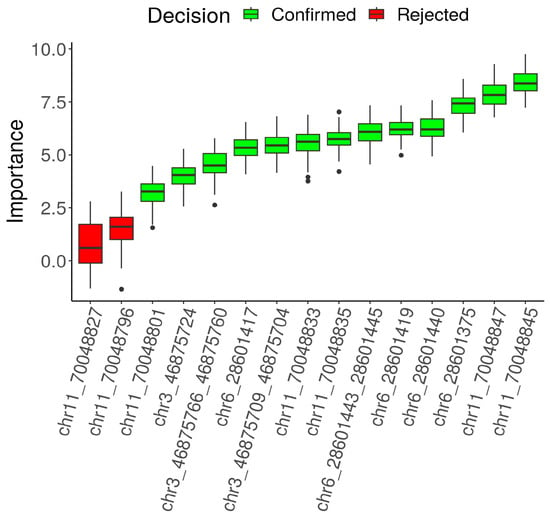
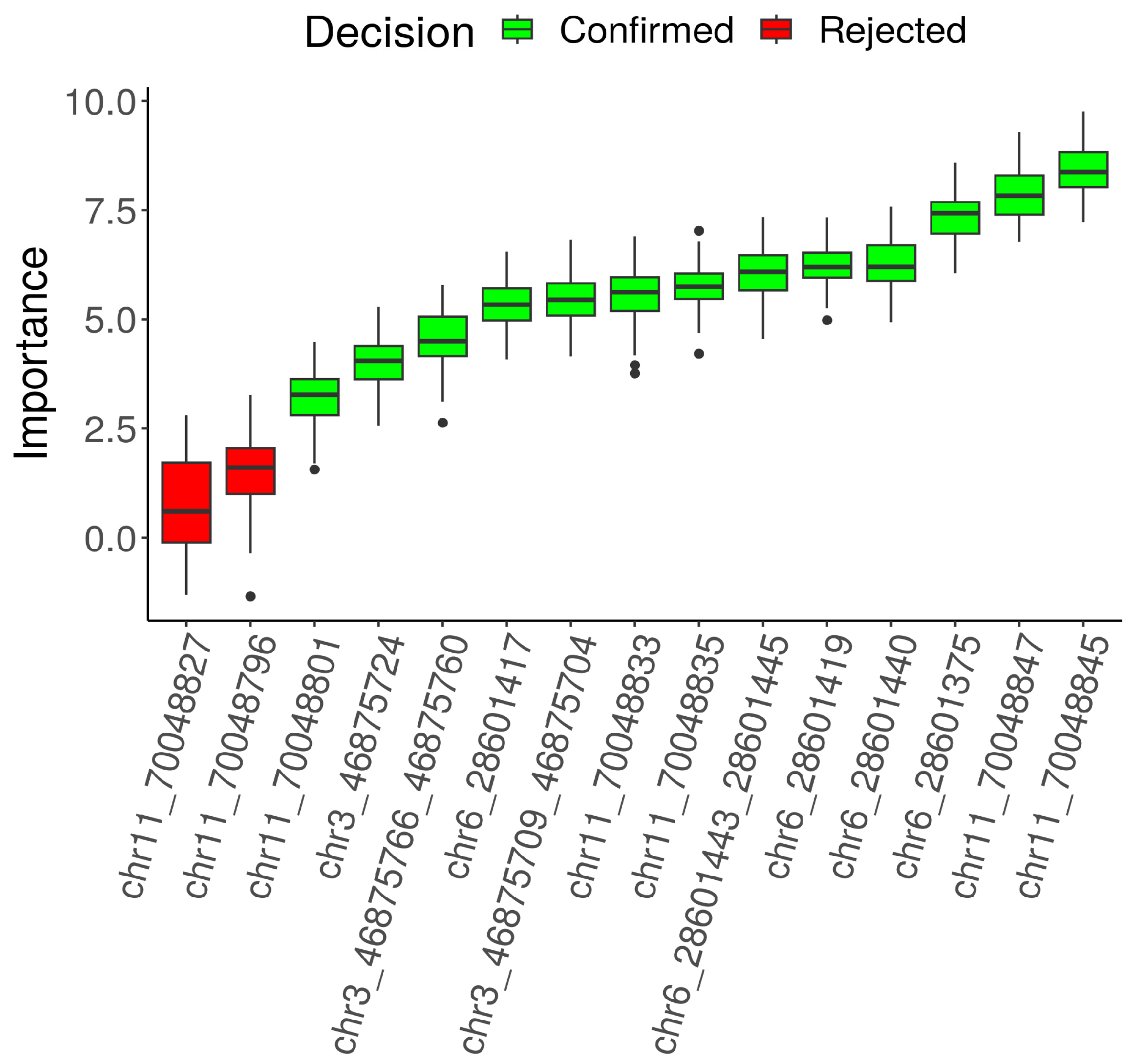
The result of the feature selection is shown in Figure 1. To account for potential fluctuations in the sequencing signal, adjacent CpG sites exhibiting similar methylation patterns were combined into a single feature using their mean methylation level; thus, CpG dinucleotides chr3:46875760 and chr3:46875766, chr3:46875704 and chr3:46875709, chr6:28601443 and chr6:28601445 were considered as one feature. CpG sites 11-845 (HumanMethylation450 cg26004810 probe), 11-847 (novel CpG dinucleotide not present in the previous studies), 6-375 (chr6:28601375), and 6-440 (chr6:28601440) showed the highest significance in terms of classifying ability among the analyzed predictors.
Figure 1.
Random Forest-derived predictor importance within the Boruta feature-selection procedure for Sotos syndrome. Original CpG predictors were assessed against randomly permuted shadow features. The predictors were considered “confirmed” (green) or “rejected” (red) in accordance with the calculations of the Boruta algorithm [13]. Five retained predictors were selected by mean importance for subsequent SVM-based subset comparison.
3.3. Classification Model
Multiple classification models were developed on the training dataset using significant predictors both individually and in various combinations. The SVM models were evaluated and ranked by their area under the ROC curve (AUC) (Table 3). The classification model based on the combination of predictors 6-375 (chr6_28601375) and 6-440 (chr6_28601440) showed the best sensitivity, specificity and accuracy values and was selected for further use; on the contrary, models based on predictors 6-375 and 6-440 individually showed lower prediction accuracy.
Table 3.
Quality assessment of the classification models for Sotos syndrome using samples from the training set. AUC, sensitivity, specificity and accuracy of each model were calculated with the Caret R package [17]. Predictors with the highest AUC value were selected for further classification model development.
Hyperparameter tuning was performed for the selected two-CpG SVM and the subsequent logistic regression classifier. The selected values were cost = 0.25 for SVM and C = 5.310526 for logistic regression. Logistic regression achieved a leave-one-out cross-validation accuracy of 0.96. The tuning results are summarized in Table 4.
Table 4.
Algorithm settings and hyperparameter selection.
3.4. Final Classification Model and Performance
After feature selection using linear SVM, the logistic regression was built with 6-375 and 6-440 predictors combined (Formula (1)). The logistic regression was chosen, because it is simple and easily interpretable for physicians. The quality of the model was evaluated on test samples using ROC analysis. The model shows perfect performance with AUC, area under precision-recall curve, sensitivity, specificity and accuracy equal 1.00 in test dataset.
The equation of logistic regression. Psotos is the probability that the sample has Sotos syndrome. M1 is methylation in chr6:28601440, M2 is methylation in chr6:28601375.
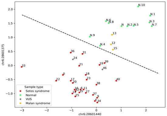
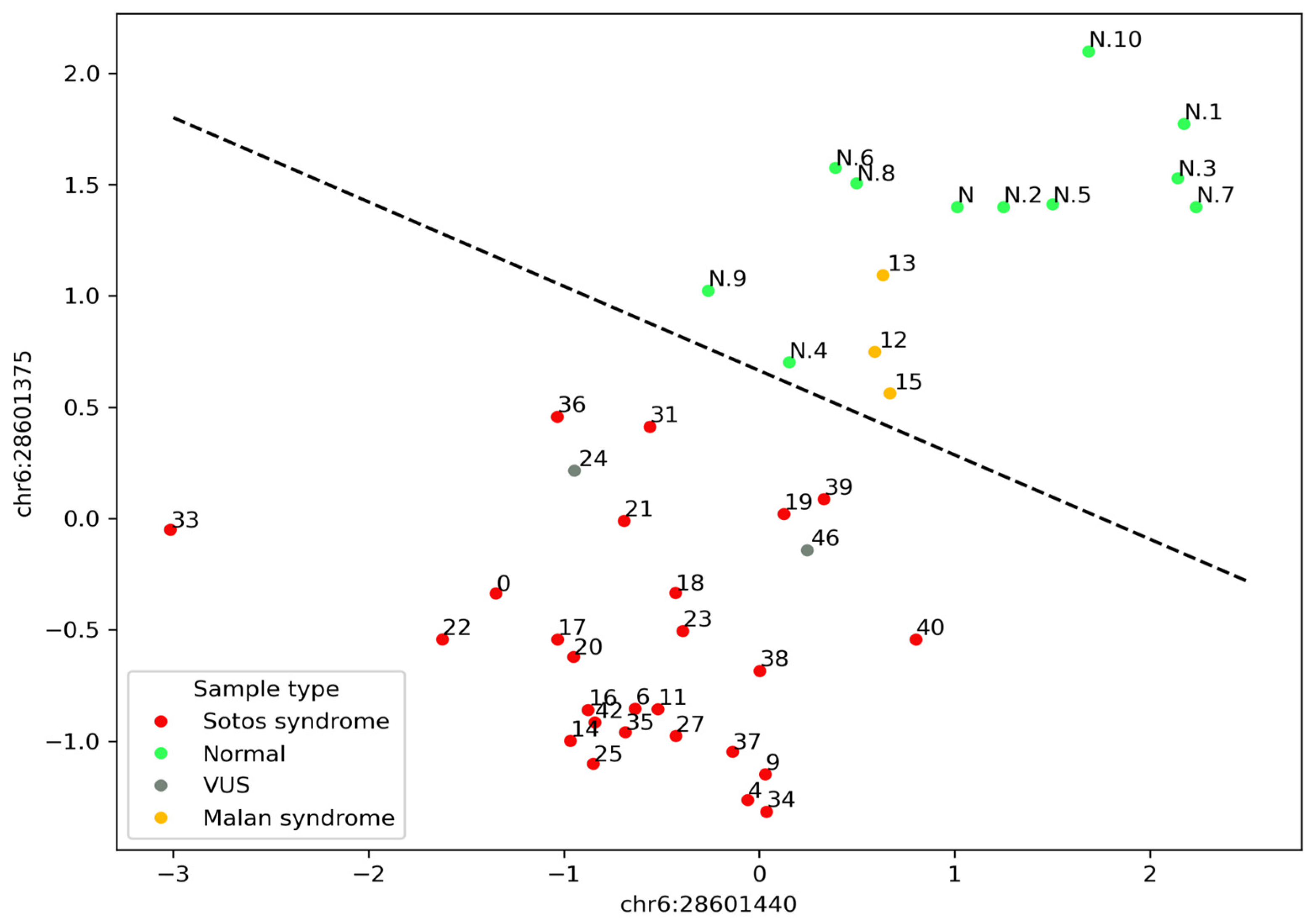
The distribution of the studied samples into the groups with Sotos syndrome (to the left and down of the decision boundary) and without Sotos syndrome (to the right and up of the decision boundary) using the selected model is shown in Figure 2. All studied samples with Sotos syndrome were correctly classified as samples with Sotos syndrome; samples with VUS in the NSD1 gene were also distributed into the group of samples with Sotos syndrome. Control samples were classified as samples without Sotos syndrome. Samples with phenotypically overlapping Malan syndrome were classified as samples without Sotos syndrome as well.
Figure 2.
Classification of the studied samples according to the model. Black dashed line represents the decision boundary; the numbers over points are numbers of the samples; the N label indicates that the sample belongs to the control group.
4. Discussion
Episignature is a unique and reproducible DNA methylation profile associated with a specific rare disease. Currently, sample classifiers designed to propose a diagnosis depending on the DNA methylation profile are based on the use of genome-wide episignatures [1,2,4,19,20]. We managed to reduce the genome-wide episignature of Sotos syndrome to the two most representative CpG dinucleotides, 6-375 and 6-440. The use of bisulfite Sanger sequencing followed by electropherogram processing makes it possible to obtain quantitative methylation values of the studied CpG dinucleotides on a par with other bisulfite sequencing methods, while maintaining a relatively low cost.
The samples derived from patients with Sotos syndrome show hypomethylation at the studied loci compared with the control samples, which corresponds to the literature data and confirms the presence of an episignature characteristic of Sotos syndrome [5]. At the same time, samples with Malan syndrome (OMIM:614753, also known as Sotos syndrome 2) showed a methylation level comparable to the control samples. Since Malan syndrome occurs due to pathogenic variants in the nuclear factor IX (NFIX) gene that is not involved in epigenetic regulation, it does not have a specific episignature characteristic of chromatin pathologies. Thus, the episignature analysis can be used for the differential diagnosis of Sotos and Malan syndromes with overlapping phenotypic manifestations. However, it is worth noting that the absence of this episignature is not a sufficient basis for establishing the diagnosis of Malan syndrome in a patient with characteristic clinical features, since this syndrome does not have its own episignature.
Recently, many researchers have considered the episignature to be a significant functional evidence and have used it for the purpose of variant interpretation [20,21,22,23,24,25]. J. Kerkhof et al. have developed recommendations for the interpretation and presentation of the results of the episignature analysis [26]. In accordance with these recommendations, the identification of a specific episignature, when accompanied by either a suspected pathogenic variant or a VUS in the associated gene, supports the application of the PS3 or PP4 criteria at a variable level of strength. The PP4 criterion is also recommended for the variant interpretation using episignature analysis by ACGS [27]. PS3 criterion indicates that a variant has been shown to have a damaging impact on the gene product, usually through reliable functional assays, whereas PP4 criterion indicates that a phenotypic manifestation highly specific for the disease is present [28]. Since the episignature is a functional consequence of pathogenic variants in the genes of the epigenetic machinery, it can be used to interpret and reclassify variants; however, caution is required. The use of unsupervised computational techniques (i.e., hierarchical clustering, multidimensional scaling) can lead to erroneous classification based on the episignature solely (for syndromes with moderate episignature) [22]. Furthermore, not all pathogenic variants within the gene can produce the same episignature. For instance, in patients with Helsmoortel–Van der Aa syndrome (OMIM:615873), the localization of pathogenic variants in different gene loci leads to the manifestation of drastically different episignatures [22]. For diseases with highly specific episignatures such as Sotos syndrome, it seems reasonable to consider the introduction of the new criteria with graded strength into the ACMG recommendations for a more accurate assessment of the pathogenicity of variants in the epigenetic regulation genes. The development of consensus recommendations, similar to those established by the Cancer Variant Interpretation Group UK (CanVIG-UK) for variant interpretation, could facilitate the standardized use of validated episignatures as evidence in variant classification, particularly for disorders with well-characterized and reproducible episignatures.
Analysis of the reduced episignature of Sotos syndrome using a classifying model showed that samples 24 and 46 obtained from patients with VUS in the NSD1 gene have the episignature of Sotos syndrome. For these patients, we performed a de novo assessment of the variant status and reclassified the variants (NSD1(NM_022455.5): c.6523T>C and c.8005del, respectively) into likely pathogenic (PS2, PM2, PP3). In this case, the additional application of the PP4 criterion would not affect the results of the interpretation of variants pathogenicity; however, the fact of the de novo occurrence of these variants and subsequent reclassification confirm the validity of the results of the reduced episignature analysis.
Previous studies explored the possibility of using episignature analysis as a first-tier tool in the chromatinopathies diagnostic algorithm [29,30]. It was stated that the identification of episignature could serve as a supportive instrument in resource-limited diagnostic settings, particularly in cases where sequencing or parental testing is delayed or inaccessible, by helping to direct subsequent diagnostic steps. Indeed, a considerable simplification and reduction in the cost in the case of a reduced episignature analysis may contribute to its introduction as a first-tier test; however, it should be emphasized that the results of the DNA methylation assessment should always be validated by molecular techniques.
This study has several limitations that should be considered when interpreting the results. First, the size of the study cohort was limited: 31 patients with a referral diagnosis of Sotos syndrome (26 with pathogenic or likely pathogenic NSD1 variants, 2 with variants of uncertain significance, and 3 with Malan syndrome) and 11 control samples. The held-out test set comprised only a small number of samples, so the reported performance metrics, despite their high values, are associated with wide confidence intervals and should be regarded as preliminary estimates rather than precise benchmarks. External validation on an independent, larger cohort will be necessary to confirm the generalizability of the reduced episignature. For the same reason, we did not perform a cross-algorithm benchmarking of classifiers: with a test set of this size, differences in accuracy between algorithms would fall within the range of sampling noise and could not support meaningful conclusions. Furthermore, confident use of the reduced episignature for the purpose of differential diagnosis requires the expansion of the sample set of patients with phenotypically overlapping syndromes. Although all three samples from patients with Malan syndrome were correctly classified as negative, a larger and more diverse comparison group would better define the specificity of the assay in a real differential-diagnostic setting. Finally, the episignature approach in general, and our reduced assay in particular, should be regarded as a supplementary rather than a self-sufficient diagnostic and research tool: the absence of the episignature does not exclude a diagnosis, and the presence of the episignature should always be interpreted alongside molecular genetic data and clinical findings.
5. Conclusions
In this study, we demonstrate that the genome-wide DNA methylation episignature of Sotos syndrome can be reduced to two CpG dinucleotides, chr6:28601375 and chr6:28601440, without loss of diagnostic accuracy. The reduced episignature can be detected by Sanger bisulfite sequencing, a method available in most genetic laboratories, and the resulting methylation values can be converted into a diagnostic probability using a simple and interpretable logistic regression model. Our findings position the reduced episignature as a cost-effective supporting tool for the diagnosis of Sotos syndrome and for the functional interpretation of NSD1 variants. At the same time, the episignature approach remains at an early stage: the list of conditions with highly specific episignatures will continue to expand, which could potentially cause a shift in the existing knowledge about episignatures. Our assay requires validation on an independent, larger cohort before it can be recommended for routine diagnostic use. The results of DNA methylation analysis should in all cases be interpreted alongside molecular landscape and clinical data rather than as a standalone diagnostic criterion.
Supplementary Materials
The following supporting information can be downloaded at: https://www.mdpi.com/article/10.3390/cimb48111254/s1.
Author Contributions
Conceptualization, I.V.V. and A.S.T.; methodology, I.V.V. and A.S.T.; software, A.I.K., A.S.S. and A.S.T.; validation, D.V.Z. and V.V.S.; investigation, A.V.E., O.A.Z.; resources, N.A.S.; data curation, A.V.E. and M.V.N.; writing—original draft preparation, A.V.E. and O.A.Z.; writing—review and editing, S.I.K. and V.V.S.; visualization, A.I.K. and A.S.S.; supervision, V.V.S.; project administration, I.V.V. and V.V.S.; funding acquisition, S.I.K. All authors have read and agreed to the published version of the manuscript.
Funding
The work was funded by a state assignment of the Ministry of Science and Higher Education of the Russian Federation for Research Centre for Medical Genetics (grant number 122032300334-3).
Institutional Review Board Statement
The clinical and molecular genetic study was performed in accordance with the Declaration of Helsinki and was approved by the Institutional Review Board of the Research Centre for Medical Genetics, Moscow, Russia (protocol 6/3, 19 April 2021), with written informed consent obtained from each participant and/or their legal representative as appropriate.
Informed Consent Statement
All research participants gave their informed consent to the clinical examination and publication of their anonymized data (for the infant probands, the responsible adult signed a consent form).
Data Availability Statement
The datasets used and/or analyzed during this study are available from the corresponding authors upon reasonable request.
Conflicts of Interest
Marina V. Nemtsova is an employee of the «Evogen LLC» company. Other authors declare that they have no competing interests.
Abbreviations
The following abbreviations are used in this manuscript:
| AUC | Area under the ROC curve |
| CpG | Cytosine-phosphate-guanine dinucleotide |
| DNA | Deoxyribonucleic acid |
| VUS | Variant of uncertain significance |
| SVM | Support vector machine |
References
- Levy, M.A.; McConkey, H.; Kerkhof, J.; Barat-Houari, M.; Bargiacchi, S.; Biamino, E.; Bralo, M.P.; Cappuccio, G.; Ciolfi, A.; Clarke, A.; et al. Novel diagnostic DNA methylation episignatures expand and refine the epigenetic landscapes of Mendelian disorders. HGG Adv. 2021, 3, 100075. [Google Scholar] [CrossRef] [Scilit] [PubMed]
- Aref-Eshghi, E.; Kerkhof, J.; Pedro, V.P.; Barat-Houari, M.; Ruiz-Pallares, N.; Andrau, J.C.; Lacombe, D.; Van-Gils, J.; Fergelot, P.; Dubourg, C.; et al. Evaluation of DNA Methylation Episignatures for Diagnosis and Phenotype Correlations in 42 Mendelian Neurodevelopmental Disorders. Am. J. Hum. Genet. 2020, 106, 356–370, Erratum in Am. J. Hum. Genet. 2021, 108, 1161–1163. https://doi.org/10.1016/j.ajhg.2021.04.022. [Google Scholar] [CrossRef] [Scilit] [PubMed]
- Husson, T.; Lecoquierre, F.; Nicolas, G.; Richard, A.C.; Afenjar, A.; Audebert-Bellanger, S.; Badens, C.; Bilan, F.; Bizaoui, V.; Boland, A.; et al. Episignatures in practice: Independent evaluation of published episignatures for the molecular diagnostics of ten neurodevelopmental disorders. Eur. J. Hum. Genet. 2024, 32, 190–199. [Google Scholar] [CrossRef] [Scilit] [PubMed]
- Levy, M.A.; Relator, R.; McConkey, H.; Pranckeviciene, E.; Kerkhof, J.; Barat-Houari, M.; Bargiacchi, S.; Biamino, E.; Palomares Bralo, M.; Cappuccio, G.; et al. Functional correlation of genome-wide DNA methylation profiles in genetic neurodevelopmental disorders. Hum. Mutat. 2022, 43, 1609–1628. [Google Scholar] [CrossRef] [Scilit] [PubMed]
- Choufani, S.; Cytrynbaum, C.; Chung, B.H.; Turinsky, A.L.; Grafodatskaya, D.; Chen, Y.A.; Cohen, A.S.; Dupuis, L.; Butcher, D.T.; Siu, M.T.; et al. NSD1 mutations generate a genome-wide DNA methylation signature. Nat. Commun. 2015, 6, 10207. [Google Scholar] [CrossRef] [Scilit] [PubMed]
- Tatton-Brown, K.; Rahman, N. Sotos syndrome. Eur. J. Hum. Genet. 2007, 15, 264–271. [Google Scholar] [CrossRef] [Scilit] [PubMed]
- Weinberg, D.N.; Papillon-Cavanagh, S.; Chen, H.; Yue, Y.; Chen, X.; Rajagopalan, K.N.; Horth, C.; McGuire, J.T.; Xu, X.; Nikbakht, H.; et al. The histone mark H3K36me2 recruits DNMT3A and shapes the intergenic DNA methylation landscape. Nature 2019, 573, 281–286. [Google Scholar] [CrossRef] [Scilit] [PubMed]
- Kernaleguen, M.; Daviaud, C.; Shen, Y.; Bonnet, E.; Renault, V.; Deleuze, J.F.; Mauger, F.; Tost, J. Whole-Genome Bisulfite Sequencing for the Analysis of Genome-Wide DNA Methylation and Hydroxymethylation Patterns at Single-Nucleotide Resolution. Methods Mol. Biol. 2018, 1767, 311–349. [Google Scholar] [CrossRef] [Scilit] [PubMed]
- Silva, C.; Machado, M.; Ferrão, J.; Sebastião Rodrigues, A.; Vieira, L. Whole human genome 5’-mC methylation analysis using long read nanopore sequencing. Epigenetics 2022, 17, 1961–1975. [Google Scholar] [CrossRef] [Scilit] [PubMed]
- Geysens, M.; Huremagic, B.; Souche, E.; Breckpot, J.; Devriendt, K.; Peeters, H.; Van Buggenhout, G.; Van Esch, H.; Van Den Bogaert, K.; Vermeesch, J.R. Clinical evaluation of long-read sequencing-based episignature detection in developmental disorders. Genome Med. 2025, 17, 1. [Google Scholar] [CrossRef] [Scilit] [PubMed]
- Breiman, L. Random Forests. Mach. Learn. 2001, 45, 5–32. [Google Scholar] [CrossRef] [Scilit]
- Nick, T.G.; Campbell, K.M. Logistic Regression. In Topics in Biostatistics. Methods in Molecular Biology; Ambrosius, W.T., Ed.; Humana Press: Totowa, NJ, USA, 2007; Volume 404, pp. 273–301. [Google Scholar]
- Noble, W. What is a support vector machine? Nat. Biotechnol. 2006, 24, 1565–1567. [Google Scholar] [CrossRef] [Scilit] [PubMed]
- Li, L.C.; Dahiya, R. MethPrimer: Designing primers for methylation PCRs. Bioinformatics 2002, 18, 1427–1431. [Google Scholar] [CrossRef] [Scilit] [PubMed]
- Zemlianaia, O.A.; Kalinkin, A.I.; Tanas, A.S.; Efremova, A.V.; Volodin, I.V.; Ismagilova, O.R.; Smirnov, A.S.; Zaletaev, D.V.; Nemtsova, M.V.; Kutsev, S.I.; et al. DNA Methylation at a Single Locus of Human Genome Accurately Recapitulates Episignature of CREBBP-Related Rubinstein-Taybi Syndrome. Int. J. Mol. Sci. 2025, 26, 9183. [Google Scholar] [CrossRef] [Scilit] [PubMed]
- Kursa, M.B.; Rudnicki, W.R. Feature Selection with the Boruta Package. J. Stat. Soft. 2010, 36, 1–13. [Google Scholar] [CrossRef] [Scilit]
- Kuhn, M. Building Predictive Models in R Using the Caret Package. J. Stat. Soft. 2008, 28, 1–26. [Google Scholar]
- Pedregosa, F.; Varoquaux, G.; Gramfort, A.; Michel, V.; Thirion, B.; Grisel, O.; Blondel, M.; Prettenhofer, P.; Weiss, R.; Dubourg, V.; et al. Scikit-learn: Machine Learning in Python. J. Mach. Learn. Res. 2011, 12, 2825–2830. [Google Scholar]
- Sadikovic, B.; Levy, M.A.; Kerkhof, J.; Aref-Eshghi, E.; Schenkel, L.; Stuart, A.; McConkey, H.; Henneman, P.; Venema, A.; Schwartz, C.E.; et al. Clinical epigenomics: Genome-wide DNA methylation analysis for the diagnosis of Mendelian disorders. Genet. Med. 2021, 23, 1065–1074, Erratum in Genet. Med. 2021, 23, 2228. https://doi.org/10.1038/s41436-021-01130-z. [Google Scholar] [CrossRef] [Scilit] [PubMed]
- Trajkova, S.; Kerkhof, J.; Rossi Sebastiano, M.; Pavinato, L.; Ferrero, E.; Giovenino, C.; Carli, D.; Di Gregorio, E.; Marinoni, R.; Mandrile, G.; et al. DNA methylation analysis in patients with neurodevelopmental disorders improves variant interpretation and reveals complexity. HGG Adv. 2024, 5, 100309. [Google Scholar] [CrossRef] [Scilit] [PubMed]
- Aref-Eshghi, E.; Rodenhiser, D.I.; Schenkel, L.C.; Lin, H.; Skinner, C.; Ainsworth, P.; Paré, G.; Hood, R.L.; Bulman, D.E.; Kernohan, K.D.; et al. Genomic DNA Methylation Signatures Enable Concurrent Diagnosis and Clinical Genetic Variant Classification in Neurodevelopmental Syndromes. Am. J. Hum. Genet. 2018, 102, 156–174. [Google Scholar] [CrossRef] [Scilit] [PubMed]
- Sadikovic, B.; Levy, M.A.; Aref-Eshghi, E. Functional annotation of genomic variation: DNA methylation episignatures in neurodevelopmental Mendelian disorders. Hum. Mol. Genet. 2020, 29, 27–32. [Google Scholar] [CrossRef] [Scilit] [PubMed]
- Coursimault, J.; Goldenberg, A.; Nicolas, G.; Saugier-Veber, P.; Coutant, S.; Vincent, A.; Pouliquen, D.; Feltin, C.; Aref-Eshghi, E.; Sadikovic, B.; et al. Contribution of DNA methylation profiling to the reclassification of a variant of uncertain significance in the KDM5C gene. Eur. J. Med. Genet. 2022, 65, 104556. [Google Scholar] [CrossRef] [Scilit] [PubMed]
- Ferilli, M.; Ciolfi, A.; Pedace, L.; Niceta, M.; Radio, F.C.; Pizzi, S.; Miele, E.; Cappelletti, C.; Mancini, C.; Galluccio, T.; et al. Genome-Wide DNA Methylation Profiling Solves Uncertainty in Classifying NSD1 Variants. Genes 2022, 13, 2163. [Google Scholar] [CrossRef] [Scilit] [PubMed]
- Man, A.; Di Scipio, M.; McConkey, H.; Hough, R.; Stein, N.; Diehl, E.; Marshall, C.R.; Sadikovic, B.; Ejaz, R. Diagnosis of TET3 -Related Beck–Fahrner Syndrome in an Individual With Chorioretinal and Iris Colobomata Using a DNA Methylation Signature. Am. J. Med. Genet. A 2025, 197, e63864. [Google Scholar] [CrossRef] [Scilit] [PubMed]
- Kerkhof, J.; Rastin, C.; Levy, M.A.; Relator, R.; McConkey, H.; Demain, L.; Dominguez-Garrido, E.; Kaat, L.D.; Houge, S.D.; DuPont, B.R.; et al. Diagnostic utility and reporting recommendations for clinical DNA methylation episignature testing in genetically undiagnosed rare diseases. Genet. Med. 2024, 26, 101075. [Google Scholar] [CrossRef] [Scilit] [PubMed]
- Durkie, M.; Cassidy, E.-J.; Berry, I.; Owens, M.; Turnbull, C.; Scott, R.H.; Taylor, R.W.; Deans, Z.C.; Ellard, S.; Baple, E.L.; et al. The Association for Clinical Genomic Science. Best Practice Guidelines for Variant Classification in Rare Disease 2024. Available online: https://www.acgs.uk.com/quality/best-practice-guidelines/ (accessed on 10 August 2026).
- Richards, S.; Aziz, N.; Bale, S.; Bick, D.; Das, S.; Gastier-Foster, J.; Grody, W.W.; Hegde, M.; Lyon, E.; Spector, E.; et al. Standards and guidelines for the interpretation of sequence variants: A joint consensus recommendation of the American College of Medical Genetics and Genomics and the Association for Molecular Pathology. Genet. Med. 2015, 17, 405–424. [Google Scholar] [CrossRef] [Scilit] [PubMed]
- Tkemladze, T.; Campbell, C.; Bregvadze, K.; Kvaratskhelia, E.; Abzianidze, E.; Demain, L.; Jenkinson, S.; Hilton, S.; Levy, M.; Kerkhof, J.; et al. Evaluating DNA methylation episignatures as a first-tier diagnostic test in individuals with suspected genetic disorders. Eur. J. Hum. Genet. 2026, 34, 296–299. [Google Scholar] [CrossRef] [Scilit] [PubMed]
- van der Laan, L.; Luijckx, A.; Lo-A.-Njoe, S.; Ecury-Goossen, G.M.; Falix, F.A.; Faries, S.; Manshande, M.E.; Philippi, P.A.E.A.C.; van der Plas, E.M.; Veenhuis, H.D.; et al. Episignature-based modelled first-tier diagnostic approach in the Dutch Caribbean: Advancing equal care through epigenetic classification. Front. Genet. 2026, 17, 1803993. [Google Scholar] [CrossRef] [Scilit] [PubMed]

Disclaimer/Publisher’s Note: The statements, opinions and data contained in all publications are solely those of the individual author(s) and contributor(s) and not of MDPI and/or the editor(s). MDPI and/or the editor(s) disclaim responsibility for any injury to people or property resulting from any ideas, methods, instructions or products referred to in the content. |
© 2026 by the authors. Licensee MDPI, Basel, Switzerland. This article is an open access article distributed under the terms and conditions of the Creative Commons Attribution (CC BY) license.