


Simple Summary
Hair follicles (HFs) are small organs in mammalian skin that constantly cycle through growth and rest. This process is driven by hair follicle stem cells (HFSCs). While researchers have studied HFSCs extensively in mice, there are major differences in how HFs develop in other mammals, especially in cashmere goats and sheep, which are important for agriculture. This review compares how HFSCs are regulated, isolated, and cultured in mice, humans, cashmere goats, sheep, and rats. We discuss how the structural differences between primary and secondary HFs affect stem cell behavior, and we summarize the signaling pathways that control their activation. A better understanding of these mechanisms could lead to new treatments for hair loss and skin wounds, and may also help improve cashmere production through molecular breeding.
Abstract
Hair follicle stem cells (HFSCs) reside mainly in the bulge region and are critical for driving the periodic regeneration of hair follicles (HFs) and maintaining skin homeostasis. Most existing studies on HFSC regulation rely on mouse models, whereas the developmental characteristics and differentiation potential of HFSCs in economically important livestock remain poorly summarized. This review compares HFSC research across mice, humans, cashmere goats, and sheep. We specifically focus on the distinct developmental trajectories and structural differences between primary and secondary HFs which are particularly prominent in ruminants. The principles, advantages, and limitations of current isolation and in vitro culture systems for HFSCs are also evaluated. Furthermore, we summarize the multilineage differentiation reported for HFSCs under specific in vitro induction, and we distinguish lineage-marker expression from functional differentiation demonstrated in vivo, and discuss their applications in regenerative medicine and tissue repair. We then detail the signaling pathways (Wnt, BMP, Shh, and Notch) and endogenous/exogenous factors that regulate the balance between HFSC activation and quiescence. Finally, we highlight the potential of emerging technologies, such as single-cell sequencing and organoid culture, to provide new insights for improving cashmere yield and advancing molecular breeding in livestock.
1. Introduction
1.1. Structure and Regenerative Basis of the Hair Follicle
Hair follicles (HFs) are miniature organs in mammalian skin that undergo periodic regeneration. They are composed of several distinct structures, including the hair matrix, inner and outer root sheaths, hair shaft, dermal papilla, and dermal sheath. The regenerative capacity of HFs relies heavily on adult stem cells residing in the bulge region, known as hair follicle stem cells (HFSCs) [1]. These cells are characterized by their unique biological properties: self-renewal, multilineage differentiation potential, and long-term quiescence. Upon receiving specific signal stimulation, quiescent HFSCs can be activated, proliferate, and differentiate to form the hair shaft and inner root sheath, thereby driving the follicle into the anagen phase and generating a new hair shaft [2].
1.2. HFSCs in Skin Homeostasis and Emerging Technologies
Beyond serving as an ideal model for studying organ regeneration and stem cell biology, HFSCs are also central to skin homeostasis, injury repair, and the treatment of related diseases. Although the majority of HFSC research has traditionally relied on mouse models, the application of emerging technologies—such as single-cell sequencing, spatial transcriptomics, and organoid culture—has provided new avenues for exploring HFSC biology. However, a cross-species understanding of HFSCs in mammals still requires further investigation.
1.3. Scope and Objectives of This Review
This review synthesizes current research on HFSCs across mammals, covering embryonic HF development, hair follicle cycle regulation, the stem cell microenvironment, isolation and culture methods, multilineage differentiation, and influencing factors. By summarizing these aspects, we aim to provide new perspectives for addressing clinical challenges, such as alopecia treatment and skin wound repair. Furthermore, this review offers a theoretical reference for the high-quality development of the wool and cashmere industry, particularly for achieving a synergistic improvement in both yield and quality.
1.4. Literature Search and Selection
This is a narrative review. We searched PubMed, Web of Science, and CNKI up to 25 October 2026, combining the following terms: “hair follicle stem cell(s)”, “HFSCs”, “bulge”, “primary hair follicle”, “secondary hair follicle”, “cashmere goat”, “sheep”, “wool”, “cashmere”, “isolation”, “culture”, “multilineage differentiation”, “Wnt”, “BMP”, “Shh”, “Notch”, and “molecular breeding”. We included peer-reviewed original studies and reviews on HFSC biology in mammals, with priority given to mice, humans, cashmere goats, sheep, and rats. We excluded non-mammalian studies, conference abstracts lacking full data, and papers unrelated to HFSC biology or hair follicle development. We also screened the reference lists of key reviews. Because the review is narrative, it is not exhaustive, and the selection of studies inevitably reflects our judgment.
2. HFs
The dermis contains loose connective tissue that includes the hair follicle layer, which comprises HFs, sebaceous glands, and other skin appendages [3]. The cells within these follicular tissues undergo continuous division, proliferation, and renewal via periodic cycles. Although the precise timing of cycle synchronization varies slightly depending on sex and strain, the morphogenesis and development of most HFs remain highly synchronized.
2.1. Conceptual Scope and Classification Boundaries
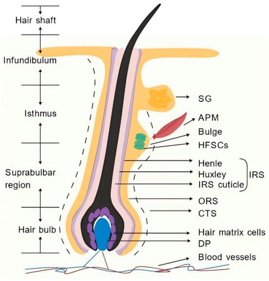
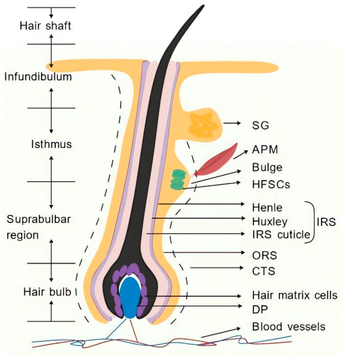
The hair follicle is composed of several distinct structures, including the connective tissue sheath (CTS), hair bulb, inner root sheath (IRS), outer root sheath (ORS), and hair shaft. HFs are classified into primary and secondary follicles. A follicle group, consisting of one primary follicle and several secondary follicles, forms a heterogeneous coat. In cashmere goats, primary follicles produce coarse hair and carry a full set of appendages, including sebaceous glands, sweat glands, and arrector pili muscles. Secondary follicles produce cashmere; they are usually smaller, lack sweat glands and arrector pili muscles, and may have reduced or absent sebaceous glands [4,5]. Sebaceous gland presence or absence should not, however, be treated as a universal defining criterion, since follicle type and appendage composition differ among species and body regions [4,6]. Primary and secondary follicles are therefore described here mainly by size, depth, and associated appendages, with species-specific qualification. The morphogenesis of HFs undergoes periodic proliferation and renewal through three stages—anagen, catagen, and telogen—in response to seasonal changes [4,7]. In cashmere goats, primary follicles remain active throughout the year, exhibiting apoptosis and regeneration across different months with minimal seasonal variation in their growth cycle. Conversely, secondary follicles display pronounced seasonal fluctuations in their growth cycle, requiring approximately one year to complete a full cycle. This characteristic is particularly prominent in ruminants, such as cashmere goats and sheep [5]. The histological structure of the HFs is illustrated in Figure 1.
Figure 1.
Hair follicle tissue structure. Figure 1 was drawn with https://BioGDP.com [8]. Abbreviations: SG, sebaceous gland; APM, arrector pili muscle; HFSCs, hair follicle stem cells; IRS, inner root sheath; ORS, outer root sheath; CTS, connective tissue sheath; DP, dermal papilla.
2.2. Morphogenetic Patterns of Embryonic HFs
Using cashmere goats as the primary research model, embryonic HF development exhibits distinct temporal heterogeneity. Specifically, the density of secondary HFs continuously increases with fetal age, whereas variations among fetuses of the same age show no regular pattern (Table 1) [9,10,11]. In mice, hair follicle morphogenesis begins around embryonic day 14 (E14). This process progresses through distinct stages—including the placode, hair germ, and hair peg—and is characterized by a short 3–4 week cycle with highly synchronized molting [12]. Furthermore, single-cell and spatial transcriptomics have revealed a specific cell subpopulation associated with hair placode formation in early pig embryonic skin, indicating that hair follicle dynamics are species-specific [13]. In fine-wool sheep, integrated histological, transcriptomic, and proteomic analyses have shown that as fetal weight increases, primary hair follicle density decreases while secondary hair follicle density increases [6,14]. In contrast, human HFs predominantly consist of continuously growing terminal hairs, with no obvious synchronization during molting [15]. In dogs, FGF5 mutations correlate with hair follicle density and coat type [16], yet whether FGF5 directly regulates HFSC activity remains to be established. Collectively, these species-specific differences form the basis for the diverse patterns of hair follicle development across mammals.
Table 1.
Sequential events in primary and secondary hair follicle morphogenesis during embryogenesis.
3. HFSCs
The stem cell pool within the bulge region of the hair follicle outer root sheath harbors HFSCs, which are essential for follicle self-renewal. Characterized by unique signal transduction pathways and robust regenerative capacity, HFSCs are multipotent adult stem cells that generate the hair follicle lineages [17,18]. Reports of broader, cross-germ-layer differentiation rest largely on specific in vitro induction protocols [3,19,20,21,22,23,24] and should be read with caution. They interact with, depend on, and communicate with various surrounding cell types to regulate hair growth and tissue regeneration [17]. HFSCs were first observed in the bulge region of mouse HFs. The formal identification of HFSCs in this location was established in 1990 by Cotsarelis et al. [25] through their proposal of the “bulge-activation hypothesis.” Since then, subsequent studies across various mammalian species, including humans [26] and cashmere goats [3], have confirmed the existence and biological characteristics of HFSCs.
3.1. Activation and Cycle Regulation of HFSCs
During the hair follicle cycle, the activation of quiescent HFSCs is a critical trigger for the transition from telogen to anagen. Upon entering the anagen phase, a subset of activated HFSCs initiates differentiation, while others remain in a quiescent state to preserve the stem cell pool [27]. When activator concentrations reach a specific threshold, HFSCs are driven to proliferate and differentiate into hair follicle, mesodermal, and ectodermal lineages. To generate new hair, signals derived from the dermal papilla (DP) activate HFSCs in the bulge region. These activated HFSCs then proliferate, migrate downward, and differentiate into various cell types of the hair follicle. Through asymmetric division and self-renewal, they maintain the stem cell population while generating new HFs [28].
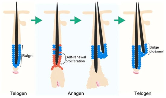
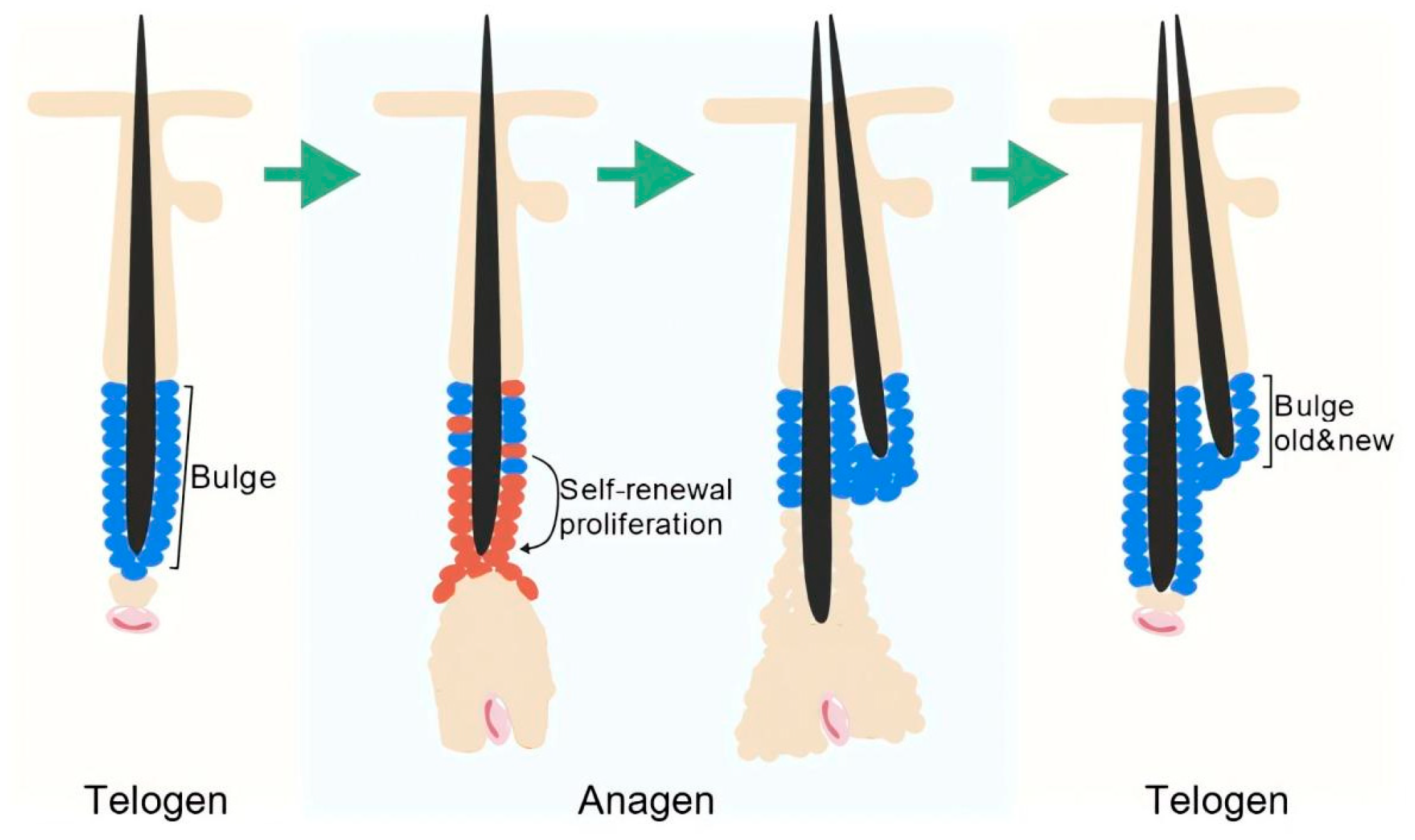
Throughout the hair follicle cycle, HFSCs continuously replenish the follicle by proliferating and differentiating. During the anagen phase, DP cells grow downward, which stimulates HFSCs to maintain their stemness while actively proliferating and differentiating. In this process, intercellular communication plays a decisive role in hair follicle development [29]. During catagen, the lower follicle (including the hair bulb, inner root sheath, and lower outer root sheath) undergoes apoptosis-driven regression, while the bulge-resident HFSC pool is largely preserved through quiescence and niche-derived survival signals. This regression of lower follicular structures is therefore distinct from stem-cell maintenance: the reservoir is retained for the next anagen. DP cells stay close to the bulge during telogen and supply activating signals at anagen onset [30]. During the telogen phase, the quiescence of HFSCs is maintained through the combined action of multiple signals and genes. Once the activation signals exceed a certain threshold, these pre-activated HFSCs are awakened and transition into anagen [31]. In mammals, the hair cycle of mice is significantly shorter than that of cashmere goats and other species, reflecting species-specific adaptations in the regulatory mechanisms of the hair follicle cycle [5,27]. The cyclical renewal of HFSCs is illustrated in Figure 2.
3.2. Regional Specificity and Microenvironmental Regulation of HFSCs
HFSCs exhibit distinct regional specificity. For instance, HFSCs located in the lower bulge can regenerate the ORS, whereas isthmus stem cells in the upper bulge lack this capacity, indicating that stem cell populations in different locations possess distinct differentiation fates [32]. Studies on label-retaining cells (LRCs) residing in the bulge have shown that quiescent stem cells with self-renewal capacity are primarily located in the upper bulge region, serving as an essential source for hair growth [33]. In vivo imaging of the mouse hair follicle niche has demonstrated that stem cells at different spatial positions within the bulge make distinct fate decisions, and that their regional location influences both their activation and differentiation direction [34]. Furthermore, comparative studies have revealed that human HFSC markers are not entirely consistent with those of mice. This suggests that the regional specificity of HFSCs in mammals is both evolutionarily conserved and species-specific [35].
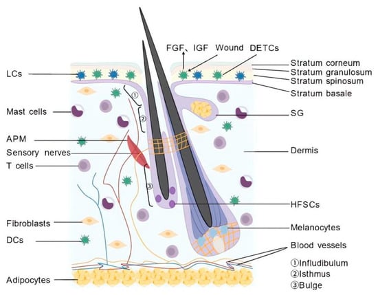
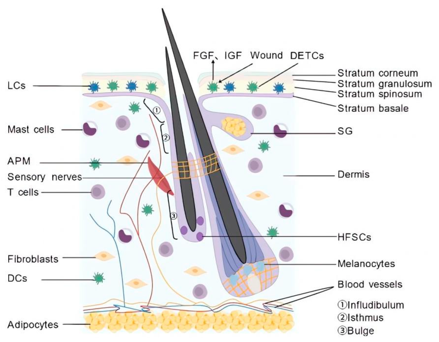
In vivo, HFSCs reside in a highly secure microenvironment characterized by favorable nutritional conditions and a rich blood supply. Upon appropriate signal stimulation, HFSCs secrete factors that act in a paracrine manner on surrounding non-stem cells, rather than on themselves, thereby participating in tissue repair and regeneration [18]. When hair follicle structures are damaged, downstream signaling centers transport DP cells to the bulge region. This alters the external microenvironment, subsequently activating HFSC proliferation and differentiation to generate new HFs and hairs [36]. When the body responds to changes in external temperature, HFSCs interact with norepinephrine—a neurotransmitter secreted by the sympathetic nervous system—to modulate their activity and maintain homeostasis, thereby promoting hair follicle regeneration [37]. Furthermore, immune cells release various signaling molecules and cytokines that interact with epithelial cells. This interaction plays a significant role in regulating HFSC activity and inducing hair follicle neogenesis [38]. The niche of HFSCs is illustrated in Figure 3.
Figure 3.
HFSC niche. Figure 3 was drawn with https://BioGDP.com [8]. Abbreviations: SG, sebaceous gland; DETCs, dendritic epidermal T cells; FGF, fibroblast growth factor; IGF, insulin-like growth factor; LCs, langerhans cells; DCs, dendritic cells; APM, arrector pili muscle.
4. Isolation and In Vitro Culture of HFSCs
The isolation and in vitro culture of HFSCs are essential for investigating their biological functions and elucidating the mechanisms of periodic hair follicle regeneration. HFSCs are usually enriched by the methods summarized in Table 2 [39,40,41,42,43,44]. Most “HFSC isolation” protocols yield follicular variable proportions of HFSCs, confirming HFSC identity requires marker-based verification (e.g., K15, CD34, K19) and functional assays such as clonogenicity and self-renewal. In practice, significant variations exist in hair follicle size and structural compactness across different species. While in vitro culture systems have been successfully established for mice, humans, and sheep, establishing similar systems for other mammalian species remains challenging, particularly in maintaining stem cell properties and simulating three-dimensional (3D) culture environments.
Table 2.
(A) Comparison of methods for HFSC isolation. (B) Comparison of methods for HFSC culture.
5. Multidirectional Differentiation Potential and Applications of HFSCs
Three levels of evidence are distinguished in this section: lineage-specific marker expression, functional assays in vitro, and in vivo validation with appropriate controls. Cross-germ-layer claims are qualified accordingly, and in vitro induction is kept separate from functional differentiation demonstrated in vivo. HFSC populations from the bulge, from nestin-positive hair follicle-associated pluripotent (HAP) cells, and from primary versus secondary follicles should not be assumed to be equivalent [3,18,33,45]. As critical players in hair follicle development, HFSCs possess unique differentiation potential and regenerative capacity, allowing them to give rise to various cell types. HFSCs are capable of differentiating not only into diverse cell types within the skin tissue, but also across multiple germ layers to contribute to cell renewal. Consequently, they play positive roles in diverse fields, including hair growth, nerve repair, and tissue engineering.
5.1. Differentiation Potential and Applications of Mouse HFSCs
Mice serve as the most extensively studied animal model for HFSC research. As ideal seed cells for tissue regeneration, HFSCs can differentiate into sebaceous gland cells following skin injury, thereby promoting sebaceous gland regeneration and contributing to the restoration of skin structure and function. Experiments utilizing bulge cells in mouse models—where sebaceous glands remained intact but hair follicle growth ceased and hairs completely disappeared—demonstrated that cells derived from the bulge possess the potential to differentiate into sebaceous gland cells under normal physiological conditions. When induced in an appropriate microenvironment, these cells can commit to the sebaceous gland lineage, subsequently promoting gland regeneration and reconstruction to repair damaged skin [46]. Furthermore, the multilineage differentiation potential of HFSCs allows them to be induced into neurons, astrocytes, and glial cells within neural tissue engineering, playing a crucial role in nerve repair and regeneration. For instance, when HFSCs isolated from mouse HFs were implanted into spinal cord transection or tibial nerve injury models, the transplanted cells differentiated into Schwann cells. This enabled mice with spinal cord contusion and tibial nerve transection to recover normal walking ability and hindlimb motor function. These findings indicate that HFSCs can survive at nerve injury sites and differentiate into Schwann cells to support neuronal regeneration and peripheral nerve repair, providing an effective and accessible novel cell source for treating peripheral nerve and spinal cord injuries [19,20].
HFSCs play a pivotal role in promoting vascular regeneration and facilitating the recovery of vascular injuries within skin wound tissues. In a study using diabetic db/db mice, cell sheets derived from HAP stem cells were transplanted onto skin wounds. The results demonstrated that after directed differentiation into vascular endothelial cells, HFSCs significantly contributed to tissue repair by promoting angiogenesis in vivo. These findings highlight HFSCs as an ideal cell source for treating ischemic diseases and accelerating skin wound healing [21]. A specific subpopulation of HFSCs expressing the neural stem cell marker nestin is termed HAP stem cells. To investigate their therapeutic potential, a mouse intracerebral hemorrhage (ICH) model was established via collagenase injection, and HAP stem cells isolated from mouse HFs were directly implanted into the hemorrhagic lesion. The transplantation led to increased expression of anti-inflammatory factors, and a portion of the HAP stem cells differentiated into neurons and glial cells. This significantly ameliorated neuroinflammation in the mice, suggesting that HFSCs can be applied to central nervous system injury repair and offer a novel therapeutic strategy for ICH [22]. Furthermore, nestin-positive cells isolated from mouse whisker HFs were induced to differentiate into cardiomyocytes in vitro. The differentiated cardiomyocytes exhibited functional activity, demonstrating that hair follicle-derived HAP stem cells possess the potential to differentiate toward the mesodermal myocardial lineage. This provides a promising new cell source for cardiovascular tissue engineering [23].
5.2. Differentiation Potential and Applications of Human HFSCs
Human HFs represent one of the most clinically valuable sources of autologous stem cells. Upon follicle damage, activated HFSCs receive stimulatory signals, proliferate, and differentiate to initiate anagen. They migrate from the bulge to the damaged site, rearrange to form hair germs, and ultimately differentiate to form new HFs, thereby participating in hair regeneration and repair [47]. HFSCs possess the potential to differentiate into skin appendage cells and are essential for appendage regeneration. Under specific induced microenvironments, HFSCs have been shown to differentiate into sweat gland secretory and ductal cells, promoting sweat gland regeneration. This capability is advantageous for restoring sweat gland function after burns and facilitating the healing of soft tissue injuries [48]. During wound healing, HFSCs differentiate into epidermal keratinocytes. These cells differentiate into epidermal lineage cells and migrate to the wound site to replace the damaged epidermis. Notably, bulge cells isolated via either enzymatic digestion or tissue explant methods can both differentiate into epidermal keratinocytes for the construction of tissue-engineered skin. This provides an efficient cell extraction technology for applications in tissue engineering and regenerative medicine [42]. Clinical evidence for HFSC-based therapy remains limited. Most available clinical data concern hair transplantation procedures, which rely on the re-implantation of intact follicular units rather than the administration of purified HFSCs; these procedures do not constitute direct evidence that HFSCs mediate the observed clinical effects. Early studies report increased hair count and density after transplantation procedures [49], but these findings require validation in larger, controlled trials.
In skin tissue engineering, HFSCs are widely applied to repair damaged tissues, serving as a primary driver of skin regeneration. Given their capacity to differentiate into multiple skin cell types, injecting or implanting HFSCs into damaged areas—such as burns, trauma, and skin lesions—can accelerate wound healing and promote tissue reconstruction and regeneration [50]. Human hair follicle-derived HAP stem cells exhibit a differentiation potential similar to that of mouse HAP stem cells, including the ability to differentiate toward dopaminergic neurons. Both in vivo and in vitro studies have demonstrated that HFSCs can successfully differentiate into neural cells under specific induction conditions. This suggests their potential utility in treating neurological disorders, such as Parkinson’s disease [51], and participating in nerve injury repair. Stem cell transplantation has emerged as a novel therapeutic strategy to promote wound repair and skin soft tissue expansion, with broad applications in both medical and cosmetic fields. Botulinum toxin type A, a legally approved material for aesthetic medicine following artificial attenuation and purification, promotes hair growth by stimulating HFSC proliferation and regulating the hair follicle growth cycle [52]. To explore the long-term preservation of hair follicle samples without compromising the cross-germ-layer differentiation potential of stem cells, HFSCs were subjected to cryopreservation and subsequently induced for multilineage differentiation. The results indicated that these cells retained the potential to differentiate into smooth muscle cells, cardiomyocytes, osteoblasts, and adipocytes. These findings deepen our understanding of the multilineage differentiation potential of HFSCs and provide technical support for future clinical translation [24].
5.3. Differentiation Potential and Applications of Cashmere Goat HFSCs
Cashmere goats serve as an optimal mammalian model for studying the seasonal regulation of hair follicle regeneration and represent a core species for livestock molecular breeding in China. By collecting skin samples across different stages of hair follicle morphogenesis, researchers identified distinct subpopulations of HFSCs through cell clustering and annotation. Pseudotime analysis was utilized to reconstruct cell differentiation trajectories, revealing the temporal dynamics of HFSC differentiation. These studies found that different subpopulations vary in their proliferation capacity, differentiation potential, and marker expression. Furthermore, HFSCs exhibit a clear differentiation trajectory toward the hair follicle lineage, providing a valuable reference for research on cell fate determination and mammalian hair follicle development [4]. The cytodifferentiation stage in cashmere goats is a critical period for the formation of various hair follicle layers. Transcriptome sequencing of skin samples from this stage identified numerous genes that are differentially expressed during cytodifferentiation. Histological analysis revealed that hair matrix cells proliferate and differentiate into the hair shaft and inner root sheath, confirming that HFSCs commit to the hair follicle lineage during this stage. This provides a theoretical basis for enhancing cashmere yield and quality through a better understanding of hair follicle development in cashmere goats [11].
Primary and secondary HFSCs were isolated from the skin of Arbas cashmere goats and cultured in vitro. Characterization revealed that these cells express multiple pluripotency markers and retain self-renewal capacity. Under neural, adipogenic, and hepatic induction, they expressed markers associated with ectodermal, mesodermal, and endodermal fates. These data indicate in vitro lineage plasticity, not functional differentiation in vivo: no study has yet shown that these cells acquire functional neural, adipocyte, or hepatocyte phenotypes. The apparent difference between primary and secondary HFSC populations also requires further validation. These findings point to new directions for studying cashmere goat HFSC biology and molecular breeding, and provide a basis for applying HFSCs from cashmere goats and other livestock in regenerative medicine [3,45].
5.4. Differentiation Potential and Applications of HFSCs in Other Mammals
Sheep and goats are economically important livestock whose tissue development and complex traits depend on the coordinated function of cells within the skin, reproductive organs, and adipose tissues. Single-cell omics technologies have elucidated the cellular heterogeneity during hair follicle development and identified key genes regulating HFSC differentiation, providing candidate genes and cellular targets for molecular breeding in sheep and goats [53]. HFSCs isolated and purified from sheep HFs were studied under specific induction conditions. The results confirmed that these HFSCs can be induced to differentiate into smooth muscle cells with normal contractile function. They exhibit regenerative potential for forming vascular wall cells and demonstrate a capacity to differentiate toward the mesodermal smooth muscle lineage. This highlights their immense potential for tissue repair and regeneration, offering a novel cell source for cardiovascular tissue engineering [54]. In Chinese Merino sheep, comparative studies under nutritional restriction revealed that nutritional factors influence HFSC differentiation by regulating gene expression, providing a basis for the exogenous regulation of HFSC fate [6]. Furthermore, TMT quantitative proteomic analysis of skin samples from fine-wool sheep at different stages of secondary hair follicle development indicated that differentially expressed proteins may regulate HFSC differentiation, offering valuable data for the molecular breeding of wool traits [14]. Although studies on sheep, goats, and fine-wool sheep have touched upon HFSC differentiation, direct evidence for cross-germ-layer differentiation of sheep HFSCs remains lacking.
HFSCs isolated from rat HFs were modified with hepatocyte growth factor (HGF) and transplanted into a rat model of cerebral ischemia–reperfusion injury. Within the brain injury microenvironment, these HFSCs differentiated into neurons and glial cells. During the acute phase, they played a role in reducing the inflammatory response, protecting blood–brain barrier (BBB) integrity, and promoting angiogenesis. Ultimately, this treatment improved neurological functional recovery, enhanced therapeutic efficacy, and promoted the regeneration of expanded skin [55]. In another study, HFSCs isolated from the hair follicle bulge were injected into a rat skin expansion model. The results showed that these HFSCs differentiated into epidermal and hair follicle cells, while their paracrine growth factors promoted angiogenesis and cell proliferation. The proliferation and differentiation of HFSCs, along with the upregulated expression of paracrine-related bioactive growth factors, not only accelerated wound healing and tissue repair but also increased skin thickness and vascular density, thereby promoting skin expansion and regeneration [32,56]. Furthermore, HFSCs isolated from rat HFs were expanded and characterized in vitro. These cells exhibited clonogenic capacity and multilineage differentiation potential. The various growth factors and cytokines secreted by HFSCs demonstrated wound healing-promoting potential by stimulating vascular endothelial cell proliferation and angiogenesis. This provides new insights for the application of HFSCs in regenerative medicine [57].
Beyond the mammalian species discussed above, other economically important animals—such as pigs, cattle, and dogs—have received comparatively less attention regarding hair follicle development and HFSC research. Future investigations could leverage integrated single-cell and spatial transcriptomics approaches to further explore HFSC differentiation. Such studies would provide novel perspectives for understanding the genetic regulation of HFSC differentiation in livestock. To compare HFSC markers, follicle features, culture status, differentiation evidence, and major knowledge gaps across species, we summarized the available data in Table 3.
Table 3.
Cross-species comparison of HFSC markers, follicle features, culture status, differentiation evidence, and knowledge gaps.
6. Factors Affecting Hair Follicle Stem Cell Activity
The role of each pathway is not fixed; the net outcome depends on developmental stage, follicular region (bulge versus matrix), and cellular state [58,59,60]. The successful activation of HFSCs is a prerequisite for the entire process of hair follicle regeneration. Fundamentally, the promotion of hair follicle regeneration relies on the interaction of various signaling factors, which exert either stimulatory or inhibitory effects on HFSCs [61]. This precise regulatory balance between tissue regeneration and homeostasis is essential for achieving periodic hair follicle regeneration and maintaining skin homeostasis.
6.1. Effects of Signaling Pathways on HFSC Activity
Signaling pathways are critical regulators of HFSC function. Specifically, the Wnt, Shh, PI3K/AKT, FGF, and TGF-β pathways primarily drive HFSC activation and promote regeneration. In contrast, BMP signaling inhibits HFSC activation and helps maintain quiescence. The Notch pathway chiefly governs the differentiation fate of HFSCs, preventing premature differentiation. Collectively, these pathways form a complex regulatory network that controls HFSC activity and cell fate determination (Table 4).
Table 4.
Comprehensive analysis of key signaling pathways related to HFSCs.
6.2. Effects of Endogenous Factors on Hair Follicle Stem Cell Activity
Stress exerts profound effects on HFSCs, largely mediated through various hormones. Cortisol, a steroid hormone produced by the adrenal glands, is widely known as the stress hormone [67]. Under chronic stress, elevated corticosterone prolongs HFSC quiescence and keeps follicles in an extended telogen phase by inhibiting GAS6 [68]. Melatonin, secreted by the pineal gland, regulates hair follicle development by interfering with key signaling pathways in HFSCs [69]. Acute stress triggers norepinephrine release, which depletes melanocyte stem cells in mice and leads to gray hair [70]. A primary factor in androgenetic alopecia is that high levels of androgens disrupt the hair follicle microenvironment and alter HFSC numbers, ultimately causing hair loss [71]. Glucocorticoids, however, can also act as positive regulators: by inducing skin-resident regulatory T cells (Tregs) to produce TGF-β3, they promote HFSC activation and proliferation. These apparently contradictory effects are reconciled by biological context. Chronic corticosterone elevation suppresses HFSC activation via GAS6 inhibition and prolongs quiescence, whereas acute or physiological glucocorticoid signaling through Treg-derived TGF-β3 promotes activation and hair regeneration [68,72].
Epidermal growth factor (EGF) is a crucial endogenous growth factor regulating cell growth, survival, and differentiation. It not only promotes the proliferation and regeneration of hair follicle-derived stem cells and inhibits inflammatory responses and apoptosis, but also preserves their multilineage differentiation potential, thereby facilitating HFSC function during wound healing [73]. Vascular endothelial growth factor (VEGF) enhances HFSC survival and repopulation via an internal autocrine loop. It improves cellular energy metabolism, reduces apoptotic cell numbers, and protects specific HFSC populations from androgen-induced damage, thereby alleviating androgen-related hair loss [74]. The transcription factors TCF3 and TCF4 also play crucial regulatory roles in HFSCs. The cellular state of HFSCs is governed by these two factors, which in turn influence periodic hair follicle development [60]. Foxp1 localizes to dynamic subcellular compartments within the hair follicle. During the telogen-to-catagen transition, oxidative stress levels in HFSCs are modulated by the Foxp1 protein. Specifically, Foxp1 regulates HFSC proliferative capacity by increasing oxidative stress during the hair cycle [75].
6.3. Effects of Exogenous Factors on Hair Follicle Stem Cell Activity
Exogenous biotin supplementation, which modulates the microbiota and its metabolism, aids in the recovery from hair loss. Lactate metabolism is crucial for maintaining in vivo biotin levels during HFSC activation and hair follicle development, thereby accelerating hair growth recovery [76]. Treatments involving traditional Chinese medicine (TCM), such as the Yiqi Buxue compound, significantly promote HFSC activation and hair growth. Specifically, serum containing the Yifa compound was shown to regulate the mRNA expression of K19 and β1 integrin, stimulating the proliferation of rat HFSCs [77]. Furthermore, a novel drug delivery system utilizing the TCM component alpinetin has been developed to promote HFSC proliferation and differentiation. This system activates HFSCs, driving HFs from telogen into anagen prematurely or prolonging the anagen phase, thus promoting normal hair follicle growth and development [78]. Additionally, in vitro studies have demonstrated that MG53 enhances the survival rate and differentiation capacity of HFSCs under oxidative stress. It also promotes the recovery of diabetic wounds, accelerating wound healing and hair follicle development in mice [79].
Obesity may act synergistically with hair cycle progression or aging-induced changes. High-fat-diet-induced obesity accelerates the decline in hair follicle number and quality, and reduces the hair growth rate by inhibiting Shh signaling and driving HFSC differentiation into epithelial keratinocytes [37]. UVB radiation upregulates the Wnt (Wingless/Integrated) signaling pathway, which prolongs both the anagen and telogen phases of HFs and reduces the number of follicle-associated stem cells [80]. Platelet-rich plasma (PRP) is rich in various growth factors and cytokines. It activates and promotes the proliferation of HFSCs, thereby stimulating hair follicle development and driving the premature transition of HFs from telogen to anagen [81]. Physical stimulation, such as electrical stimulation, promotes the secretion of vascular endothelial growth factor (VEGF) and keratinocyte growth factor (KGF), increasing hair follicle numbers and ultimately promoting hair growth. Furthermore, low-level laser therapy (LLLT) has been shown to significantly regulate stem cell proliferation, migration, and protein expression. It activates HFSCs to enhance wound healing, with the most pronounced recovery observed approximately 10 days post-treatment. This approach holds promise as a powerful tool for manipulating stem cell biological behavior [82].
7. Conclusions and Prospects
HFSCs are essential adult stem cells that contribute to hair follicle structure and participate in skin and follicle metabolism during embryonic development. Characterized by slow cycling, self-renewal, rapid proliferation, and multilineage differentiation potential, HFSCs are fundamental to maintaining skin barrier function and systemic homeostasis. They represent a promising alternative strategy in the field of hair follicle development. We now know that HFSCs are not a uniform population: they differ by location within the follicle, by species, and by follicle type, and these differences shape how they respond to signals and how they behave in culture. Three points stand out from the literature reviewed here. First, findings from mice cannot be transferred to livestock without direct validation; marker sets, cycle timing, and appendage composition differ markedly among species. Second, most claims of multilineage differentiation rest on in vitro induction, and in vivo functional evidence remains scarce. Third, the field still lacks standardized isolation and culture protocols, which makes it difficult to compare purity, viability, and stemness across studies. These gaps, rather than any single technical hurdle, currently limit progress in both regenerative medicine and livestock breeding.
In future work, these gaps need to be addressed on several fronts. Cross-species validation of HFSC markers and regulatory mechanisms is needed, especially in livestock. Differentiation claims should be tested in vivo rather than inferred from in vitro induction alone. Isolation and culture protocols need common standards for purity, viability, and stemness. Single-cell and spatial transcriptomics should be combined with organoid and 3D culture systems, which remain better at approximating the niche than conventional monolayers. Biomimetic platforms—layer-by-layer self-assembled nanocoatings, microfluidics, 3D bioprinting, and microcarrier bioreactors—may help preserve stemness during expansion, although their long-term benefit is still unproven. For molecular breeding, candidate genes associated with fiber traits (e.g., FGF5 and other coat-related variants, and follicle-development genes in cashmere goats) will require a stepwise path: association analysis in large populations, functional validation in vitro and in animal models, replication in independent populations, and field evaluation before any breeding application. Whether these genes act through HFSC regulation remains to be determined. Currently, the direct contribution of HFSC biology to commercial cashmere breeding remains a theoretical framework rather than a practical reality, and future efforts must bridge this gap through large-scale population validation and field trials.
Author Contributions
Conceptualization, H.Z.; literature search and synthesis, H.Z., P.L., Y.Y. and J.L.; writing—original draft preparation, H.Z.; visualization, H.Z.; validation and reference checking, J.Q.; funding acquisition, X.A.; writing—review and editing, X.A., J.P., Y.Z. and F.S.; supervision, Y.Z. and F.S. All authors have read and agreed to the published version of the manuscript.
Funding
This research was funded by the National Natural Science Foundation of China, grant number 32402721, and the Conservation and Innovative Utilization of Sheep Genetic Resources, grant number BR261201. The funder had no role in the design of the review; collection, analysis, or interpretation of the literature; writing of the manuscript; or decision to submit the manuscript for publication.
Institutional Review Board Statement
Not applicable.
Informed Consent Statement
Not applicable.
Data Availability Statement
No new data were created or analyzed in this study. Data sharing is not applicable to this article.
Acknowledgments
During the preparation of this manuscript, the authors used DeepSeek web version for the purpose of language polishing. The authors reviewed and edited the content as needed and take full responsibility for the content of the publication.
Conflicts of Interest
The authors declare no conflicts of interest.
References
- Shwartz, Y.; Gonzalez-Celeiro, M.; Chen, C.-L.; Pasolli, H.A.; Sheu, S.-H.; Fan, S.M.-Y.; Shamsi, F.; Assaad, S.; Lin, E.T.-Y.; Zhang, B.; et al. Cell types promoting goosebumps form a niche to regulate hair follicle stem cells. Cell 2020, 182, 578–593. [Google Scholar] [CrossRef] [Scilit]
- Zhang, B.; Chen, T. Local and systemic mechanisms that control the hair follicle stem cell niche. Nat. Rev. Mol. Cell Biol. 2023, 25, 87–100. [Google Scholar] [CrossRef] [Scilit]
- Yan, W.; Hao, F.; Zhe, X.; Wang, Y.; Liu, D. Neural, adipocyte and hepatic differentiation potential of primary and secondary hair follicle stem cells isolated from Arbas Cashmere goats. BMC Vet. Res. 2022, 18, 313. [Google Scholar] [CrossRef] [Scilit]
- Liu, Y.; Wang, M.; Wei, Y.; Liang, X.; Yu, H.; Li, H.; Liu, B.; Zhang, J.; Zhang, X.; Bai, W.; et al. Molecular characterization and functional insights of secondary hair follicles across the cashmere growth cycle. BMC Genom. 2026, 27, 539. [Google Scholar] [CrossRef] [Scilit]
- Zhang, J.; Yu, Z.; Deng, X. Advances in hair follicle growth and regulation mechanism of cashmere goat. China Anim. Husb. J. 2021, 57, 35–40. [Google Scholar] [CrossRef]
- Lv, X.; Chen, L.; He, S.; Liu, C.; Han, B.; Liu, Z.; Yusupu, M.; Blair, H.; Kenyon, P.; Morris, S.; et al. Effect of nutritional restriction on the hair follicles development and skin transcriptome of Chinese Merino sheep. Animals 2020, 10, 1058. [Google Scholar] [CrossRef] [Scilit]
- Song, W.; Peng, M.; Ma, Q.; Han, X.; Gao, C.; Zhang, W.; Liu, D. The skin microenvironment: A dynamic regulator of hair follicle development, cycling and disease. Biomolecules 2025, 15, 1335. [Google Scholar] [CrossRef] [Scilit]
- Jiang, S.; Li, H.; Zhang, L.; Mu, W.; Zhang, Y.; Chen, T.; Wu, J.; Tang, H.; Zheng, S.; Liu, Y.; et al. Generic Diagramming Platform (GDP): A comprehensive database of high-quality biomedical graphics. Nucleic Acids Res. 2025, 53, D1670–D1676. [Google Scholar] [CrossRef] [Scilit]
- Wu, Z.; Hai, E.; Di, Z.; Ma, R.; Shang, F.; Wang, Y.; Wang, M.; Liang, L.; Rong, Y.; Pan, J.; et al. Using WGCNA (weighted gene co-expression network analysis) to identify the hub genes of skin hair follicle development in fetus stage of Inner Mongolia cashmere goat. PLoS ONE 2020, 15, e0243507. [Google Scholar] [CrossRef] [Scilit]
- Gao, Y.; Duo, L.; Zhe, X.; Hao, L.; Song, W.; Gao, L.; Cai, J.; Liu, D. Developmental mapping of hair follicles in the embryonic stages of cashmere goats using proteomic and metabolomic construction. Animals 2023, 13, 3076. [Google Scholar] [CrossRef] [Scilit]
- Li, M.; Hao, X.; Cheng, Z.; Du, J.; Wang, X.; Wang, N.; Zhang, T.; Zhong, Z.; Wang, X. The molecular anatomy of cashmere goat hair follicle during cytodifferentiation stage. BMC Genom. 2024, 25, 961. [Google Scholar] [CrossRef] [Scilit]
- Plikus, M.V.; Chuong, C.M. Complex hair cycle domain patterns and regenerative hair waves in living rodents. J. Investig. Dermatol. 2008, 128, 1071–1080. [Google Scholar] [CrossRef] [Scilit]
- Wang, Y.; Jiang, Y.; Ni, G.; Li, S.; Balderson, B.; Zou, Q.; Liu, H.; Jiang, Y.; Sun, J.; Ding, X. Integrating single-cell and spatial transcriptomics reveals heterogeneity of early pig skin development and a subpopulation with hair placode formation. Adv. Sci. 2024, 11, e2306703. [Google Scholar] [CrossRef] [Scilit]
- Qiu, L.-X.; Yu, Q.; Zhou, H.-Q.; Fan, W.-H.; Zheng, J.-J.; Yang, Y.-L.; Zhang, W.-Z.; Cao, X.; Yang, H. TMT-based quantitative proteomics reveals the genetic mechanisms of secondary hair follicle development in fine-wool sheep. PLoS ONE 2020, 15, e0315637. [Google Scholar] [CrossRef] [Scilit]
- Oh, J.W.; Kloepper, J.; Langan, E.A.; Kim, Y.; Yeo, J.; Kim, M.J.; Hsi, T.-C.; Rose, C.; Yoon, G.S.; Lee, S.-J.; et al. A guide to studying human hair follicle cycling in vivo. J. Investig. Dermatol. 2016, 136, 34–44, Erratum in J. Investig. Dermatol. 2016, 136, 883. https://doi.org/10.1016/j.jid.2016.02.023. [Google Scholar] [CrossRef] [Scilit]
- Dierks, C.; Mömke, S.; Philipp, O.; Distl, O. Allelic heterogeneity of FGF5 mutations causes the long-hair phenotype in dogs. Anim. Genet. 2013, 44, 425–431. [Google Scholar] [CrossRef] [Scilit]
- Gao, X.; Wang, Q.; Yuan, L.; Jiao, C.; Yu, Y.; Wang, X.; Xu, P.; Ma, Y.; Wu, Y.; Wu, Z.; et al. REGγ regulates hair cycle by activating Lgr5 positive hair follicle stem cells. J. Dermatol. Sci. 2021, 102, 101–108. [Google Scholar] [CrossRef] [Scilit]
- Blanpain, C.; Lowry, W.E.; Geoghegan, A.; Polak, L.; Fuchs, E. Self-renewal, multipotency and the existence of two cell populations within an epithelial stem cell niche. Cell 2004, 118, 635–648. [Google Scholar] [CrossRef] [Scilit]
- Amoh, Y.; Li, L.; Campillo, R.; Kawahara, K.; Katsuoka, K.; Penman, S.; Hoffman, R.M. Implanted hair follicle stem cells form Schwann cells that support repair of severed peripheral nerves. Proc. Natl. Acad. Sci. USA 2005, 102, 17734–17738. [Google Scholar] [CrossRef] [Scilit]
- Amoh, Y.; Li, L.; Katsuoka, K.; Hoffman, R.M. Multipotent hair follicle stem cells promote repair of spinal cord injury and recovery of walking function. Cell Cycle 2008, 7, 1865–1869. [Google Scholar] [CrossRef] [Scilit]
- Hasegawa-Haruki, A.; Obara, K.; Takaoka, N.; Shirai, K.; Hamada, Y.; Arakawa, N.; Aki, R.; Hoffman, R.M.; Amoh, Y. Hair-follicle associated pluripotent (HAP)-cell-sheet implantation enhanced wound healing in diabetic db/db mice. PLoS ONE 2024, 19, e0304676. [Google Scholar] [CrossRef] [Scilit]
- Obara, K.; Shirai, K.; Hamada, Y.; Arakawa, N.; Hasegawa, A.; Takaoka, N.; Aki, R.; Hoffman, R.M.; Amoh, Y. Direct implantation of hair-follicle-associated pluripotent (HAP) stem cells repairs intracerebral hemorrhage and reduces neuroinflammation in mouse model. PLoS ONE 2023, 18, e0280304. [Google Scholar] [CrossRef] [Scilit]
- Yashiro, M.; Mii, S.; Aki, R.; Hamada, Y.; Arakawa, N.; Kawahara, K.; Hoffman, R.M.; Amoh, Y. From hair to heart: Nestin-expressing hair-follicle-associated pluripotent (HAP) stem cells differentiate to beating cardiac muscle cells. Cell Cycle 2015, 14, 2362–2366. [Google Scholar] [CrossRef] [Scilit]
- Obara, K.; Tohgi, N.; Mii, S.; Hamada, Y.; Arakawa, N.; Aki, R.; Singh, S.R.; Hoffman, R.M.; Amoh, Y. Hair-follicle-associated pluripotent stem cells derived from cryopreserved intact human hair follicles sustain multilineage differentiation potential. Sci. Rep. 2019, 9, 9326. [Google Scholar] [CrossRef] [Scilit]
- Cotsarelis, G.; Sun, T.-T.; Lavker, R.M. Label-retaining cells reside in the bulge area of pilosebaceous unit: Implications for follicular stem cells, hair cycle, and skin carcinogenesis. Cell 1990, 61, 1329–1337. [Google Scholar] [CrossRef] [Scilit]
- Rochat, A.; Kobayashi, K.; Barrandon, Y. Location of stem cells of human hair follicles by clonal analysis. Cell 1994, 76, 1063–1073. [Google Scholar] [CrossRef] [Scilit]
- Ji, S.; Zhu, Z.; Sun, X.; Fu, X. Functional hair follicle regeneration: An updated review. Signal Transduct. Target. Ther. 2021, 6, 66. [Google Scholar] [CrossRef] [Scilit]
- Castro, A.R.; Logarinho, E. Tissue engineering strategies for human hair follicle regeneration: How far from a hairy goal? Stem Cells Transl. Med. 2020, 9, 342–350. [Google Scholar] [CrossRef] [Scilit]
- Ge, W.; Tan, S.-J.; Wang, S.-H.; Li, L.; Sun, X.-F.; Shen, W.; Wang, X. Single-cell transcriptome profiling reveals dermal and epithelial cell fate decisions during embryonic hair follicle development. Theranostics 2020, 10, 7581–7598. [Google Scholar] [CrossRef] [Scilit]
- Hsu, Y.C.; Fuchs, E. Building and maintaining the skin. Cold Spring Harb. Perspect. Biol. 2022, 14, a040840. [Google Scholar] [CrossRef] [Scilit]
- Lee, J.H.; Choi, S. Deciphering the molecular mechanisms of stem cell dynamics in hair follicle regeneration. Exp. Mol. Med. 2024, 56, 110–117. [Google Scholar] [CrossRef] [Scilit]
- Zhang, Y.; Cui, J.; Cang, Z.; Pei, J.; Zhang, X.; Song, B.; Fan, X.; Ma, X.; Li, Y. Hair follicle stem cells promote epidermal regeneration under expanded condition. Front. Physiol. 2024, 15, 1306011. [Google Scholar] [CrossRef] [Scilit]
- Tumbar, T.; Guasch, G.; Greco, V.; Blanpain, C.; Lowry, W.E.; Rendl, M.; Fuchs, E. Defining the epithelial stem cell niche in skin. Science 2004, 303, 359–363. [Google Scholar] [CrossRef] [Scilit]
- Rompolas, P.; Mesa, K.R.; Greco, V. Spatial organization within a niche as a determinant of stem-cell fate. Nature 2013, 502, 513–518. [Google Scholar] [CrossRef] [Scilit]
- Purba, T.S.; Haslam, I.S.; Poblet, E.; Jiménez, F.; Gandarillas, A.; Izeta, A.; Paus, R. Human epithelial hair follicle stem cells and their progeny: Current state of knowledge, the widening gap in translational research and future challenges. BioEssays 2014, 36, 513–525. [Google Scholar] [CrossRef] [Scilit]
- Fuchs, E. Scratching the surface of skin development. Nature 2007, 445, 834–842. [Google Scholar] [CrossRef] [Scilit]
- Morinaga, H.; Mohri, Y.; Grachtchouk, M.; Asakawa, K.; Matsumura, H.; Oshima, M.; Takayama, N.; Kato, T.; Nishimori, Y.; Sorimachi, Y.; et al. Obesity accelerates hair thinning by stem cell-centric converging mechanisms. Nature 2021, 595, 266–271. [Google Scholar] [CrossRef] [Scilit]
- Wu, W.; Zhou, W.; Jiang, J.; Wang, M.; Zhang, J.; Yang, J.; Tang, Q.; Liu, H.; Liu, D.; Xu, W.; et al. Mechanical stimuli-induced CCL2 restores adult mouse cells to regenerate hair follicles. Mol. Ther. Nucleic Acids 2023, 32, 94–110. [Google Scholar] [CrossRef] [Scilit]
- He, N.; Dong, Z.; Tao, L.; Zhao, S.; Bou, S.; Liu, D. Isolation and characterization of hair follicle stem cells from Arbas Cashmere goat. Cytotechnology 2016, 68, 2579–2588. [Google Scholar] [CrossRef] [Scilit]
- Tan, T.; Hu, Z.; Zhou, H. Separation and purification of human hair follicle stem cells by micromanipulation and magnetic cell sorting. China J. Reconstr. Surg. 2008, 22, 202–205. [Google Scholar]
- Nowak, J.A.; Fuchs, E. Isolation and culture of epithelial stem cells. Methods Mol. Biol. 2009, 482, 215–232. [Google Scholar] [CrossRef] [Scilit]
- Cattier, B.; Guignard, R.; Martel, I.; Martel, C.; Simard-Bisson, C.; Larouche, D.; Guiraud, B.; Bessou-Touya, S.; Germain, L. Bulge-derived epithelial cells isolated from human hair follicles using enzymatic digestion or explants result in comparable tissue-engineered skin. Int. J. Mol. Sci. 2025, 26, 1852. [Google Scholar] [CrossRef] [Scilit]
- Lisse, T.S. Isolating stem cells from mouse hair follicles: A tool for regenerative research. Protocols.io 2024. [Google Scholar] [CrossRef] [Scilit]
- Shi, M.; Yang, X.; Dou, Z. Study on the isolation and culture of goat hair follicle stem cells. J. Anim. Husb. Vet. Med. 2006, 37, 436–440. [Google Scholar]
- He, N.; Dong, Z.; Zhu, B.; Nuo, M.; Bou, S.; Liu, D. Expression of pluripotency markers in Arbas Cashmere goat hair follicle stem cells. Vitr. Cell Dev. Biol. Anim. 2016, 52, 782–788. [Google Scholar] [CrossRef] [Scilit]
- Panteleyev, A.A.; Rosenbach, T.; Paus, R.; Christiano, A.M. The bulge is the source of cellular renewal in the sebaceous gland of mouse skin. Arch. Dermatol. Res. 2000, 292, 573–576. [Google Scholar] [CrossRef] [Scilit]
- Xu, M.; Zhang, R.; Gong, G.; Gan, S.; Zheng, W. Hair follicles as micro-organs: MicroRNA-mediated control of growth, cycling, and fiber traits. Biomolecules 2026, 16, 504. [Google Scholar] [CrossRef] [Scilit]
- Wang, Y.; Liu, Z.-Y.; Zhao, Q.; Sun, T.-Z.; Ma, K.; Fu, X.-B. Future application of hair follicle stem cells: Capable in differentiation into sweat gland cells. Chin. Med. J. 2013, 126, 3545–3552. [Google Scholar] [CrossRef] [Scilit]
- Gentile, P. Autologous cellular method using micrografts of human adipose tissue derived follicle stem cells in androgenic alopecia. Int. J. Mol. Sci. 2019, 20, 3446. [Google Scholar] [CrossRef] [Scilit]
- Xu, K.; Yu, E.; Wu, M.; Wei, P.; Yin, J. Cells, growth factors and biomaterials used in tissue engineering for hair follicles regeneration. Regen. Ther. 2022, 21, 596–610. [Google Scholar] [CrossRef] [Scilit]
- Yamane, M.; Takaoka, N.; Obara, K.; Shirai, K.; Aki, R.; Hamada, Y.; Arakawa, N.; Hoffman, R.M.; Amoh, Y. Hair-follicle-associated pluripotent (HAP) stem cells can extensively differentiate to tyrosine-hydroxylase-expressing dopamine-secreting neurons. Cells 2021, 10, 864. [Google Scholar] [CrossRef] [Scilit]
- Yu, L.; Moorthy, S.; Li, X.; Peng, L.; Zhang, Z.; Shen, L.; Han, Y.; Huang, X. Assessing the efficacy and quality of life improvements of botulinum toxin type A with topical minoxidil versus topical minoxidil in male androgenetic alopecia: A randomized controlled trial. Arch. Dermatol. Res. 2024, 316, 532. [Google Scholar] [CrossRef] [Scilit]
- Wang, J.; Ma, H.; Jilo, D.D.; Kuraz, A.B.; Guo, J.; Li, Y.; Diao, X.; Badaoui, B.; Su, R.; Liu, Y. Single-cell omics advances in understanding tissue development and complex trait formation in sheep and goats. Animals 2026, 16, 1948. [Google Scholar] [CrossRef] [Scilit]
- Liu, J.; Peng, H.; Andreadis, S.T. Contractile smooth muscle cells derived from hair-follicle stem cells. Cardiovasc. Res. 2008, 79, 24–33. [Google Scholar] [CrossRef] [Scilit]
- Park, B.-Y.; Wu, D.; Kwon, K.-R.; Kim, M.-J.; Kim, T.-G.; Lee, J.-H.; Park, D.Y.; Kim, I.-K. Hepatocyte growth factor-modified hair follicle stem cells ameliorate cerebral ischemia/reperfusion injury in rats. Stem Cell Res. Ther. 2023, 14, 25. [Google Scholar] [CrossRef] [Scilit]
- Cheng, X.; Yu, Z.; Song, Y.; Zhang, Y.; Du, J.; Su, Y.; Ma, X. Hair follicle bulge-derived stem cells promote tissue regeneration during skin expansion. Biomed. Pharmacother. 2020, 132, 110805. [Google Scholar] [CrossRef] [Scilit]
- Sousa, P.; Lopes, B.; Sousa, A.C.; Coelho, A.; Moreira, A.d.S.; Rêma, A.; Gonçalves-Maia, M.; Amorim, I.; Alvites, R.; Alves, N.; et al. Isolation, expansion, and characterization of rat hair follicle stem cells and their secretome: Insights into wound healing potential. Biomedicines 2024, 12, 2854. [Google Scholar] [CrossRef] [Scilit]
- Zou, Y.; Tang, F.; Li, P.; Qiu, W.; Lei, M. Wnt10b regulation of hair follicle development, regeneration, and skin diseases. Stem Cell Rev. Rep. 2025, 21, 1728–1737. [Google Scholar] [CrossRef] [Scilit]
- Genander, M.; Cook, P.J.; Ramsköld, D.; Keyes, B.E.; Mertz, A.F.; Sandberg, R.; Fuchs, E. BMP signaling and its pSMAD1/5 target genes differentially regulate hair follicle stem cell lineages. Cell Stem Cell 2014, 15, 619–633. [Google Scholar] [CrossRef] [Scilit]
- Lien, W.-H.; Polak, L.; Lin, M.; Lay, K.; Zheng, D.; Fuchs, E. In vivo transcriptional governance of hair follicle stem cells by canonical Wnt regulators. Nat. Cell Biol. 2014, 16, 179–190. [Google Scholar] [CrossRef] [Scilit]
- Claudinot, S.; Sakabe, J.-I.; Oshima, H.; Gonneau, C.; Mitsiadis, T.; Littman, D.; Bonfanti, P.; Martens, G.; Nicolas, M.; Rochat, A.; et al. Tp63-expressing adult epithelial stem cells cross lineages boundaries revealing latent hairy skin competence. Nat. Commun. 2020, 11, 5645. [Google Scholar] [CrossRef] [Scilit]
- Zhang, X.; Lei, T.; Chen, P.; Wang, L.; Wang, J.; Wang, D.; Guo, W.; Zhou, Y.; Li, Q.; Du, H. Stem cells from human exfoliated deciduous teeth promote hair regeneration in mouse. Cell Transpl. 2021, 30, 9636897211042927. [Google Scholar] [CrossRef] [Scilit]
- Massagué, J.; Sheppard, D. TGF-β signaling in health and disease. Cell 2023, 186, 4007–4037. [Google Scholar] [CrossRef] [Scilit]
- Wang, X.; Liu, Y.; He, J.; Wang, J.; Chen, X.; Yang, R. Regulation of signaling pathways in hair follicle stem cells. Burns Trauma 2022, 10, tkac022. [Google Scholar] [CrossRef] [Scilit]
- Chen, Y.; Fan, Z.; Wang, X.; Mo, M.; Bin Zeng, S.; Xu, R.-H.; Wang, X.; Wu, Y. PI3K/Akt signaling pathway is essential for de novo hair follicle regeneration. Stem Cell Res. Ther. 2020, 11, 144. [Google Scholar] [CrossRef] [Scilit]
- Cadieu, E.; Neff, M.W.; Quignon, P.; Walsh, K.; Chase, K.; Parker, H.G.; VonHoldt, B.M.; Rhue, A.; Boyko, A.; Byers, A.; et al. Coat variation in the domestic dog is governed by variants in three genes. Science 2009, 326, 150–153. [Google Scholar] [CrossRef] [Scilit]
- Kinlein, S.A.; Phillips, D.J.; Keller, C.R.; Karatsoreos, I.N. Role of corticosterone in altered neurobehavioral responses to acute stress in a model of compromised hypothalamic pituitary-adrenal axis function. Psychoneuroendocrinology 2018, 102, 248–255. [Google Scholar] [CrossRef] [Scilit]
- Choi, S.; Zhang, B.; Ma, S.; Gonzalez-Celeiro, M.; Stein, D.; Jin, X.; Kim, S.T.; Kang, Y.-L.; Besnard, A.; Rezza, A.; et al. Corticosterone inhibits GAS6 to govern hair follicle stem-cell quiescence. Nature 2021, 592, 428–432. [Google Scholar] [CrossRef] [Scilit]
- Ge, W.; Wang, S.-H.; Sun, B.; Zhang, Y.-L.; Shen, W.; Khatib, H.; Wang, X. Melatonin promotes Cashmere goat (Capra hircus) secondary hair follicle growth: A view from integrated analysis of long non-coding and coding RNAs. Cell Cycle 2018, 17, 1255–1267. [Google Scholar] [CrossRef] [Scilit]
- Zhang, B.; Ma, S.; Rachmin, I.; He, M.; Baral, P.; Choi, S.; Gonçalves, W.A.; Shwartz, Y.; Fast, E.M.; Su, Y.; et al. Hyperactivation of sympathetic nerves drives depletion of melanocyte stem cells. Nature 2020, 577, 676–681. [Google Scholar] [CrossRef] [Scilit]
- Garza, L.A.; Yang, C.-C.; Zhao, T.; Blatt, H.B.; Lee, M.; He, H.; Stanton, D.C.; Carrasco, L.; Spiegel, J.H.; Tobias, J.W.; et al. Bald scalp in men with androgenetic alopecia retains hair follicle stem cells but lacks CD200-rich and CD34-positive hair follicle progenitor cells. J. Clin. Investig. 2011, 121, 613–622. [Google Scholar] [CrossRef] [Scilit]
- Liu, Z.; Hu, X.; Liang, Y.; Yu, J.; Li, H.; Shokhirev, M.N.; Zheng, Y. Glucocorticoid signaling and regulatory T cells cooperate to maintain the hair-follicle stem-cell niche. Nat. Immunol. 2022, 23, 1086–1097. [Google Scholar] [CrossRef] [Scilit]
- Zhang, X.; Wang, Y.; Gao, Y.; Liu, X.; Bai, T.; Li, M.; Li, L.; Chi, G.; Xu, H.; Liu, F.; et al. Maintenance of high proliferation and multipotent potential of human hair follicle-derived mesenchymal stem cells by growth factors. Int. J. Mol. Med. 2013, 31, 913–921. [Google Scholar] [CrossRef] [Scilit]
- Zhang, X.; Zhou, D.; Ma, T.; Liu, Q. Vascular endothelial growth factor protects CD200-rich and CD34-positive hair follicle stem cells against androgen-induced apoptosis through the phosphoinositide 3-kinase/Akt pathway in patients with androgenic alopecia. Dermatol. Surg. 2020, 46, 358–368. [Google Scholar] [CrossRef] [Scilit]
- Zhao, J.; Li, H.; Zhou, R.; Ma, G.; Dekker, J.D.; Tucker, H.O.; Yao, Z.; Guo, X. Foxp1 regulates the proliferation of hair follicle stem cells in response to oxidative stress during hair cycling. PLoS ONE 2015, 10, e0131674. [Google Scholar] [CrossRef] [Scilit]
- Flores, A.; Schell, J.; Krall, A.S.; Jelinek, D.; Miranda, M.; Grigorian, M.; Braas, D.; White, A.C.; Zhou, J.L.; Graham, N.A.; et al. Lactate dehydrogenase activity drives hair follicle stem cell activation. Nat. Cell Biol. 2017, 19, 1017–1026. [Google Scholar] [CrossRef] [Scilit]
- Zhu, P.; Hou, Z.; Zhang, X.; Xie, T.; Liu, W.; Chen, X. Regulatory effects of YiFa compound medicated serum on proliferation of follicle stem cells and mRNA expression of K19 and β1 integrin. China J. Integr. Med. Dermatol. Venereol. 2013, 12, 356–358. [Google Scholar]
- Fan, X.; Chen, J.; Zhang, Y.; Wang, S.; Zhong, W.; Yuan, H.; Wu, X.; Wang, C.; Zheng, Y.; Wei, Y.; et al. Alpinetin promotes hair regeneration via activating hair follicle stem cells. Chin. Med. 2022, 17, 63. [Google Scholar] [CrossRef] [Scilit]
- Niu, H.; Li, H.; Guan, Y.; Zhou, X.; Li, Z.; Zhao, S.L.; Chen, P.; Tan, T.; Zhu, H.; Bergdall, V.; et al. Sustained delivery of rhMG53 promotes diabetic wound healing and hair follicle development. Bioact. Mater. 2022, 18, 104–115. [Google Scholar] [CrossRef] [Scilit]
- Zhai, X.; Gong, M.; Peng, Y.; Yang, D. Effects of UV induced-photoaging on the hair follicle cycle of C57BL6/J mice. Clin. Cosmet. Investig. Dermatol. 2021, 14, 527–539. [Google Scholar] [CrossRef] [Scilit]
- Gentile, P.; Garcovich, S. Systematic review of platelet-rich plasma use in androgenetic alopecia compared with minoxidil®, finasteride®, and adult stem cell-based therapy. Int. J. Mol. Sci. 2020, 21, 2702. [Google Scholar] [CrossRef] [Scilit]
- Chen, Y.H. Experimental Study on the Effect and Mechanism of Low-Level Laser on Hair Follicle Stem Cells in Skin Wound Healing. Ph.D. Thesis, Peking Union Medical College, Beijing, China, 2022. [Google Scholar] [CrossRef]

Disclaimer/Publisher’s Note: The statements, opinions and data contained in all publications are solely those of the individual author(s) and contributor(s) and not of MDPI and/or the editor(s). MDPI and/or the editor(s) disclaim responsibility for any injury to people or property resulting from any ideas, methods, instructions or products referred to in the content. |
© 2026 by the authors. Licensee MDPI, Basel, Switzerland. This article is an open access article distributed under the terms and conditions of the Creative Commons Attribution (CC BY) license.