


Abstract
Cancer cell plasticity—the capacity of tumor cells to alter phenotypic and functional states under microenvironmental and therapeutic pressure—contributes to invasion, metastasis, and therapy resistance, but is poorly captured by endpoint assays. This review asks which manifestations of plasticity current imaging and spatially resolved technologies can detect, and which conclusions their signals support. We critically reviewed studies using fluorescent and bioluminescent reporters, intravital microscopy, tissue clearing and light-sheet microscopy, spatial transcriptomics, multiplexed imaging, MRI, PET, liquid biopsy, and computational image analysis, prioritizing work that combined imaging with lineage tracing, functional perturbation, or clinical outcomes, and compared approaches using a state–space–time–causality–translation framework. Each approach resolves a defined cellular state, spatial scale, and temporal window. Reporters and intravital microscopy capture cellular dynamics but remain largely preclinical; spatial profiling provides molecularly rich snapshots without temporal history; MRI, PET, and liquid biopsy are clinically accessible but indirect. Lineage-tracing studies show that observed phenotypes must be distinguished from demonstrated state transitions, and few imaging-derived signals have been validated as biomarkers of plasticity. Integrating longitudinal imaging with spatial profiling, lineage tracing, and targeted perturbation is required to separate correlates of metastasis and therapy resistance from causal drivers and to develop clinically validated biomarkers.
1. Introduction: Why Visualize Tumor Plasticity?
Tumor cell plasticity refers to the capacity of cancer cells to acquire, maintain, or reverse phenotypic and functional states in response to microenvironmental and therapeutic pressures. Its manifestations include epithelial–mesenchymal transitions, drug-tolerant persistence, lineage switching, dormancy, and metabolic reprogramming [1,2,3,4,5]. Importantly, plasticity as a cellular capacity should be distinguished from the presence of a particular phenotypic state and from a demonstrated transition between states. Among the best-characterized examples are epithelial–mesenchymal transition (EMT) and its reverse, mesenchymal–epithelial transition (MET). EMT-associated programs can promote migratory and invasive properties, whereas partial or complete MET may facilitate proliferative outgrowth at distant sites [6,7,8,9]. However, EMT is neither a universal prerequisite for metastasis nor a comprehensive representation of tumor plasticity, and other adaptive processes require distinct biological definitions and measurement strategies (Table 1).
Table 1.
Major manifestations of tumor-cell plasticity and their imaging-relevant features.
EMT itself is not a binary switch. Tumor cells can occupy intermediate or hybrid epithelial–mesenchymal states arising from context-dependent transcriptional and signaling programs [6,7,8,9]. These states have been associated with stem-like properties, treatment tolerance, and metastatic competence, although their functional significance varies across tumor types and experimental models [2,6,10,11]. Beyond EMT, cancer cells can dynamically reconfigure metabolic pathways in response to environmental or therapeutic stress [1,3,12]. Similarly, treatment can select for or induce drug-tolerant persister (DTP) populations that survive therapeutic exposure and may resume proliferation following drug withdrawal or contribute to the emergence of more stable resistance [3,4]. These processes may interact but should not be treated as interchangeable manifestations of a single cellular program [1,3,4,5].
A central challenge in studying tumor plasticity is distinguishing a static phenotypic observation from evidence of a dynamic state transition. Conventional tissue sampling provides molecular and morphological information at defined time points but cannot independently establish the trajectory, reversibility, or cellular history underlying an observed phenotype. Intravital microscopy can partially address these limitations by enabling repeated observation of cellular behavior within living tissues [13], whereas three-dimensional models and spatially resolved molecular approaches reveal heterogeneity and microenvironmental organization that may be obscured by conventional two-dimensional or bulk analyses [14,15,16,17,18,19,20]. However, spatial molecular profiling generally provides snapshots of cellular organization rather than direct evidence of temporal transitions. Combining these approaches with longitudinal imaging, lineage tracing, or functional perturbation is therefore essential when the objective is to distinguish phenotypic association from cellular history or mechanistic causality.
Imaging strategies interrogate tumor plasticity at different biological scales and generate fundamentally different types of evidence. Optical reporters and intravital microscopy can resolve selected molecular activities and cellular behaviors with high spatial and temporal resolution, while three-dimensional tissue imaging reveals tumor architecture and the spatial organization of tumor–microenvironment interactions. MRI, PET, and related modalities extend longitudinal assessment to whole tumors and patients, offering clinically accessible measurements of anatomical, physiological, and tracer-dependent molecular changes [21,22,23]. Nevertheless, spatial resolution, molecular specificity, temporal accessibility, and clinical applicability are not interchangeable advantages. A highly resolved imaging signal may lack biological specificity, whereas a lower-resolution measurement may provide clinically relevant information about treatment response. Crucially, no imaging readout constitutes a universal measure of tumor plasticity, and detection of a plasticity-associated signal does not, by itself, establish a functionally meaningful state transition.
This review evaluates imaging approaches using a state–space–time–causality–translation framework to answer five practical questions: what biological feature is measured, where is it located, can its change be followed over time, what evidence supports a causal interpretation, and can the readout inform clinical decisions? By applying the same questions to optical imaging, spatial profiling, whole-tumor imaging, and liquid biopsy, we identify which combinations of methods can discriminate phenotypic heterogeneity from dynamic adaptation and which claims remain unsupported (Figure 1 and Figure 2; Table 2). The framework is intended as a critical guide to study design and interpretation, not as a validated scoring system or a substitute for functional experiments.
Figure 1.
Imaging tumor plasticity across space and time: from cell state to metastatic dissemination. Complementary imaging and molecular approaches reveal different aspects of tumor-cell plasticity across biological scales and stages of cancer progression. The schematic illustrates molecular and phenotypic states at the single-cell level (1), tumor–microenvironment interactions within local niches (2), spatial heterogeneity and three-dimensional tumor organization (3), longitudinal monitoring of tumor progression and therapeutic response at the organism level (4), and circulating tumor cells, metastatic dissemination, and colonization at distant sites (5). These complementary measurements must be integrated to relate cellular phenotypes to spatial context, temporal dynamics, and metastatic behavior. Imaging readouts alone do not establish cellular state transitions or their causal contribution to cancer progression.
Figure 2.
From imaging data to mechanistic and clinical insight: a state–space–time–causality–translation framework. The framework integrates five complementary dimensions for interpreting tumor-plasticity measurements: state (observed molecular and phenotypic features), space (anatomical and microenvironmental context), time (temporal evolution of cellular features and behavior), causality (functional mechanisms tested by perturbation), and translation (potential clinical relevance). Imaging, spatial molecular profiling, longitudinal tracking, lineage tracing, and functional perturbation provide complementary evidence. Computational analysis and artificial intelligence support multimodal integration but do not independently establish causality. Mechanistic conclusions require experimental validation, and clinical applications require reproducibility and validation in patient cohorts.
Table 2.
Imaging and analytical approaches used to study tumor-cell plasticity across biological scales.
The literature was identified through searches of PubMed and Scopus using combinations of terms related to tumor plasticity, epithelial–mesenchymal transition, intravital and molecular imaging, spatial transcriptomics, and circulating tumor cells. Emphasis was placed on studies published within the past decade, supplemented by seminal earlier work. Original studies that combined imaging with lineage tracing, functional perturbation, or clinical outcome data were prioritized, and reviews were cited mainly for background and methodological context.
2. Tracking EMT and Other Tumor-Cell State Transitions
Tumor-cell state transitions must be distinguished from current molecular activity and clonal history, especially for transient or hybrid epithelial–mesenchymal states [6,7,8,9,11,24]. Imaging interpretation therefore depends on whether a method measures a phenotype, its evolution, or evidence of a prior event.
Genetically encoded reporters, longitudinal live-cell imaging, and lineage-tracing systems provide complementary, but distinct, information. Fluorescent and bioluminescent reporters enable monitoring of selected molecular activities or labeled cell populations, whereas repeated live-cell imaging can reveal the timing and sequence of observable phenotypic changes. Lineage-tracing approaches, in contrast, can preserve information about prior activation of defined genetic programs or reconstruct clonal relationships, depending on the recording strategy [10,23,25,26]. Each approach is constrained by its molecular specificity, detection threshold, temporal resolution, and experimental design. Accordingly, the absence of reporter activation or lineage labeling should not be interpreted as evidence that tumor-cell plasticity has not occurred, but rather that the particular event targeted by the system was not detected.
2.1. Reporter-Based Imaging and Lineage Recording
Fluorescent reporter systems enable longitudinal monitoring of predefined molecular programs associated with epithelial–mesenchymal plasticity. Depending on their design, these systems report promoter activity, protein abundance, or selected signaling events, which should not be considered biologically equivalent readouts [9,25,26]. Dual-reporter approaches provide additional information by simultaneously visualizing epithelial- and mesenchymal-associated features. Esquer et al. used a dual-fluorescent EMT reporter strategy to identify and isolate epithelial-like, hybrid epithelial/mesenchymal, and mesenchymal-like cancer-cell populations [25]. The reporter readouts reflect selected epithelial- and mesenchymal-associated regulatory activities rather than directly recording complete state-transition histories. These populations displayed distinct molecular and functional characteristics, illustrating the utility of reporter-based approaches for resolving phenotypic heterogeneity. Nevertheless, simultaneous reporter expression establishes the coexistence of selected regulatory activities rather than demonstrating a specific EMT trajectory. Moreover, reporter-defined states are influenced by marker selection, promoter activity, fluorescence thresholds, and experimental context, limiting their direct comparability across tumor models.
Permanent genetic lineage recording provides information that cannot be recovered from conventional fluorescent state reporters alone. Cre/lox-based systems can irreversibly label cells following activation of a selected promoter, preserving a record of that event even when the associated molecular program is no longer detectable [27,28]. The biological significance of this distinction was demonstrated by Fischer et al., who used an EMT lineage-tracing system in mouse models of breast cancer. Although EMT-labeled cells constituted a minority of the primary tumor and were not the predominant contributors to lung metastases, they exhibited increased resistance to chemotherapy and contributed to metastatic recurrence following treatment [27]. These findings illustrate how permanent lineage recording can reveal associations between prior activation of EMT-associated programs and subsequent therapeutic response that might be missed by endpoint phenotypic analysis. However, promoter-dependent lineage tracing remains susceptible to incomplete detection of transient or alternative EMT programs, and negative labeling cannot definitively exclude their occurrence.
CRISPR-Cas9-based evolving-barcode systems address a complementary question by reconstructing clonal ancestry rather than directly recording predefined phenotypic transitions. Using macsGESTALT in a mouse model of pancreatic cancer, Simeonov et al. combined lineage barcoding with single-cell transcriptomic profiling to reconstruct the dissemination of approximately 28,000 tumor cells across multiple metastatic sites. Their analysis revealed that metastatic potential was particularly associated with rare late-hybrid EMT states selected from a predominantly epithelial ancestral population [10]. Importantly, this approach connected transcriptionally defined states with clonal history but did not directly visualize individual EMTs or establish that the late-hybrid program was itself causally responsible for metastasis. Longitudinal live-cell imaging therefore remains complementary, providing direct information about observable changes in cellular phenotype over time [26].
Bioluminescence imaging (BLI) extends longitudinal monitoring to larger biological scales. Constitutive luciferase expression permits sensitive tracking of labeled tumor populations and metastatic burden, whereas promoter-dependent constructs can provide indirect information about selected transcriptional programs in vivo [23]. These applications should be distinguished because changes in bioluminescent intensity may reflect differences in viable cell number, reporter activity, substrate availability, or tissue physiology. Moreover, photon absorption and scattering limit spatial resolution and generally prevent discrimination of heterogeneous plasticity states at the single-cell level [23]. BLI is therefore particularly useful for monitoring the temporal evolution of tumor burden or predefined reporter activity but requires complementary molecular or cellular measurements when specific phenotypic transitions are investigated.
Together, fluorescent reporters identify selected molecular features, live imaging captures observable changes, lineage recording preserves aspects of cellular history, and BLI monitors larger-scale dynamics. Their combined interpretation depends on the specific biological event measured.
2.2. Multiphoton and Intravital Microscopy
Intravital microscopy enables direct visualization of tumor-cell behavior and tumor–microenvironment interactions within living tissues, providing access to dynamic processes that cannot be reliably reconstructed from static histological observations [13,29,30]. Multiphoton microscopy is particularly useful for imaging cell migration, tissue architecture, and interactions with stromal and extracellular-matrix components at greater depths than conventional confocal microscopy, while limiting out-of-focus excitation and photodamage [29]. Implantable imaging windows permit repeated observation of selected tumor regions, allowing changes in cellular behavior and local interactions to be examined over time [13,30]. However, repeated imaging of the same region does not necessarily ensure continuous tracking of individual cells, particularly when they migrate beyond the field of view.
The biological value of intravital imaging is illustrated by studies revealing that tumor-cell migration is both heterogeneous and dynamically regulated. Giampieri et al. combined intravital imaging with measurements of Smad2 localization and TGF-β-dependent transcriptional activity in breast cancer models. They demonstrated that TGF-β signaling was transiently activated in individually migrating cells and that its inhibition suppressed single-cell, but not collective, motility. Moreover, TGF-β signaling was required for intravasation and blood-borne, but not lymphatic, metastasis, suggesting that its transient activation is important during dissemination [31]. In a complementary orthotopic breast cancer model, intravital microscopy revealed plasticity between collective and single-cell invasion modes within the native tumor microenvironment [32]. Together, these findings demonstrate that invasive behavior can change within the tumor microenvironment and that its regulation cannot be inferred from static morphology alone.
Nevertheless, changes in migratory behavior should not automatically be interpreted as molecular EMT. Collectively invading cells may retain epithelial characteristics, whereas acquisition of individual motility does not necessarily establish a complete epithelial-to-mesenchymal transition. Even when intravital microscopy is combined with signaling reporters, the resulting observations must be interpreted according to the molecular process actually measured. Technical limitations, including restricted imaging depth, optical accessibility, tissue motion, phototoxicity, and potential effects of surgical preparation, further constrain the duration and representativeness of longitudinal observations [29,30]. The limited field of view may also overlook spatially distinct plasticity programs. Accordingly, combining intravital imaging with validated molecular reporters, targeted perturbations, and complementary molecular profiling provides a stronger basis for distinguishing coordinated changes in cell behavior from causally established tumor-cell state transitions.
2.3. Imaging Non-EMT Plasticity States
Tumor-cell plasticity extends beyond epithelial–mesenchymal transitions and encompasses changes in metabolic activity, drug tolerance, proliferative behavior, dormancy, and lineage identity. These processes present distinct imaging challenges because they are not defined by universal molecular markers and may emerge only under specific microenvironmental or therapeutic conditions. Their investigation therefore requires approaches that distinguish physiological changes associated with cellular adaptation from direct evidence of reversible or persistent state transitions.
Metabolic plasticity can be investigated using label-free optical approaches. In particular, endogenous NADH and FAD fluorescence provides information about cellular metabolic activity, while fluorescence lifetime measurements can further characterize metabolic heterogeneity [33,34]. Walsh et al. demonstrated the potential of optical metabolic imaging in breast cancer organoids, where treatment-associated changes in NADH/FAD-derived measurements were detectable early after treatment and distinguished responsive from resistant experimental models [34]. These observations were supported by comparisons with tumor growth and complementary proliferation and apoptosis measurements. However, optical redox signals integrate multiple metabolic processes and do not uniquely identify a specific metabolic pathway or establish metabolic state switching. Consequently, their interpretation as evidence of plasticity requires longitudinal measurements and independent molecular or functional validation.
Drug-tolerant persister (DTP) states require approaches capable of distinguishing reversible adaptation from stable resistance and transient growth arrest. Although many persister cells exhibit reduced proliferative activity, a subset can maintain or regain proliferative capacity during treatment. Oren et al. addressed this heterogeneity using Watermelon, an expressed-barcode system integrating clonal lineage information with proliferative and transcriptional states. Their findings demonstrated that cycling and non-cycling persister populations can arise from distinct cellular lineages and exhibit different transcriptional and metabolic programs [35]. Importantly, this approach provides lineage-resolved molecular evidence rather than direct optical visualization of every state transition. Longitudinal live-cell imaging can complement such analyses by following survival and proliferation during drug exposure and withdrawal, while functional drug-rechallenge experiments are required to establish whether tolerance is reversible [3,4].
Dormancy similarly requires longitudinal assessment rather than classification based solely on low proliferative activity. In experimental breast cancer models, Ghajar et al. demonstrated that the perivascular microenvironment regulates the balance between tumor-cell quiescence and metastatic outgrowth. Stable microvascular endothelium supported growth arrest, whereas actively sprouting vascular regions promoted tumor-cell proliferation [36]. These findings illustrate how spatially resolved observations, combined with functional manipulation of the microenvironment, can reveal mechanisms regulating dormant and proliferative states. Nevertheless, a non-expanding tumor population cannot automatically be classified as dormant, because reduced growth may also reflect cell death, insufficient detection sensitivity, or other environmental constraints. Establishing cellular dormancy therefore requires evidence of viable persistence over time and, where relevant, subsequent reactivation.
Lineage switching represents another form of plasticity in which phenotypic measurements must be interpreted alongside cellular history. In prostate cancer, Ku et al. demonstrated that combined loss of RB1 and TP53 promotes lineage plasticity, metastatic progression, and resistance to antiandrogen therapy [37]. This study provides mechanistic evidence that alterations in lineage-regulatory pathways can facilitate treatment-associated phenotypic adaptation, although molecular and histological characterization alone should not be equated with direct visualization of lineage transitions. Genetic lineage tracing and evolving-barcode approaches can provide complementary information by linking acquired phenotypes to prior cellular identity or clonal ancestry [10].
For non-EMT plasticity, the choice of readout should follow the biological definition: metabolic adaptation calls for repeated metabolic measurements, drug tolerance for treatment–withdrawal and rechallenge experiments, dormancy for evidence of viable persistence, and lineage switching for ancestry-resolved phenotyping. Imaging provides the temporal or spatial context; the decisive validation differs for each process. Within the proposed framework, optical metabolic imaging primarily addresses the state dimension and, when repeated in the same experimental system, the time dimension; spatial information depends on imaging configuration. Causal attribution requires targeted metabolic perturbation and independent functional validation.
3. Imaging Tumor–Microenvironment Interactions
3.1. Labeling and Tracking Stromal and Immune Cells
Cancer-associated fibroblasts (CAFs) comprise heterogeneous stromal populations characterized by context-dependent secretory, contractile, and extracellular-matrix remodeling programs [38]. Fibroblast activation protein (FAP), frequently expressed by activated fibroblasts in epithelial malignancies, provides a molecular target for imaging tumor-associated stroma [39]. However, FAP expression is not restricted to malignant tissues and may also occur during fibrosis, inflammation, and tissue repair. Accordingly, fibroblast activation protein inhibitor positron emission tomography (FAPI-PET) should be interpreted primarily as a measure of tracer-accessible FAP expression rather than a direct readout of tumor-cell plasticity. Although spatially heterogeneous FAPI uptake may reflect differences in stromal activation, it cannot independently establish CAF subtype identity or demonstrate fibroblast-driven phenotypic transitions in neighboring cancer cells. Representative markers and imaging readouts for tumor and stromal populations are summarized in Table 3.
Table 3.
Representative markers, reporters and probes for tumor-cell states and microenvironmental components.
Tumor-associated macrophages (TAMs) constitute heterogeneous myeloid populations capable of influencing invasion, angiogenesis, and immune regulation [41]. CSF1R-based reporters can facilitate visualization of selected mononuclear phagocyte populations and their interactions with tumor cells [42,43], although CSF1R expression does not uniquely identify a functionally defined TAM state. The mechanistic value of intravital imaging was demonstrated by Wyckoff et al., who combined multiphoton microscopy with in vivo invasion assays in mammary tumors. They observed coordinated macrophage–tumor-cell migration and showed that inhibition of either CSF-1R or EGFR signaling reduced the migration of both populations, supporting a functional role for reciprocal CSF-1/EGF signaling in tumor-cell invasion [44]. These findings provide evidence for a microenvironment-dependent mechanism regulating invasive behavior, although they do not establish that macrophage interactions directly induce a molecular EMT.
Fluorescent immune-cell reporters and multicolor intravital imaging can resolve the spatial distribution and dynamic behavior of lymphocyte populations within tumors. CX3CR1-associated labeling may identify selected antigen-experienced CD8+ T-cell populations, but CX3CR1 is also expressed by subsets of monocytes and macrophages, making additional markers necessary for reliable cellular identification [45]. Spatially resolved imaging can distinguish immune-cell accumulation from effective penetration into tumor-cell regions and reveal whether lymphocytes remain confined to stromal compartments [45,47]. In a multicolor-coded tumor model, regulatory T cells accumulated around tumor regions in a ring-like distribution, whereas cyclophosphamide-mediated Treg depletion was followed by increased cytotoxic T-cell access and tumor-cell killing [48]. Although this observation links immune organization to a functional response, its spatial architecture should not be generalized across tumor types without independent validation.
Intravital microscopy also reveals early immune interactions during metastatic dissemination that are difficult to reconstruct from endpoint tissue analysis. Using intravital two-photon imaging of the mouse lung, Headley et al. observed the formation of tumor-derived microparticles shortly after circulating tumor-cell arrival and their subsequent uptake by distinct myeloid populations. These interactions occurred in a temporally organized sequence, while resident conventional dendritic cells exerted protective effects against metastatic establishment [49]. This study illustrates how imaging can identify spatially and temporally distinct immune responses during metastatic seeding, although these observations primarily concern immune–tumor interactions rather than direct changes in tumor-cell identity.
Neutrophil–tumor interactions represent another mechanism through which the microenvironment can influence metastatic competence. In a liver metastasis model, Spicer et al. used intravital microscopy and functional perturbations to show that neutrophil Mac-1 binding to tumor-cell ICAM-1 facilitates early vascular adhesion [46]. In addition, Szczerba et al. identified circulating tumor-cell–neutrophil clusters in breast cancer patients and experimental models. Combining cellular isolation, single-cell transcriptomics, and functional analyses, they demonstrated that neutrophil association promoted tumor-cell-cycle progression and enhanced metastatic potential [50]. Other experimental models implicate neutrophil extracellular traps in tumor-cell capture and metastatic niche remodeling [51]. These findings highlight the contribution of immune-cell interactions to metastatic behavior, but do not establish that neutrophils necessarily induce EMT or other defined tumor-cell state transitions.
These examples identify two complementary roles for microenvironmental imaging: mapping the cellular interactions and matrix structures associated with invasive behavior, and guiding perturbation experiments that test their functional importance. A useful next step is to image the same niche before and after intervention while measuring a molecularly defined tumor-cell state, rather than treating altered cell movement or stromal organization as a surrogate for EMT.
3.2. Extracellular Matrix Remodeling and Architecture
Second-harmonic generation (SHG) microscopy is a label-free nonlinear optical technique that enables visualization of non-centrosymmetric structures, particularly fibrillar collagen. It generates light at twice the excitation frequency through a coherent nonlinear optical process rather than fluorescence emission. Commonly integrated into multiphoton microscopy platforms, SHG can be combined with two-photon fluorescence to relate collagen organization to the distribution and behavior of tumor and stromal cells [52,53,54]. Its principal advantage is the visualization of fibrillar collagen without exogenous labeling, although signal intensity depends on collagen organization, fibril properties, and imaging conditions. SHG does not comprehensively characterize extracellular-matrix composition and cannot independently determine tissue stiffness or tumor-cell phenotype.
The biological significance of collagen architecture was demonstrated by Provenzano et al., who used multiphoton microscopy and SHG to identify distinct tumor-associated collagen signatures (TACS) at the tumor–stromal interface. In particular, TACS-3, characterized by collagen fibers oriented approximately perpendicular to the tumor boundary, was associated with local invasion, with tumor cells migrating along aligned collagen structures [55]. In a subsequent study using a transgenic mammary tumor model with increased stromal collagen density, the same group observed approximately threefold increases in tumor formation and lung metastasis, accompanied by earlier collagen reorganization and enhanced invasion [56]. Together, these findings demonstrate that extracellular-matrix organization can provide spatial information about invasive behavior, while experimental modulation of collagen abundance offers stronger evidence for its functional contribution to tumor progression. Nevertheless, collagen density, fiber orientation, and matrix stiffness represent distinct properties, and their individual effects cannot be inferred from SHG measurements alone.
Extracellular-matrix architecture also influences immune-cell accessibility. Using real-time imaging of viable human lung-tumor slices, Salmon et al. demonstrated that T-cell migration was preferentially directed along stromal collagen and fibronectin networks, whereas dense matrix regions restricted access to tumor-cell areas. Enzymatic reduction of collagen increased the frequency of direct T-cell contacts with cancer cells by approximately twofold [47]. These findings provide functional evidence that extracellular-matrix organization can influence the spatial distribution and migratory behavior of lymphocytes. However, collagenase treatment modifies multiple physical and biochemical properties of the extracellular environment, preventing attribution of the observed effects exclusively to collagen-fiber orientation. Moreover, increased T-cell contact does not by itself establish improved antitumor cytotoxicity.
Thus, SHG-based imaging provides a spatially resolved assessment of fibrillar collagen architecture within the microenvironment in which tumor-cell plasticity occurs, rather than a direct measure of plasticity itself. Integrating collagen imaging with longitudinal tumor-cell reporters, immune-cell tracking, molecular profiling, and targeted matrix perturbation can help determine whether specific extracellular-matrix features merely accompany invasive phenotypes or functionally contribute to their emergence. In framework terms, SHG primarily resolves space, whereas repeated imaging adds time and controlled matrix perturbation can test causality.
3.3. Spatial Transcriptomics and Multiplexed Molecular Imaging
Spatial transcriptomics and multiplexed molecular imaging provide complementary approaches for characterizing tumor-cell states within their native tissue architecture. Unlike conventional single-cell RNA sequencing, which requires tissue dissociation and consequently loses information about cellular localization, spatially resolved methods link molecular profiles to anatomical regions, neighboring cell populations, and microenvironmental structures. Sequencing-based spatial transcriptomics enables the mapping of gene-expression programs across tissue sections, whereas imaging-based RNA detection and highly multiplexed protein imaging can provide cellular or subcellular localization of selected molecular targets. These approaches are particularly relevant to tumor plasticity because epithelial–mesenchymal, stress-response, and other adaptive programs may be distributed heterogeneously across invasive fronts, vascular niches, and immune-rich or stromal compartments. However, the detection of spatially distinct transcriptional states does not independently establish that individual cells have transitioned between them.
The biological value of integrating spatial and molecular information was demonstrated by Ji et al., who combined single-cell RNA sequencing, spatial transcriptomics, and multiplexed ion beam imaging in human cutaneous squamous cell carcinoma. Their analysis identified a tumor-specific keratinocyte population preferentially localized within a fibrovascular niche at the tumor–stromal interface. Integration of spatial and single-cell datasets further identified candidate ligand–receptor communication networks involving malignant and stromal populations. Complementary functional experiments, including in vivo CRISPR-based screening, supported the biological relevance of selected tumor-cell regulatory programs [57]. This study illustrates how spatial multi-omics can connect transcriptionally defined cancer-cell populations with their microenvironmental context and prioritize candidate mechanisms for functional investigation. Nevertheless, spatial co-localization and computationally inferred ligand–receptor interactions do not independently demonstrate direct signaling or establish that the fibrovascular niche induces a particular phenotypic transition.
The relationship between cancer-cell states and their surrounding microenvironment was further examined by Barkley et al. through pan-cancer single-cell transcriptomic analysis integrated with spatial profiling and experimental perturbation. Across 15 cancer types, the authors identified recurrent malignant-cell expression programs, including epithelial–mesenchymal transition, cellular stress, and interferon response. Spatial analysis associated interferon-response programs in cancer cells with neighboring immune populations, while experiments in mouse models demonstrated that the expression of this program depended partly on anatomical context and lymphocyte presence [58]. These findings provide stronger evidence for microenvironment-dependent regulation of a tumor-cell transcriptional program than spatial association alone. However, the observed changes in gene-expression programs should not automatically be interpreted as reversible transitions within individual cells, because changes in population composition and selection may contribute to the measured differences.
Multiplexed protein imaging offers a complementary perspective by resolving cellular identity and tissue organization through simultaneous detection of numerous protein markers. Techniques such as co-detection by indexing (CODEX), imaging mass cytometry, and multiplexed ion beam imaging can characterize immune and stromal neighborhoods and relate their spatial organization to tumor architecture. In colorectal cancer, Schürch et al. used highly multiplexed CODEX imaging to identify coordinated cellular neighborhoods at the invasive tumor front and demonstrated associations between their organization and clinical outcomes [59]. Such findings reveal spatial relationships that cannot be inferred from bulk molecular measurements or isolated cell counts. Nevertheless, protein-marker panels capture selected phenotypic features rather than complete cellular states, and spatial proximity does not necessarily imply direct communication or functional regulation. In particular, the identification of an immune or stromal neighborhood associated with invasion should not be equated with evidence that it induces EMT in adjacent cancer cells.
Despite their advantages, spatial molecular technologies differ substantially in resolution, molecular coverage, and analytical requirements. Spot-based transcriptomic platforms may capture transcripts from multiple neighboring cells, requiring computational deconvolution, whereas higher-resolution imaging-based approaches can provide more precise localization but are constrained by probe design, transcript detection efficiency, or the number of molecular targets measured. Multiplexed protein imaging adds information about protein abundance and cellular organization, although antibody specificity, segmentation accuracy, tissue processing, and batch effects can influence phenotypic assignment. Moreover, most spatial transcriptomic and multiplexed tissue-imaging workflows analyze fixed specimens and therefore provide molecularly rich spatial snapshots rather than direct observations of cellular dynamics. Computational trajectory inference or comparisons between spatially distinct populations cannot substitute for longitudinal tracking or lineage recording.
Spatial multi-omics can nominate state–niche relationships for experimental testing. Candidate programs identified at invasive fronts, for example, can be investigated through local longitudinal imaging and perturbation of proposed niche signals.
4. 3D and Whole-Tumor Imaging Platforms
Three-dimensional imaging approaches enable reconstruction of volumetric relationships that cannot be reliably inferred from individual two-dimensional tissue sections, revealing the spatial distribution of tumor cells, stromal populations, vascular structures, and metastatic lesions across large tissue volumes [20,60,61,62,63]. These approaches are particularly valuable for identifying spatially heterogeneous patterns of invasion and dissemination that may be underrepresented by conventional histological sampling. However, three-dimensional reconstruction should not be equated with direct observation of dynamic cellular transitions or with preservation of tissue in an entirely native state. Fixation, labeling, tissue clearing, and refractive-index matching may alter tissue dimensions, morphology, and molecular signal [60,61,62].
4.1. Tissue Clearing and Light-Sheet Microscopy
Tissue-clearing methods, including CLARITY, iDISCO, uDISCO, and CUBIC, reduce optical scattering and enable volumetric imaging of large tissue specimens without conventional serial histological sectioning [60,61,64,65,66,67]. These protocols are frequently combined with light-sheet fluorescence microscopy, which provides efficient optical sectioning of large cleared volumes while limiting out-of-focus excitation and photobleaching relative to conventional point-scanning approaches [60,61]. When integrated with genetically encoded reporters or molecular labeling, these methods can reconstruct the three-dimensional distribution of tumor cells and their spatial relationships with stromal and vascular structures [20,60,61,62,64].
The biological value of whole-organ imaging was demonstrated by Kubota et al., who developed CUBIC-cancer to characterize metastatic dissemination across multiple experimental cancer models. Using tissue clearing and volumetric imaging, the authors quantified metastatic tumor-cell distributions at single-cell resolution in whole organs and examined the contribution of TGF-β-associated EMT programs to metastatic progression [68]. Their findings suggested that TGF-β-associated phenotypic changes can influence both extravasation and tumor-cell survival at distant sites. Importantly, however, these experiments did not directly visualize EMTs in individual living cells, and the contribution of TGF-β signaling cannot be attributed exclusively to EMT. The study therefore illustrates how whole-organ imaging can strengthen the spatial assessment of metastasis while requiring complementary functional evidence for mechanistic interpretation.
The integration of tissue clearing with computational analysis further extends the scope of volumetric imaging. Pan et al. developed DeepMACT, combining vDISCO-based signal enhancement, whole-body light-sheet microscopy, and deep-learning-assisted detection of metastatic lesions. Across several experimental cancer models, this approach enabled quantitative mapping of metastatic burden and assessment of the spatial distribution of tumor-targeting therapeutic antibodies throughout cleared mouse bodies [69]. These findings demonstrate the potential of large-scale imaging to overcome sampling limitations and characterize heterogeneous dissemination patterns. Nevertheless, automated detection of metastatic cells or therapeutic antibody localization does not establish their molecular plasticity state or demonstrate effective therapeutic response.
Tissue clearing and light-sheet microscopy are affected by probe penetration, autofluorescence, signal attenuation, and method-dependent tissue deformation [60,61,62,64]. Because most workflows use fixed specimens, quantitative comparisons require checks of labeling efficiency and morphometric distortion; temporal or mechanistic questions call for complementary in vivo measurements.
4.2. MRI and PET for Whole-Tumor and Systemic Imaging
MRI and PET extend the assessment of tumor heterogeneity from microscopic tissue volumes to whole tumors and intact organisms, enabling repeated measurements during disease progression and treatment [21,22,23,70]. Beyond anatomical imaging, functional MRI techniques, including diffusion-weighted imaging (DWI) and dynamic contrast-enhanced MRI (DCE-MRI), provide information about tissue water mobility and contrast-agent kinetics, respectively. These measurements can reveal spatial heterogeneity and treatment-associated physiological changes, although they remain indirect indicators of cellular state. The clinical potential and limitations of DWI were illustrated by the prospective multicenter ACRIN 6698 trial, in which Partridge et al. evaluated serial apparent diffusion coefficient (ADC) measurements during neoadjuvant chemotherapy for breast cancer. Among 242 evaluable patients, changes in ADC at 12 weeks showed modest discrimination of pathological complete response (AUC = 0.60), with substantial variation across molecular subtypes [71]. These findings demonstrate that quantitative MRI can provide clinically relevant information about treatment response, but its predictive performance is context dependent and does not establish which cellular programs underlie the observed changes.
PET provides complementary information determined by the molecular properties and biodistribution of the administered radiotracer. [18F]FDG PET measures tracer accumulation related to glucose utilization and can reveal spatially heterogeneous metabolic activity and treatment-associated changes, but uptake is influenced by tumor-cell metabolism, inflammation, immune-cell activity, and tissue physiology [70]. Targeted PET tracers offer greater molecular selectivity but remain dependent on the biological specificity of their targets. For example, in a prospective translational study involving patients with solid cancers, [68Ga]FAPi-46 PET uptake correlated with tissue FAP expression assessed by immunohistochemistry (r approximately 0.78), supporting the use of this tracer to assess FAP-expressing tissue [40]. This supports tracer–target correspondence rather than establishing CAF subtype identity or tumor-cell plasticity.
The complementary value of PET and functional MRI was further illustrated by Mayerhoefer et al., who performed serial [18F]FDG PET/MR in 58 patients with lymphoma. Across 166 lesions, changes in FDG uptake and ADC were detectable within 48–72 h after treatment initiation, preceding substantial changes in morphological tumor volume [72]. These findings demonstrate that multimodal imaging can capture distinct aspects of early treatment response within the same lesions. However, these imaging changes cannot be attributed to specific adaptive cell populations without molecular validation.
Thus, the principal contribution of MRI and PET to tumor-plasticity research lies in their ability to monitor heterogeneous tumor physiology longitudinally at clinically relevant scales. Their clinical accessibility offers a translational advantage over genetically encoded reporters and most intravital imaging approaches, but at the expense of cellular resolution and mechanistic specificity. Integrating regional imaging measurements with spatially matched tissue sampling, molecular profiling, and longitudinal clinical outcomes may help establish whether particular imaging patterns correspond to biologically defined plasticity states. Cross-scale validation should pair spatially registered MRI or PET measurements with image-guided tissue sampling and single-cell or spatial molecular profiling. Regional ADC changes should be assessed alongside cellularity, stromal composition, and independently defined adaptive phenotypes; FAPI-PET uptake should be compared with spatial FAP expression and fibroblast phenotypes. Intratumoral heterogeneity, biopsy-to-image registration error, partial-volume effects, and treatment-related physiological changes must be considered.
5. Visualizing Tumor Dissemination and Metastatic Colonization
Metastatic dissemination and colonization involve spatially and temporally distinct events, including local invasion, intravasation, survival in the circulation, vascular arrest, extravasation, persistence at distant sites, and metastatic outgrowth [73,74]. Tumor-cell plasticity may contribute to several of these stages, although no single phenotypic trajectory is obligatory across tumor types or experimental models. Importantly, the detection of disseminated tumor cells does not establish their capacity to form metastases. This distinction was demonstrated by Luzzi et al., who combined in vivo videomicroscopy with quantitative analysis of experimental liver metastasis. Although most injected melanoma cells survived in the hepatic microcirculation and extravasated, only a small proportion formed micrometastases, and even fewer progressed to macroscopic lesions [75]. These findings identified post-extravasation survival and growth as major barriers to successful metastatic colonization.
Circulating tumor cells (CTCs) offer a clinically accessible view of disseminated populations, but ex vivo sampling cannot establish the fate of individual cells. Breast cancer studies have shown that oligoclonal CTC clusters can retain intercellular adhesion and contribute disproportionately to metastatic dissemination [76]. These observations challenge the assumption that complete EMT is obligatory for dissemination; the experimental basis and measurement limitations of CTC analysis are considered in Section 5.1.
At distant sites, longitudinal intravital microscopy can resolve vascular arrest, extravasation, persistence, and metastatic growth. In experimental brain metastasis models, Kienast et al. tracked individual tumor cells in relation to cerebral vessels over extended periods and demonstrated that arrival and extravasation do not guarantee metastatic outgrowth [77]. Section 5.2 examines the interpretation of these fate measurements and their relationship to the metastatic niche.
Together, circulating-cell analysis and imaging of distant organs capture distinct phases of the metastatic cascade. Connecting them to plasticity requires molecularly defined states, evidence of cellular ancestry, and perturbation experiments rather than inference from dissemination or colonization alone.
5.1. Tracking Circulating Tumor Cells and CTC Clusters
Fluorescence- and microfluidics-based platforms provide complementary approaches for detecting and phenotyping circulating tumor cells (CTCs) and CTC clusters. Most microfluidic “lab-on-a-chip” systems analyze blood ex vivo, enriching rare tumor cells and coupling their capture to fluorescence imaging, molecular phenotyping, or other sensing strategies [73,78]. Because blood can be sampled repeatedly, these approaches provide a longitudinal clinical window into changes in disseminated tumor populations during disease progression or therapy. However, they analyze cells recovered at discrete sampling time points and should not be interpreted as real-time visualization of tumor cells within the circulation. Sarioglu et al. developed the Cluster-Chip to capture intact CTC clusters from unprocessed blood without requiring tumor-specific surface markers, enabling phenotypic and molecular characterization while preserving cluster architecture [79].
CTC clusters are particularly relevant to plasticity because metastatic dissemination does not necessarily require complete acquisition of a single-cell mesenchymal phenotype. In breast cancer models, CTC clusters arise as oligoclonal groups of tumor cells maintained by intercellular adhesion and display substantially greater metastatic potential than individual CTCs [76]. Experimental models of collective invasion further demonstrate that disseminating cell groups can retain epithelial adhesion while displaying hybrid epithelial–mesenchymal features; in three-dimensional invasion assays, leader cancer stem-like cells frequently co-expressed epithelial and mesenchymal markers while directing collective invasion [80]. Together, these observations argue against a simple model in which complete EMT and loss of cell–cell cohesion are prerequisites for metastatic dissemination. Gkountela et al. demonstrated that CTC clustering is associated with a distinct DNA methylation landscape and that experimental cluster dissociation can suppress metastasis in breast cancer models [81]. These findings support a functional link between collective organization and metastatic seeding, without implying that every clustered CTC has undergone EMT.
Preclinical in vivo flow cytometry and related optical approaches address a different temporal question by detecting fluorescently labeled circulating cells directly within blood vessels of living animals [82,83]. Depending on the experimental configuration, these methods can enable repeated or continuous detection of individual circulating cells and, in some settings, CTC clusters. When combined with intravital microscopy or appropriate optical-window models, they can additionally relate circulating-cell dynamics to events such as vascular entry, arrest, or interaction with the vessel wall. Their application is nevertheless largely preclinical and commonly depends on fluorescent labeling and optical accessibility. In an early proof-of-principle study, Georgakoudi et al. quantified the circulation kinetics of labeled prostate cancer cells in live rodents using in vivo flow cytometry [84]. A distinct, clinically investigated approach is the Cytophone platform, which uses label-free photoacoustic detection of melanin-bearing CTCs through intact skin in patients with melanoma [85]; its contrast mechanism is tumor-type dependent and does not directly resolve EMT states.
CTC behavior in the circulation is influenced by mechanical stress and interactions with platelets, immune cells, and other blood components. Platelet–tumor-cell interactions, for example, can protect circulating cells from hostile conditions in the bloodstream and expose them to platelet-derived signals, including TGF-β, that can promote EMT-associated programs [74]. Labelle et al. showed experimentally that platelet-derived TGF-β and direct platelet–cancer-cell contact cooperate to activate TGF-β/Smad and NF-κB signaling, promote an EMT-like invasive phenotype, and enhance metastasis [86]. This perturbation-based evidence is stronger than an association between platelet coating and a CTC marker profile.
These approaches offer complementary views of dissemination: ex vivo assays characterize recovered CTCs, whereas in vivo methods monitor cells passing through accessible vessels. In the state–space–time–causality–translation framework, CTC phenotyping primarily characterizes sampled states, while serial blood collection adds population-level time information without tracking the same cells. Functional perturbation is needed for causal claims.
5.2. Visualizing Metastatic Seeding and Colonization
Only a minority of disseminated cells establish growing metastases. Longitudinal intravital microscopy can distinguish vascular arrest, extravasation, persistence, and outgrowth, providing temporal information that endpoint histology cannot recover. Identifying the molecular determinants of these fates requires complementary state measurements.
The local microenvironment can influence the early fate of disseminated tumor cells. As discussed in Section 3.1, intravital lung imaging by Headley et al. resolved temporally organized interactions between newly arrived tumor cells and resident myeloid populations [49]. In the context of metastatic seeding, the central methodological advantage is the ability to assign these interactions to defined periods after tumor-cell arrival. Such observations identify candidate niche-associated processes, but do not demonstrate that individual tumor cells undergo phenotypic reprogramming.
A separate challenge is distinguishing transient survival from metastatic dormancy. The perivascular niche experiments of Ghajar et al., discussed in Section 2.3, implicated stable versus sprouting vascular environments in the regulation of tumor-cell growth arrest and outgrowth [36]. For colonization studies, the key interpretive requirement is longitudinal evidence that disseminated cells remain viable without expansion and, where relevant, later reactivate. An isolated cell in a fixed section or a low proliferation-marker signal alone is insufficient to establish dormancy.
Longitudinal observation can distinguish vascular arrest, extravasation, loss of detectable cells, persistent solitary cells, and expanding lesions, as illustrated by the brain metastasis imaging study introduced above [77]. Yet these different fates cannot be attributed automatically to reversible adaptation: pre-existing clonal differences, selective survival, and local niche conditions may generate similar trajectories. State-specific reporters, lineage-resolved measurements, and functional perturbation are therefore needed to test whether plasticity contributes to successful colonization.
Together, these studies illustrate that imaging metastatic seeding and colonization provides a critical link between dissemination and clinically relevant metastatic progression. Intravital microscopy can establish the timing and location of cellular events, while three-dimensional tissue imaging and spatial molecular profiling can characterize the organization of metastatic niches. Their integration with lineage recording and functional perturbation is necessary to determine whether microenvironment-associated changes represent adaptive tumor-cell plasticity or selection of populations already capable of metastatic growth. This distinction is essential for identifying the cellular and microenvironmental mechanisms that enable disseminated tumor cells to survive, reactivate, and establish metastases.
6. Computational and AI-Driven Image Analysis
The increasing scale and complexity of cancer imaging datasets have made computational methods essential for quantifying cellular morphology, spatial organization, and dynamic behavior [21,22,87,88]. Deep-learning approaches such as Cellpose enable automated segmentation across diverse microscopy images, including three-dimensional datasets [89]. Similarly, Greenwald et al. developed Mesmer, a tissue-oriented segmentation framework trained using the TissueNet dataset, which contains more than one million manually annotated cells [90]. These tools facilitate the extraction of cell-level measurements and the construction of spatial maps, but segmentation accuracy does not establish the biological identity of the segmented cells. Morphology-based machine learning can subsequently identify phenotypic patterns associated with epithelial–mesenchymal plasticity [91], although assigning these patterns to molecularly defined EMT states requires independent validation.
Computational analysis enables reconstruction of migration trajectories and quantification of cell–cell and cell–matrix relationships in time-lapse imaging [13,23]. For fixed tissues, automated cell segmentation, spatial graph analysis, and neighborhood classification can extract quantitative features from multiplexed images. The CODEX study by Schürch et al., discussed in Section 3.3, illustrates the biological relevance of spatial neighborhoods [59]; the distinct contribution of computational analysis is to define and compare these neighborhoods reproducibly across specimens. Such classifications remain sensitive to segmentation errors, marker panels, spatial scale, and statistical assumptions, and cannot by themselves establish causal cell–cell signaling or temporal state transitions.
Integration of imaging with spatial transcriptomics offers a complementary route to connecting tissue morphology with molecularly defined states. He et al. developed a deep-learning approach that used histopathology images paired with spatial gene-expression measurements from breast tumors to predict localized transcriptional patterns, including genes associated with tumor heterogeneity and immune activity [92]. This study demonstrates that tissue morphology contains information predictive of selected molecular features. However, computationally inferred gene expression is not equivalent to direct molecular measurement, and predictive performance may vary across genes, tissue regions, and tumor types. More broadly, combining image-derived morphology, reporter activity, motility, extracellular-matrix architecture, and molecular profiles could help characterize plasticity-associated phenotypes across complementary scales.
For computational analysis, the limiting factor is often the biological ground truth rather than model complexity. In longitudinal studies, segmentation and tracking errors must also be quantified because they can mimic transitions. Generalizability is further limited by domain shift arising from differences in staining, tissue processing, scanners, imaging settings, and tumor types between training and deployment datasets. Training cohorts may underrepresent rare hybrid or treatment-induced phenotypes, biasing predictions toward common states. When morphology-based models use single EMT markers or reporter activity as surrogate labels, they can reproduce the biological assumptions and detection limits of those labels rather than independently establish plasticity. Evaluation should therefore include external datasets, patient- or model-level separation, subgroup performance analyses, and orthogonal molecular or functional validation.
7. Translational Relevance and Future Perspectives
Clinical translation of tumor-plasticity imaging requires approaches that can be applied repeatedly in patients and provide information beyond conventional measurements of tumor burden. MRI and PET enable longitudinal assessment of whole-tumor anatomy, physiology, and tracer-specific molecular targets, whereas liquid-biopsy approaches characterize selected features of circulating tumor cells (CTCs) or tumor-derived nucleic acids [21,22,23,73,78]. These measurements are complementary rather than interchangeable: imaging preserves anatomical and spatial context, while circulating biomarkers can reveal molecular or phenotypic changes that may not be apparent from tumor size alone. Their relevance to plasticity nevertheless depends on whether the measured features can be linked to biologically defined adaptive states.
Longitudinal CTC studies illustrate both the potential and limitations of phenotype-based monitoring. Yu et al. demonstrated that epithelial and mesenchymal marker expression varied among CTCs from patients with breast cancer, with serial samples suggesting associations between mesenchymal CTC enrichment and disease progression [93]. EMT-related CTC phenotypes have been associated with unfavorable treatment outcomes [94]. Broader applications of CTC profiling in precision oncology have been reviewed by Labib and Kelley [95]. Separately, changes in CTC and circulating tumor stem-cell abundance have been linked to neoadjuvant chemotherapy response in locally advanced triple-negative breast cancer [96]. However, changes in the composition of sampled CTC populations do not establish that individual cells underwent reversible phenotypic transitions, and prognostic associations do not necessarily translate into clinical utility. This distinction was highlighted by the randomized SWOG S0500 trial, in which switching chemotherapy early on the basis of persistently elevated CTC counts did not improve overall survival in patients with metastatic breast cancer [97]. Although this trial assessed CTC enumeration rather than plasticity-specific phenotypes, it demonstrates why biomarker-guided treatment strategies require prospective interventional validation.
Circulating tumor DNA (ctDNA) provides a complementary example of molecular monitoring. In a proof-of-concept study involving 30 patients with metastatic breast cancer, Dawson et al. found that serial ctDNA measurements reflected changes in tumor burden and frequently provided an earlier indication of treatment response than other circulating biomarkers [98]. More recently, the randomized phase III SERENA-6 trial demonstrated that detecting an emerging ESR1 mutation in ctDNA before radiological progression could guide a treatment change in estrogen receptor-positive, HER2-negative advanced breast cancer. Switching from an aromatase inhibitor to camizestrant while continuing CDK4/6 inhibition prolonged median progression-free survival from 9.2 to 16.0 months [99]. These findings provide clinical evidence that longitudinal molecular monitoring can identify actionable treatment-associated changes before conventional progression. However, ESR1 mutation detection reflects a defined mechanism of endocrine resistance rather than a direct measurement of tumor-cell phenotypic plasticity.
The development of clinically useful plasticity biomarkers will require more than demonstrating associations between imaging signals and treatment outcomes. A candidate biomarker must have a reproducible analytical definition, a validated relationship to the biological state or process of interest, and evidence that it provides information beyond established clinical measurements. For MRI and PET, this may involve spatially matched tissue sampling, molecular profiling, and assessment of whether regional imaging changes correspond to specific adaptive programs. For circulating biomarkers, longitudinal sampling should distinguish changes in population composition from demonstrated transitions within individual cellular lineages. Prospective studies must then determine whether biomarker-guided interventions improve clinically relevant outcomes rather than merely detect treatment failure earlier.
A clinically useful strategy may characterize tumor adaptation as a treatment-associated trajectory rather than a universal plasticity score. Translating experimental findings into such measurements faces several specific hurdles: genetic lineage recording is generally not feasible in patients; microscopy and MRI/PET operate at markedly different spatial resolutions; biopsies sample only limited regions and may not align with imaging voxels; and acquisition, segmentation, and molecular assays vary between centers. Priorities therefore include image-guided, spatially registered sampling, harmonized multimodal protocols, independent molecular reference standards, and longitudinal cohorts with prespecified clinical endpoints.
Key Challenges in CTC-Based Translation
The clinical translation of circulating tumor cell (CTC)-based plasticity measurements is limited by differences in analytical platforms, biological representativeness, and functional validation. Imaging flow cytometry, conventional immunofluorescence, and label-free image cytometry differ in marker coverage, throughput, morphological resolution, and cell recovery [100,101]. Importantly, enrichment strategies can systematically alter the populations available for downstream analysis. In a comparative study of CellSearch and Parsortix, epithelial CTC recovery was broadly comparable under selected experimental conditions, whereas the marker-independent enrichment strategy improved recovery of mesenchymal-like cells [102]. Thus, differences in detected epithelial–mesenchymal phenotypes may reflect platform-specific selection rather than genuine biological differences between patient cohorts. Standardization must therefore address both cell enrichment and the molecular criteria used to classify CTC states [73,78,103].
Functional models provide complementary information but introduce additional selection biases. Baccelli et al. demonstrated that a subset of CTCs isolated from patients with luminal breast cancer could initiate metastases in immunodeficient mice, identifying an EpCAM-positive population co-expressing CD44, CD47, and MET [104]. Similarly, Hodgkinson et al. generated CTC-derived xenografts from patients with small-cell lung cancer and found that the resulting models reflected the donors’ responses to platinum–etoposide chemotherapy [105]. These studies establish that selected circulating populations can retain clinically relevant tumorigenic and treatment-response characteristics. However, successful engraftment depends on sample yield and cellular fitness, while immunodeficient hosts cannot reproduce the full human immune environment or treatment history [106,107,108]. Consequently, CTC-derived models provide functional evidence for selected populations rather than an unbiased representation of all circulating cells.
Serial sampling reveals treatment-associated changes in CTC abundance, clustering, and phenotype [109,110], but differences may reflect shedding, survival, anatomical source, or recovery efficiency rather than within-cell switching. Sampling also provides limited information about the cells’ tissue origin and history.
Matching CTC phenotypes to tissue imaging and spatial molecular profiles [19] may clarify their biological context. Spatial perturbation mapping offers experimental support for candidate mechanisms [18], while lineage recording and evolving barcodes can validate ancestry-related interpretations in preclinical models [10,27,28].
For clinical CTC monitoring, standardized recovery, independently validated phenotype definitions, and prospective outcome studies are essential. Changes in CTC composition may suggest adaptation, but treatment-guiding utility requires interventional validation.
8. Conclusions
The central contribution of this review is an evidence-oriented framework for choosing and interpreting complementary measurements of tumor plasticity. A method should not be judged solely by resolution or throughput: its value depends on whether it identifies a biologically defined state, locates that state in tissue, captures its change over time, supports a mechanistic test, or yields a clinically useful measurement. These five dimensions—state, space, time, causality, and translation—define distinct questions rather than interchangeable levels of technological sophistication (Figure 2).
State refers to the biological feature actually measured. EMT reporters, metabolic readouts, proliferation markers, and lineage-associated proteins sample different aspects of cellular identity or physiology (Table 1). Phenotypic assignment should therefore use process-specific definitions and, where possible, orthogonal molecular measurements.
Space situates candidate states within invasive fronts, vascular niches, and stromal or immune compartments (Figure 1). Spatial profiling and multiplexed imaging identify state–niche associations, which can then be tested through longitudinal observation and targeted microenvironmental perturbation.
Time addresses whether a state changes within a defined observation window. Live imaging follows observable behavior; lineage tracing records selected past events or clonal ancestry. EMT lineage-tracing studies illustrate both the value and reporter-dependent limits of such records [27,28,111].
Causality requires testing whether a candidate regulator or niche signal changes both the proposed state and a relevant functional outcome. Perturbations should be interpreted alongside effects on proliferation, survival, and other processes that could independently influence invasion or treatment response.
Translation requires connecting experimentally defined adaptive processes with feasible patient measurements. MRI, PET, and liquid biopsy offer repeated assessment but operate at different biological scales from lineage tracing. Independent validation and prospective studies must establish added predictive value and clinical utility.
Researchers can apply the framework as a sequence of design decisions. For example, to investigate treatment-associated EMT-like adaptation, they could first define epithelial, hybrid, and mesenchymal phenotypes using complementary markers (state), then map their distribution relative to invasive fronts and stromal niches by multiplexed imaging or spatial profiling (space). Repeated live-cell or intravital imaging in a matched experimental model would test whether the phenotypes change during treatment and withdrawal (time), while targeted perturbation of a candidate regulator would assess its functional contribution (causality). Finally, a feasible imaging or blood-based correlate could be compared with spatially matched tissue measurements and evaluated prospectively for clinical utility (translation). The choice of modalities should follow the hypothesis: not every study requires all five dimensions, but each claim should be supported by the relevant level of evidence.
Thus, technological sophistication should not be confused with evidentiary strength. The proposed framework is an interpretive guide, not a validated scoring instrument; its utility should be tested in studies with prespecified biological and clinical endpoints.
Author Contributions
Conceptualization, K.B.-R.; investigation, K.L., K.S. and M.T.; writing—original draft preparation, K.L., K.S., M.T. and A.D.-K.; writing—review and editing, K.B.-R.; supervision, K.B.-R. All authors have read and agreed to the published version of the manuscript.
Funding
This research was funded by the Medical University of Silesia, grant number BNW-1-017/N/6/I.
Institutional Review Board Statement
Not applicable.
Informed Consent Statement
Not applicable.
Data Availability Statement
No new data were created or analyzed in this study. Data sharing is not applicable to this article.
Acknowledgments
During the preparation of this manuscript, the authors used Claude (https://claude.com, Anthropic) for language editing, and ChatGPT (OpenAI, GPT-6) to generate Figure 1 and Figure 2 and the graphical abstract based on author-defined content. The authors have reviewed and edited the output and take full responsibility for the content of this publication.
Conflicts of Interest
The authors declare no conflicts of interest.
References
- Bhat, G.R.; Sethi, I.; Sadida, H.Q.; Rah, B.; Mir, R.; Algehainy, N.; Albalawi, I.A.; Masoodi, T.; Subbaraj, G.K.; Jamal, F.; et al. Cancer cell plasticity: From cellular, molecular, and genetic mechanisms to tumor heterogeneity and drug resistance. Cancer Metastasis Rev. 2024, 43, 197–228. [Google Scholar] [CrossRef] [Scilit] [PubMed]
- Marjanovic, N.D.; Weinberg, R.A.; Chaffer, C.L. Cell plasticity and heterogeneity in cancer. Clin. Chem. 2013, 59, 168–179. [Google Scholar] [CrossRef] [Scilit] [PubMed]
- Niu, X.; Liu, W.; Zhang, Y.; Liu, J.; Zhang, J.; Li, B.; Qiu, Y.; Zhao, P.; Wang, Z.; Wang, Z. Cancer plasticity in therapy resistance: Mechanisms and novel strategies. Drug Resist. Updat. 2024, 76, 101114. [Google Scholar] [CrossRef] [Scilit] [PubMed]
- Shi, Z.D.; Pang, K.; Wu, Z.X.; Dong, Y.; Hao, L.; Qin, J.X.; Wang, W.; Chen, Z.-S.; Han, C.-H. Tumor cell plasticity in targeted therapy-induced resistance: Mechanisms and new strategies. Signal Transduct. Target. Ther. 2023, 8, 113. [Google Scholar] [CrossRef] [Scilit] [PubMed]
- Rosenbaum, S.R.; Fields, K.M.; Ford, H.L. Masters of adaptation: How cancer and immune cell plasticity mediates tumor progression. PLoS Biol. 2025, 23, e3003301. [Google Scholar] [CrossRef] [Scilit] [PubMed]
- Pastushenko, I.; Blanpain, C. EMT transition states during tumor progression and metastasis. Trends Cell Biol. 2019, 29, 212–226. [Google Scholar] [CrossRef] [Scilit] [PubMed]
- Nieto, M.A.; Huang, R.Y.J.; Jackson, R.A.; Thiery, J.P. EMT: 2016. Cell 2016, 166, 21–45. [Google Scholar] [CrossRef] [Scilit] [PubMed]
- Brabletz, S.; Schuhwerk, H.; Brabletz, T.; Stemmler, M.P. Dynamic EMT: A multi-tool for tumor progression. EMBO J. 2021, 40, e108647. [Google Scholar] [CrossRef] [Scilit] [PubMed]
- Yang, J.; Antin, P.; Berx, G.; Blanpain, C.; Brabletz, T.; Bronner, M.; Campbell, K.; Cano, A.; Casanova, J.; Christofori, G.; et al. Guidelines and definitions for research on epithelial-mesenchymal transition. Nat. Rev. Mol. Cell Biol. 2020, 21, 341–352, Erratum in Nat. Rev. Mol. Cell Biol. 2021, 22, 834. https://doi.org/10.1038/s41580-021-00428-9. [Google Scholar] [CrossRef] [Scilit] [PubMed]
- Simeonov, K.P.; Byrns, C.N.; Clark, M.L.; Norgard, R.J.; Martin, B.; Stanger, B.Z.; Shendure, J.; McKenna, A.; Lengner, C.J. Single-cell lineage tracing of metastatic cancer reveals selection of hybrid EMT states. Cancer Cell 2021, 39, 1150–1162.e9. [Google Scholar] [CrossRef] [Scilit] [PubMed]
- Lüönd, F.; Sugiyama, N.; Bill, R.; Bornes, L.; Hager, C.; Tang, F.; Santacroce, N.; Beisel, C.; Ivanek, R.; Bürglin, T.; et al. Distinct contributions of partial and full EMT to breast cancer malignancy. Dev. Cell 2021, 56, 3203–3221.e11. [Google Scholar] [CrossRef] [Scilit] [PubMed]
- Masoud, G.N.; Li, W. HIF-1α pathway: Role, regulation and intervention for cancer therapy. Acta Pharm. Sin. B 2015, 5, 378–389. [Google Scholar] [CrossRef] [Scilit] [PubMed]
- van Rheenen, J.; Scheele, C.L.G.J. Intravital microscopy to illuminate cell state plasticity during metastasis. Curr. Opin. Cell Biol. 2021, 72, 28–35. [Google Scholar] [CrossRef] [Scilit] [PubMed]
- Antunes, N.; Kundu, B.; Kundu, S.C.; Reis, R.L.; Correlo, V. In Vitro Cancer Models: A Closer Look at Limitations on Translation. Bioengineering 2022, 9, 166. [Google Scholar] [CrossRef] [Scilit] [PubMed]
- Chen, C.; Zhao, Z.; Seah, D.H.; Wu, K.Z.; Mohanaselvi, S.; Fong, E.L.S. Engineering tumor spatial heterogeneity in vitro. Adv. Drug Deliv. Rev. 2026, 229, 115757. [Google Scholar] [CrossRef] [Scilit] [PubMed]
- Afewerki, S.; Stocco, T.D.; Rosa da Silva, A.D.; Aguiar Furtado, A.S.; Fernandes de Sousa, G.; Ruiz-Esparza, G.U.; Webster, T.J.; Marciano, F.R.; Strømme, M.; Zhang, Y.S.; et al. In vitro high-content tissue models to address precision medicine challenges. Mol. Asp. Med. 2023, 91, 101108. [Google Scholar] [CrossRef] [Scilit] [PubMed]
- Anandi, L.; Garcia, J.; Ros, M.; Janská, L.; Liu, J.; Carmona-Fontaine, C. Direct visualization of emergent metastatic features within an ex vivo model of the tumor microenvironment. Life Sci. Alliance 2025, 8, e202403053. [Google Scholar] [CrossRef] [Scilit] [PubMed]
- Breinig, M.; Lomakin, A.; Heidari, E.; Ritter, M.; Rukhovich, G.; Böse, L.; Butthof, L.; Wendler-Link, L.; Wiethoff, H.; Poth, T.; et al. Integrated in vivo combinatorial functional genomics and spatial transcriptomics of tumours to decode genotype-to-phenotype relationships. Nat. Biomed. Eng. 2026, 10, 125–143. [Google Scholar] [CrossRef] [Scilit] [PubMed]
- Park, Y.M.; Lin, D.C. Moving closer towards a comprehensive view of tumor biology and microarchitecture using spatial transcriptomics. Nat. Commun. 2023, 14, 7017. [Google Scholar] [CrossRef] [Scilit] [PubMed]
- Dos Santos, N.; Aquino, A.; Preußer, F.; Rojas Rusak, F.; Farias Jandrey, E.H.; Uno, M.; Furuya, T.K.; Lancellotti, C.L.P.; Maldaun, M.V.C.; Chammas, R.; et al. Innovative In Vivo Imaging and Single Cell Expression from Tumor Bulk and Corpus Callosum Reveal Glioma Stem Cells with Unique Regulatory Programs. Cancers 2025, 17, 3851. [Google Scholar] [CrossRef] [Scilit] [PubMed]
- Vermeulen, I.; Isin, E.M.; Barton, P.; Cillero-Pastor, B.; Heeren, R.M.A. Multimodal molecular imaging in drug discovery and development. Drug Discov. Today 2022, 27, 2086–2099. [Google Scholar] [CrossRef] [Scilit] [PubMed]
- Wang, X.; Shen, W.; Yao, L.; Li, C.; You, H.; Guo, D. Current status and future prospects of molecular imaging in targeting the tumor immune microenvironment. Front. Immunol. 2025, 16, 1518555. [Google Scholar] [CrossRef] [Scilit] [PubMed]
- Condeelis, J.; Weissleder, R. In vivo imaging in cancer. Cold Spring Harb. Perspect. Biol. 2010, 2, a003848. [Google Scholar] [CrossRef] [Scilit] [PubMed]
- Aiello, N.M.; Maddipati, R.; Norgard, R.J.; Balli, D.; Li, J.; Yuan, S.; Yamazoe, T.; Black, T.; Sahmoud, A.; Furth, E.E.; et al. EMT subtype influences epithelial plasticity and mode of cell migration. Dev. Cell 2018, 45, 681–695.e4. [Google Scholar] [CrossRef] [Scilit] [PubMed]
- Esquer, H.; Zhou, Q.; Nemkov, T.; Abraham, A.D.; Rinaldetti, S.; Chen, Y.C.; Zhang, X.; Orman, M.V.; D’aLessandro, A.; Ferrer, M.; et al. Isolating and targeting the real-time plasticity and malignant properties of epithelial-mesenchymal transition in cancer. Oncogene 2021, 40, 2884–2897. [Google Scholar] [CrossRef] [Scilit] [PubMed]
- Wang, W.; Douglas, D.; Zhang, J.; Kumari, S.; Enuameh, M.S.; Dai, Y.; Wallace, C.T.; Watkins, S.C.; Shu, W.; Xing, J. Live-cell imaging and analysis reveal cell phenotypic transition dynamics inherently missing in snapshot data. Sci. Adv. 2020, 6, eaba9319. [Google Scholar] [CrossRef] [Scilit] [PubMed]
- Fischer, K.R.; Durrans, A.; Lee, S.; Sheng, J.; Li, F.; Wong, S.T.C.; Choi, H.; El Rayes, T.; Ryu, S.; Troeger, J.; et al. Epithelial-to-mesenchymal transition is not required for lung metastasis but contributes to chemoresistance. Nature 2015, 527, 472–476. [Google Scholar] [CrossRef] [Scilit] [PubMed]
- Zheng, X.; Carstens, J.L.; Kim, J.; Scheible, M.; Kaye, J.; Sugimoto, H.; Wu, C.-C.; LeBleu, V.S.; Kalluri, R. Epithelial-to-mesenchymal transition is dispensable for metastasis but induces chemoresistance in pancreatic cancer. Nature 2015, 527, 525–530. [Google Scholar] [CrossRef] [Scilit] [PubMed]
- Beerling, E.; Ritsma, L.; Vrisekoop, N.; Derksen, P.W.B.; van Rheenen, J. Intravital microscopy: New insights into metastasis of tumors. J. Cell Sci. 2011, 124, 299–310. [Google Scholar] [CrossRef] [Scilit] [PubMed]
- Entenberg, D.; Oktay, M.H.; Condeelis, J.S. Intravital imaging to study cancer progression and metastasis. Nat. Rev. Cancer 2023, 23, 25–42. [Google Scholar] [CrossRef] [Scilit] [PubMed]
- Giampieri, S.; Manning, C.; Hooper, S.; Jones, L.; Hill, C.S.; Sahai, E. Localized and reversible TGFβ signalling switches breast cancer cells from cohesive to single cell motility. Nat. Cell Biol. 2009, 11, 1287–1296. [Google Scholar] [CrossRef] [Scilit] [PubMed]
- Ilina, O.; Campanello, L.; Gritsenko, P.G.; Vullings, M.; Wang, C.; Bult, P.; Losert, W.; Friedl, P. Intravital microscopy of collective invasion plasticity in breast cancer. Dis. Models Mech. 2018, 11, dmm034330. [Google Scholar] [CrossRef] [Scilit] [PubMed]
- Skala, M.C.; Riching, K.M.; Gendron-Fitzpatrick, A.; Eickhoff, J.; Eliceiri, K.W.; White, J.G.; Ramanujam, N. In vivo multiphoton microscopy of NADH and FAD redox states, fluorescence lifetimes, and cellular morphology in precancerous epithelia. Proc. Natl. Acad. Sci. USA 2007, 104, 19494–19499. [Google Scholar] [CrossRef] [Scilit] [PubMed]
- Walsh, A.J.; Cook, R.S.; Sanders, M.E.; Aurisicchio, L.; Ciliberto, G.; Arteaga, C.L.; Skala, M.C. Quantitative optical imaging of primary tumor organoid metabolism predicts drug response in breast cancer. Cancer Res. 2014, 74, 5184–5194. [Google Scholar] [CrossRef] [Scilit] [PubMed]
- Oren, Y.; Tsabar, M.; Cuoco, M.S.; Amir-Zilberstein, L.; Cabanos, H.F.; Hütter, J.C.; Hu, B.; Thakore, P.I.; Tabaka, M.; Fulco, C.P.; et al. Cycling cancer persister cells arise from lineages with distinct programs. Nature 2021, 596, 576–582. [Google Scholar] [CrossRef] [Scilit] [PubMed]
- Ghajar, C.M.; Peinado, H.; Mori, H.; Matei, I.R.; Evason, K.J.; Brazier, H.; Almeida, D.; Koller, A.; Hajjar, K.A.; Stainier, D.Y.; et al. The perivascular niche regulates breast tumour dormancy. Nat. Cell Biol. 2013, 15, 807–817. [Google Scholar] [CrossRef] [Scilit] [PubMed]
- Ku, S.Y.; Rosario, S.; Wang, Y.; Mu, P.; Seshadri, M.; Goodrich, Z.W.; Goodrich, M.M.; Labbé, D.P.; Gomez, E.C.; Wang, J.; et al. Rb1 and Trp53 cooperate to suppress prostate cancer lineage plasticity, metastasis, and antiandrogen resistance. Science 2017, 355, 78–83. [Google Scholar] [CrossRef] [Scilit] [PubMed]
- Öhlund, D.; Handly-Santana, A.; Biffi, G.; Elyada, E.; Almeida, A.S.; Ponz-Sarvise, M.; Corbo, V.; Oni, T.E.; Hearn, S.A.; Lee, E.J.; et al. Distinct populations of inflammatory fibroblasts and myofibroblasts in pancreatic cancer. J. Exp. Med. 2017, 214, 579–596. [Google Scholar] [CrossRef] [Scilit] [PubMed]
- Privé, B.M.; Boussihmad, M.A.; Timmermans, B.; van Gemert, W.A.; Peters, S.M.B.; Derks, Y.H.W.; van Lith, S.A.M.; Mehra, N.; Nagarajah, J.; Heskamp, S.; et al. Fibroblast activation protein-targeted radionuclide therapy: Background, opportunities, and challenges of first (pre)clinical studies. Eur. J. Nucl. Med. Mol. Imaging 2023, 50, 1906–1918. [Google Scholar] [CrossRef] [Scilit] [PubMed]
- Mona, C.E.; Benz, M.R.; Hikmat, F.; Grogan, T.R.; Lueckerath, K.; Razmaria, A.; Riahi, R.; Slavik, R.; Girgis, M.D.; Carlucci, G.; et al. Correlation of 68Ga-FAPi-46 PET Biodistribution with FAP Expression by Immunohistochemistry in Patients with Solid Cancers: Interim Analysis of a Prospective Translational Exploratory Study. J. Nucl. Med. 2022, 63, 1021–1026. [Google Scholar] [CrossRef] [Scilit] [PubMed]
- Noy, R.; Pollard, J.W. Tumor-associated macrophages: From mechanisms to therapy. Immunity 2014, 41, 49–61, Erratum in Immunity 2014, 41, 866. [Google Scholar] [CrossRef] [Scilit] [PubMed]
- Lohela, M.; Casbon, A.J.; Olow, A.; Bonham, L.; Branstetter, D.; Weng, N.; Smith, J.; Werb, Z. Intravital imaging reveals distinct responses of depleting dynamic tumor-associated macrophage and dendritic cell subpopulations. Proc. Natl. Acad. Sci. USA 2014, 111, E5086–E5095. [Google Scholar] [CrossRef] [Scilit] [PubMed]
- Wyckoff, J.B.; Wang, Y.; Lin, E.Y.; Li, J.F.; Goswami, S.; Stanley, E.R.; Segall, J.E.; Pollard, J.W.; Condeelis, J. Direct visualization of macrophage-assisted tumor cell intravasation in mammary tumors. Cancer Res. 2007, 67, 2649–2656. [Google Scholar] [CrossRef] [Scilit] [PubMed]
- Wyckoff, J.; Wang, W.; Lin, E.Y.; Wang, Y.; Pixley, F.; Stanley, E.R.; Graf, T.; Pollard, J.W.; Segall, J.; Condeelis, J. A paracrine loop between tumor cells and macrophages is required for tumor cell migration in mammary tumors. Cancer Res. 2004, 64, 7022–7029. [Google Scholar] [CrossRef] [Scilit] [PubMed]
- Gerlach, C.; Moseman, E.A.; Loughhead, S.M.; Alvarez, D.; Zwijnenburg, A.J.; Waanders, L.; Garg, R.; de la Torre, J.C.; von Andrian, U.H. The Chemokine Receptor CX3CR1 Defines Three Antigen-Experienced CD8 T Cell Subsets with Distinct Roles in Immune Surveillance and Homeostasis. Immunity 2016, 45, 1270–1284. [Google Scholar] [CrossRef] [Scilit] [PubMed]
- Spicer, J.D.; McDonald, B.; Cools-Lartigue, J.J.; Chow, S.C.; Giannias, B.; Kubes, P.; Ferri, L.E. Neutrophils promote liver metastasis via Mac-1—Mediated interactions with circulating tumor cells. Cancer Res. 2012, 72, 3919–3927. [Google Scholar] [CrossRef] [Scilit] [PubMed]
- Salmon, H.; Franciszkiewicz, K.; Damotte, D.; Dieu-Nosjean, M.C.; Validire, P.; Trautmann, A.; Mami-Chouaib, F.; Donnadieu, E. Matrix architecture defines the preferential localization and migration of T cells into the stroma of human lung tumors. J. Clin. Investig. 2012, 122, 899–910. [Google Scholar] [CrossRef] [Scilit] [PubMed]
- Qi, S.; Li, H.; Lu, L.; Qi, Z.; Liu, L.; Chen, L.; Shen, G.; Fu, L.; Luo, Q.; Zhang, Z. Long-term intravital imaging of the multicolor-coded tumor microenvironment during combination immunotherapy. eLife 2016, 5, e14756. [Google Scholar] [CrossRef] [Scilit] [PubMed]
- Headley, M.B.; Bins, A.; Nip, A.; Roberts, E.W.; Looney, M.R.; Gerard, A.; Krummel, M.F. Visualization of immediate immune responses to pioneer metastatic cells in the lung. Nature 2016, 531, 513–517. [Google Scholar] [CrossRef] [Scilit] [PubMed]
- Szczerba, B.M.; Castro-Giner, F.; Vetter, M.; Krol, I.; Gkountela, S.; Landin, J.; Scheidmann, M.C.; Donato, C.; Scherrer, R.; Singer, J.; et al. Neutrophils escort circulating tumour cells to enable cell cycle progression. Nature 2019, 566, 553–557. [Google Scholar] [CrossRef] [Scilit] [PubMed]
- Cools-Lartigue, J.; Spicer, J.; McDonald, B.; Gowing, S.; Chow, S.; Giannias, B.; Bourdeau, F.; Kubes, P.; Ferri, L. Neutrophil extracellular traps sequester circulating tumor cells and promote metastasis. J. Clin. Investig. 2013, 123, 3446–3458. [Google Scholar] [CrossRef] [Scilit] [PubMed]
- Song, K.; Yu, Z.; Zu, X.; Li, G.; Hu, Z.; Xue, Y. Collagen Remodeling along Cancer Progression Providing a Novel Opportunity for Cancer Diagnosis and Treatment. Int. J. Mol. Sci. 2022, 23, 10509. [Google Scholar] [CrossRef] [Scilit] [PubMed]
- Golaraei, A.; Mostaço-Guidolin, L.B.; Raja, V.; Navab, R.; Wang, T.; Sakashita, S.; Yasufuku, K.; Tsao, M.-S.; Wilson, B.C.; Barzda, V. Polarimetric second-harmonic generation microscopy of the hierarchical structure of collagen in stage I-III non-small cell lung carcinoma. Biomed. Opt. Express 2020, 11, 1851–1863. [Google Scholar] [CrossRef] [Scilit] [PubMed]
- Campbell, K.R.; Chaudhary, R.; Montano, M.; Iozzo, R.V.; Bushman, W.A.; Campagnola, P.J. Second-harmonic generation microscopy analysis reveals proteoglycan decorin is necessary for proper collagen organization in prostate. J. Biomed. Opt. 2019, 24, 066501. [Google Scholar] [CrossRef] [Scilit] [PubMed]
- Provenzano, P.P.; Eliceiri, K.W.; Campbell, J.M.; Inman, D.R.; White, J.G.; Keely, P.J. Collagen reorganization at the tumor-stromal interface facilitates local invasion. BMC Med. 2006, 4, 38. [Google Scholar] [CrossRef] [Scilit] [PubMed]
- Provenzano, P.P.; Inman, D.R.; Eliceiri, K.W.; Knittel, J.G.; Yan, L.; Rueden, C.T.; White, J.G.; Keely, P.J. Collagen density promotes mammary tumor initiation and progression. BMC Med. 2008, 6, 11. [Google Scholar] [CrossRef] [Scilit] [PubMed]
- Ji, A.L.; Rubin, A.J.; Thrane, K.; Jiang, S.; Reynolds, D.L.; Meyers, R.M.; Guo, M.G.; George, B.M.; Mollbrink, A.; Bergenstråhle, J.; et al. Multimodal Analysis of Composition and Spatial Architecture in Human Squamous Cell Carcinoma. Cell 2020, 182, 497–514.e22, Erratum in Cell 2020, 182, 1661–1662. https://doi.org/10.1016/j.cell.2020.08.043. [Google Scholar] [CrossRef] [Scilit] [PubMed]
- Barkley, D.; Moncada, R.; Pour, M.; Liberman, D.A.; Dryg, I.; Werba, G.; Wang, W.; Baron, M.; Rao, A.; Xia, B.; et al. Cancer cell states recur across tumor types and form specific interactions with the tumor microenvironment. Nat. Genet. 2022, 54, 1192–1201. [Google Scholar] [CrossRef] [Scilit] [PubMed]
- Schürch, C.M.; Bhate, S.S.; Barlow, G.L.; Phillips, D.J.; Noti, L.; Zlobec, I.; Chu, P.; Black, S.; Demeter, J.; McIlwain, D.R.; et al. Coordinated Cellular Neighborhoods Orchestrate Antitumoral Immunity at the Colorectal Cancer Invasive Front. Cell 2020, 182, 1341–1359.e19, Erratum in Cell 2020, 183, 838. https://doi.org/10.1016/j.cell.2020.10.021. [Google Scholar] [CrossRef] [Scilit] [PubMed]
- Takahashi-Yamashiro, K.; Miyazono, K. Tissue clearing method in visualization of cancer progression and metastasis. Upsala J. Med. Sci. 2024, 129, 10634. [Google Scholar] [CrossRef] [Scilit] [PubMed]
- Almagro, J.; Messal, H.A. Volume imaging to interrogate cancer cell-tumor microenvironment interactions in space and time. Front. Immunol. 2023, 14, 1176594. [Google Scholar] [CrossRef] [Scilit] [PubMed]
- Taranda, J.; Turcan, S. 3D whole-brain imaging approaches to study brain tumors. Cancers 2021, 13, 1897. [Google Scholar] [CrossRef] [Scilit] [PubMed]
- Haag, F.; Hertel, A.; Tharmaseelan, H.; Kuru, M.; Haselmann, V.; Brochhausen, C.; Schönberg, S.O.; Froelich, M.F. Imaging-based characterization of tumoral heterogeneity for personalized cancer treatment. Rofo 2024, 196, 262–272, Erratum in Rofo 2024, 196, e1. https://doi.org/10.1055/a-2227-7184. [Google Scholar] [CrossRef] [Scilit] [PubMed]
- Roostalu, U.; Hansen, H.H.; Hecksher-Sørensen, J. 3D light-sheet fluorescence microscopy in preclinical and clinical drug discovery. Drug Discov. Today 2024, 29, 104196. [Google Scholar] [CrossRef] [Scilit] [PubMed]
- Epp, J.R.; Niibori, Y.; Hsiang, H.L.; Mercaldo, V.; Deisseroth, K.; Josselyn, S.A.; Frankland, P.W. Optimization of CLARITY for Clearing Whole-Brain and Other Intact Organs. eNeuro 2015, 2, ENEURO.0022-15.2015. [Google Scholar] [CrossRef] [Scilit] [PubMed]
- Chung, K.; Wallace, J.; Kim, S.Y.; Kalyanasundaram, S.; Andalman, A.S.; Davidson, T.J.; Mirzabekov, J.J.; Zalocusky, K.A.; Mattis, J.; Denisin, A.K.; et al. Structural and molecular interrogation of intact biological systems. Nature 2013, 497, 332–337. [Google Scholar] [CrossRef] [Scilit] [PubMed]
- Renier, N.; Wu, Z.; Simon, D.J.; Yang, J.; Ariel, P.; Tessier-Lavigne, M. IDISCO: A simple, rapid method to immunolabel large tissue samples for volume imaging. Cell 2014, 159, 896–910. [Google Scholar] [CrossRef] [Scilit] [PubMed]
- Kubota, S.I.; Takahashi, K.; Nishida, J.; Morishita, Y.; Ehata, S.; Tainaka, K.; Miyazono, K.; Ueda, H.R. Whole-Body Profiling of Cancer Metastasis with Single-Cell Resolution. Cell Rep. 2017, 20, 236–250. [Google Scholar] [CrossRef] [Scilit] [PubMed]
- Pan, C.; Schoppe, O.; Parra-Damas, A.; Cai, R.; Todorov, M.I.; Gondi, G.; von Neubeck, B.; Böğürcü-Seidel, N.; Seidel, S.; Sleiman, K.; et al. Deep Learning Reveals Cancer Metastasis and Therapeutic Antibody Targeting in the Entire Body. Cell 2019, 179, 1661–1676.e19. [Google Scholar] [CrossRef] [Scilit] [PubMed]
- Pantel, A.R.; Bae, S.W.; Li, E.J.; O’Brien, S.R.; Manning, H.C. PET imaging of metabolism, perfusion, and hypoxia: FDG and beyond. Cancer J. 2024, 30, 159–169. [Google Scholar] [CrossRef] [Scilit] [PubMed]
- Partridge, S.C.; Zhang, Z.; Newitt, D.C.; Gibbs, J.E.; Chenevert, T.L.; Rosen, M.A.; Bolan, P.J.; Marques, H.S.; Romanoff, J.; Cimino, L.; et al. Diffusion-Weighted MRI Findings Predict Pathologic Response in Neoadjuvant Treatment of Breast Cancer: The ACRIN 6698 Multicenter Trial. Radiology 2018, 289, 618–627. [Google Scholar] [CrossRef] [Scilit] [PubMed]
- Mayerhoefer, M.E.; Raderer, M.; Jaeger, U.; Staber, P.; Kiesewetter, B.; Senn, D.; Gallagher, F.A.; Brindle, K.; Porpaczy, E.; Weber, M.; et al. Ultra-early response assessment in lymphoma treatment: [18F]FDG PET/MR captures changes in glucose metabolism and cell density within the first 72 hours of treatment. Eur. J. Nucl. Med. Mol. Imaging 2018, 45, 931–940. [Google Scholar] [CrossRef] [Scilit] [PubMed]
- Lin, D.; Shen, L.; Luo, M.; Zhang, K.; Li, J.; Yang, Q.; Zhu, F.; Zhou, D.; Zheng, S.; Chen, Y.; et al. Circulating tumor cells: Biology and clinical significance. Signal Transduct. Target. Ther. 2021, 6, 404. [Google Scholar] [CrossRef] [Scilit] [PubMed]
- Kurma, K.; Alix-Panabières, C. Mechanobiology and survival strategies of circulating tumor cells: A process towards the invasive and metastatic phenotype. Front. Cell Dev. Biol. 2023, 11, 1188499. [Google Scholar] [CrossRef] [Scilit] [PubMed]
- Luzzi, K.J.; MacDonald, I.C.; Schmidt, E.E.; Kerkvliet, N.; Morris, V.L.; Chambers, A.F.; Groom, A.C. Multistep nature of metastatic inefficiency: Dormancy of solitary cells after successful extravasation and limited survival of early micrometastases. Am. J. Pathol. 1998, 153, 865–873. [Google Scholar] [CrossRef] [Scilit] [PubMed]
- Aceto, N.; Bardia, A.; Miyamoto, D.T.; Donaldson, M.C.; Wittner, B.S.; Spencer, J.A.; Yu, M.; Pely, A.; Engstrom, A.; Zhu, H.; et al. Circulating Tumor Cell Clusters Are Oligoclonal Precursors of Breast Cancer Metastasis. Cell 2014, 158, 1110–1122. [Google Scholar] [CrossRef] [Scilit] [PubMed]
- Kienast, Y.; von Baumgarten, L.; Fuhrmann, M.; Klinkert, W.E.F.; Goldbrunner, R.; Herms, J.; Winkler, F. Real-time imaging reveals the single steps of brain metastasis formation. Nat. Med. 2010, 16, 116–122. [Google Scholar] [CrossRef] [Scilit] [PubMed]
- Vidlařová, M.; Řehulková, A.; Stejskal, P.; Prokopova, A.; Slavík, H.; Hajdůch, M.; Srovnal, J. Recent Advances in Methods for Circulating Tumor Cell Detection. Int. J. Mol. Sci. 2023, 24, 3902. [Google Scholar] [CrossRef] [Scilit] [PubMed]
- Sarioglu, A.F.; Aceto, N.; Kojic, N.; Donaldson, M.C.; Zeinali, M.; Hamza, B.; Engstrom, A.; Zhu, H.; Sundaresan, T.K.; Miyamoto, D.T.; et al. A microfluidic device for label-free, physical capture of circulating tumor cell clusters. Nat. Methods 2015, 12, 685–691. [Google Scholar] [CrossRef] [Scilit] [PubMed]
- Quan, Q.; Wang, X.; Lu, C.; Ma, W.; Wang, Y.; Xia, G.; Wang, C.; Yang, G. Cancer stem-like cells with hybrid epithelial/mesenchymal phenotype leading the collective invasion. Cancer Sci. 2020, 111, 467–476. [Google Scholar] [CrossRef] [Scilit] [PubMed]
- Gkountela, S.; Castro-Giner, F.; Szczerba, B.M.; Vetter, M.; Landin, J.; Scherrer, R.; Krol, I.; Scheidmann, M.C.; Beisel, C.; Stirnimann, C.U.; et al. Circulating tumor cell clustering shapes DNA methylation to enable metastasis seeding. Cell 2019, 176, 98–112.e14. [Google Scholar] [CrossRef] [Scilit] [PubMed]
- Williams, A.L.; Scorzo, A.V.; Strawbridge, R.R.; Davis, S.C.; Niedre, M. Two-color diffuse in vivo flow cytometer. J. Biomed. Opt. 2024, 29, 065003. [Google Scholar] [CrossRef] [Scilit] [PubMed]
- Tan, X.; Patil, R.; Bartosik, P.; Runnels, J.M.; Lin, C.P.; Niedre, M. In Vivo Flow Cytometry of Extremely Rare Circulating Cells. Sci. Rep. 2019, 9, 3366. [Google Scholar] [CrossRef] [Scilit] [PubMed]
- Georgakoudi, I.; Solban, N.; Novak, J.; Rice, W.L.; Wei, X.; Hasan, T.; Lin, C.P. In vivo flow cytometry: A new method for enumerating circulating cancer cells. Cancer Res. 2004, 64, 5044–5047. [Google Scholar] [CrossRef] [Scilit] [PubMed]
- Galanzha, E.I.; Menyaev, Y.A.; Yadem, A.C.; Sarimollaoglu, M.; Juratli, M.A.; Nedosekin, D.A.; Foster, S.R.; Jamshidi-Parsian, A.; Siegel, E.R.; Makhoul, I.; et al. In vivo liquid biopsy using Cytophone platform for photoacoustic detection of circulating tumor cells in patients with melanoma. Sci. Transl. Med. 2019, 11, eaat5857, Erratum in Sci. Transl. Med. 2019, 11, eaaz4393. https://doi.org/10.1126/scitranslmed.aaz4393. [Google Scholar] [CrossRef] [Scilit] [PubMed]
- Labelle, M.; Begum, S.; Hynes, R.O. Direct signaling between platelets and cancer cells induces an epithelial-mesenchymal-like transition and promotes metastasis. Cancer Cell 2011, 20, 576–590. [Google Scholar] [CrossRef] [Scilit] [PubMed]
- Zahedi, R.; Ghamsari, R.; Argha, A.; Macphillamy, C.; Beheshti, A.; Alizadehsani, R.; Lovell, N.H.; Lotfollahi, M.; Alinejad-Rokny, H. Deep learning in spatially resolved transcriptomics: A comprehensive technical view. Brief. Bioinform. 2024, 25, bbae082. [Google Scholar] [CrossRef] [Scilit] [PubMed]
- Riendeau, J.M.; Gillette, A.A.; Contreras Guzman, E.; Costa Cruz, M.; Kralovec, A.; Udgata, S.; Schmitz, A.; Deming, D.A.; Cimini, B.A.; Skala, M.C. Cellpose as a reliable method for single-cell segmentation of autofluorescence microscopy images. Sci. Rep. 2025, 15, 5548. [Google Scholar] [CrossRef] [Scilit] [PubMed]
- Stringer, C.; Wang, T.; Michaelos, M.; Pachitariu, M. Cellpose: A generalist algorithm for cellular segmentation. Nat. Methods 2021, 18, 100–106. [Google Scholar] [CrossRef] [Scilit] [PubMed]
- Greenwald, N.F.; Miller, G.; Moen, E.; Kong, A.; Kagel, A.; Dougherty, T.; Fullaway, C.C.; McIntosh, B.J.; Leow, K.X.; Schwartz, M.S.; et al. Whole-cell segmentation of tissue images with human-level performance using large-scale data annotation and deep learning. Nat. Biotechnol. 2022, 40, 555–565. [Google Scholar] [CrossRef] [Scilit] [PubMed]
- Slager, J.; Gatto, F.; Frey, B.; Shi, W.; Porebski, B.; Carreras-Puigvert, J.; Parniewska, M.M.; Fuxe, J. A morphology-based machine learning model for scoring epithelial-mesenchymal plasticity using organelle dynamics. Commun. Biol. 2026, 9, 59. [Google Scholar] [CrossRef] [Scilit] [PubMed]
- He, B.; Bergenstråhle, L.; Stenbeck, L.; Abid, A.; Andersson, A.; Borg, Å.; Maaskola, J.; Lundeberg, J.; Zou, J. Integrating spatial gene expression and breast tumour morphology via deep learning. Nat. Biomed. Eng. 2020, 4, 827–834. [Google Scholar] [CrossRef] [Scilit] [PubMed]
- Yu, M.; Bardia, A.; Wittner, B.S.; Stott, S.L.; Smas, M.E.; Ting, D.T.; Isakoff, S.J.; Ciciliano, J.C.; Wells, M.N.; Shah, A.M.; et al. Circulating breast tumor cells exhibit dynamic changes in epithelial and mesenchymal composition. Science 2013, 339, 580–584, Erratum in Science 2019, 363, eaaw7579. https://doi.org/10.1126/science.aaw7579. [Google Scholar] [CrossRef] [Scilit] [PubMed]
- Cohen, E.N.; Jayachandran, G.; Gao, H.; Peabody, P.; McBride, H.B.; Alvarez, F.D.; Kai, M.; Song, J.; Shen, Y.; Willey, J.S.; et al. Phenotypic Plasticity in Circulating Tumor Cells Is Associated with Poor Response to Therapy in Metastatic Breast Cancer Patients. Cancers 2023, 15, 1616. [Google Scholar] [CrossRef] [Scilit] [PubMed]
- Labib, M.; Kelley, S.O. Circulating tumor cell profiling for precision oncology. Mol. Oncol. 2021, 15, 1622–1646. [Google Scholar] [CrossRef] [Scilit] [PubMed]
- Wang, C.; Li, J.; Luo, T.; Zhu, S.; Zhao, M.; Jia, Y.; Liu, Y. Detection of circulating tumor cells that predicts the efficacy of neoadjuvant chemotherapy for locally advanced triple-negative breast cancer. Front. Med. 2025, 12, 1536971. [Google Scholar] [CrossRef] [Scilit] [PubMed]
- Smerage, J.B.; Barlow, W.E.; Hortobagyi, G.N.; Winer, E.P.; Leyland-Jones, B.; Srkalovic, G.; Tejwani, S.; Schott, A.F.; O’Rourke, M.A.; Lew, D.L.; et al. Circulating tumor cells and response to chemotherapy in metastatic breast cancer: SWOG S0500. J. Clin. Oncol. 2014, 32, 3483–3489. [Google Scholar] [CrossRef] [Scilit] [PubMed]
- Dawson, S.J.; Tsui, D.W.Y.; Murtaza, M.; Biggs, H.; Rueda, O.M.; Chin, S.F.; Dunning, M.J.; Gale, D.; Forshew, T.; Mahler-Araujo, B.; et al. Analysis of circulating tumor DNA to monitor metastatic breast cancer. N. Engl. J. Med. 2013, 368, 1199–1209. [Google Scholar] [CrossRef] [Scilit] [PubMed]
- Bidard, F.C.; Mayer, E.L.; Park, Y.H.; Janni, W.; Ma, C.; Cristofanilli, M.; Bianchini, G.; Kalinsky, K.; Iwata, H.; Chia, S.; et al. First-line camizestrant for emerging ESR1-mutated advanced breast cancer. N. Engl. J. Med. 2025, 393, 569–580. [Google Scholar] [CrossRef] [Scilit] [PubMed]
- Muchlińska, A.; Smentoch, J.; Żaczek, A.J.; Bednarz-Knoll, N. Detection and Characterization of Circulating Tumor Cells Using Imaging Flow Cytometry—A Perspective Study. Cancers 2022, 14, 4178. [Google Scholar] [CrossRef] [Scilit] [PubMed]
- Du, Z.; Li, Y.; Chen, B.; Wang, L.; Hu, Y.; Wang, X.; Zhang, W.; Yang, X. Label-free detection and enumeration of rare circulating tumor cells by bright-field image cytometry and multi-frame image correlation analysis. Lab Chip 2022, 22, 3390–3401. [Google Scholar] [CrossRef] [Scilit] [PubMed]
- Kitz, J.; Goodale, D.; Postenka, C.; Lowes, L.E.; Allan, A.L. EMT-independent detection of circulating tumor cells in human blood samples and pre-clinical mouse models of metastasis. Clin. Exp. Metastasis 2021, 38, 97–108. [Google Scholar] [CrossRef] [Scilit] [PubMed]
- Welsch, E.; Schuster, E.; Krainer, M.; Marhold, M.; Bartsch, R.; Fischer, M.B.; Hermann, M.; Hastermann, G.; Uher, H.; Sliutz, G.; et al. Comparison of RNA Marker Panels for Circulating Tumor Cells and Evaluation of Their Prognostic Relevance in Breast Cancer. Cancers 2023, 15, 1271. [Google Scholar] [CrossRef] [Scilit] [PubMed]
- Baccelli, I.; Schneeweiss, A.; Riethdorf, S.; Stenzinger, A.; Schillert, A.; Vogel, V.; Klein, C.; Saini, M.; Bäuerle, T.; Wallwiener, M.; et al. Identification of a population of blood circulating tumor cells from breast cancer patients that initiates metastasis in a xenograft assay. Nat. Biotechnol. 2013, 31, 539–544. [Google Scholar] [CrossRef] [Scilit] [PubMed]
- Hodgkinson, C.L.; Morrow, C.J.; Li, Y.; Metcalf, R.L.; Rothwell, D.G.; Trapani, F.; Polanski, R.; Burt, D.J.; Simpson, K.L.; Morris, K.; et al. Tumorigenicity and genetic profiling of circulating tumor cells in small-cell lung cancer. Nat. Med. 2014, 20, 897–903. [Google Scholar] [CrossRef] [Scilit] [PubMed]
- Kahounová, Z.; Pícková, M.; Drápela, S.; Bouchal, J.; Szczyrbová, E.; Navrátil, J.; Souček, K. Circulating tumor cell-derived preclinical models: Current status and future perspectives. Cell Death Dis. 2023, 14, 530, Erratum in Cell Death Dis. 2024, 15, 472. https://doi.org/10.1038/s41419-024-06825-0. [Google Scholar] [CrossRef] [Scilit] [PubMed]
- Tayoun, T.; Faugeroux, V.; Oulhen, M.; Aberlenc, A.; Pawlikowska, P.; Farace, F. CTC-Derived Models: A Window into the Seeding Capacity of Circulating Tumor Cells (CTCs). Cells 2019, 8, 1145. [Google Scholar] [CrossRef] [Scilit] [PubMed]
- Long, Y.; Xie, B.; Shen, H.C.; Wen, D. Translation Potential and Challenges of In Vitro and Murine Models in Cancer Clinic. Cells 2022, 11, 3868. [Google Scholar] [CrossRef] [Scilit] [PubMed]
- Szostakowska-Rodzos, M.; Fabisiewicz, A.; Wakula, M.; Tabor, S.; Szafron, L.; Jagiello-Gruszfeld, A.; Grzybowska, E.A. Longitudinal analysis of circulating tumor cell numbers improves tracking metastatic breast cancer progression. Sci. Rep. 2024, 14, 12924. [Google Scholar] [CrossRef] [Scilit] [PubMed]
- Larsson, A.M.; Jansson, S.; Bendahl, P.O.; Jörgensen, C.L.T.; Loman, N.; Graffman, C.; Lundgren, L.; Aaltonen, K.; Rydén, L. Longitudinal enumeration and cluster evaluation of circulating tumor cells improve prognostication for patients with newly diagnosed metastatic breast cancer in a prospective observational trial. Breast Cancer Res. 2018, 20, 48. [Google Scholar] [CrossRef] [Scilit] [PubMed]
- Jolly, M.K.; Ware, K.E.; Gilja, S.; Somarelli, J.A.; Levine, H. EMT and MET: Necessary or permissive for metastasis? Mol. Oncol. 2017, 11, 755–769. [Google Scholar] [CrossRef] [Scilit] [PubMed]
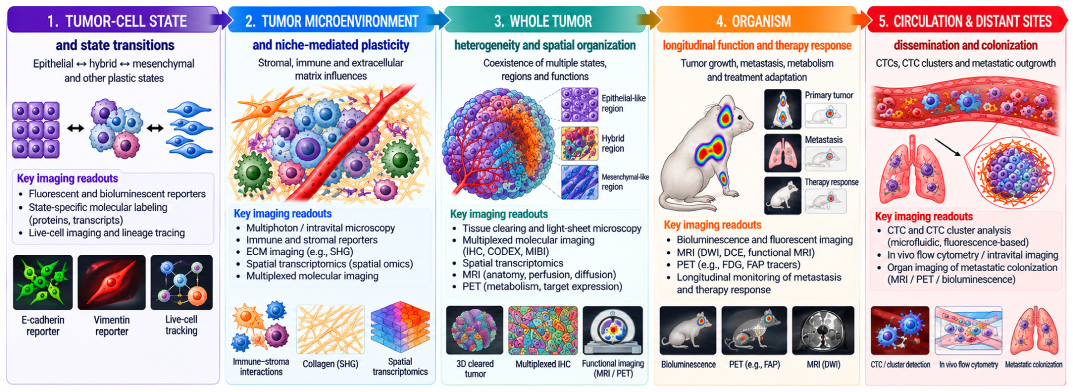
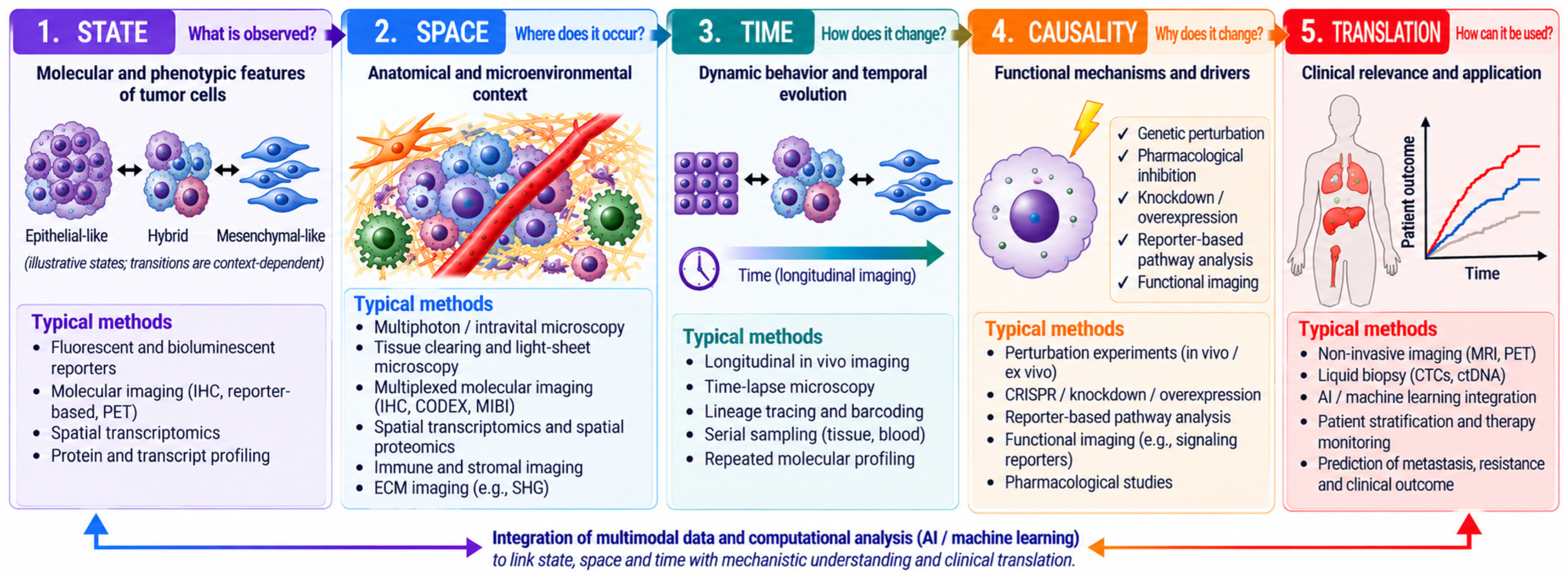

Disclaimer/Publisher’s Note: The statements, opinions and data contained in all publications are solely those of the individual author(s) and contributor(s) and not of MDPI and/or the editor(s). MDPI and/or the editor(s) disclaim responsibility for any injury to people or property resulting from any ideas, methods, instructions or products referred to in the content. |
© 2026 by the authors. Licensee MDPI, Basel, Switzerland. This article is an open access article distributed under the terms and conditions of the Creative Commons Attribution (CC BY) license.