


Abstract
Head and neck Kaposi sarcoma (HNKS) is a vascular neoplasm, associated with human herpesvirus 8 (HHV-8) infection and immunosuppression; it is one of the most common malignancies among people living with human immunodeficiency virus (PLWHIV). Comprehensive characterisation of HNKS aetiopathogenesis and clinicopathological critical for timeous diagnosis and prognosis is essential. A retrospective, descriptive cross-sectional study was conducted on forty-one histopathologically confirmed HNKS cases diagnosed between 2004 and 2024. Formalin-fixed, paraffin-embedded (FFPE) sections were retrieved and stained for immunohistochemical (IHC) biomarkers. Associations between variables were evaluated using Fisher’s exact test. The study cases showed a male: female ratio of 1.05:1 and a mean age of 33.4 ± 11.6 years. HIV positivity was documented in 85.4% of patients. Nodular stage predominated (63.4%); classical KS was the most common histopathological variant (82.9%). The CD34 endothelial biomarker was positive in all 41 cases (100%). HHV-8, D2-40, and p53 were positive in 51.2%, 45%, and 47.5% of cases respectively; high Ki-67 (≥15%) was observed in 25% of cases. No statistically significant associations were identified between stage and clinicopathological features (all p > 0.05). The epidemic KS was most common, predominantly affecting young adults. The combination of CD34, D2-40 and HHV8 may improve diagnostic accuracy. The prognostic/diagnostic roles of p53 and Ki-67 remain unelucidated.
Keywords:
Kaposi sarcoma; head and neck cancer; HHV-8; immunohistochemistry; HIV; CD34; D2-40; p53; Ki-67; FFPE 1. Introduction
Kaposi sarcoma (KS) tumours arise from endothelial cells of blood and lymphatic vessels; its aetiopathogenesis is primarily associated with human herpesvirus 8 (HHV-8), also known as Kaposi sarcoma-associated herpesvirus (KSHV) infection and immune suppression [1,2]. Four clinical-epidemiological subtypes are recognised, namely, classic, African (endemic), iatrogenic, and AIDS-related (epidemic) KS, all sharing consistent histopathological features regardless of subtype or anatomical location [3]. A fifth epidemiological subtype has been increasingly recognised in Human immunodeficiency virus (HIV)-negative men who have sex with men (MSM), presenting with features resembling classic KS [4,5].
Clinically, KS typically presents as single or multiple well-defined violaceous macules, papules, or patches that may progress into plaques and nodules, occasionally ulcerating and causing pain, haemorrhage, or lymphoedema anywhere on the head and neck region [6]. Kaposi sarcoma incidence has declined dramatically in high-income countries following widespread antiretroviral therapy (ART) availability [1], but it remains one of the most prevalent cancers in sub-Saharan Africa (SSA), due to the intersecting epidemics of HIV and HHV-8 co-infection that are sustaining a disproportionate global burden [7,8,9]. The latest Global Cancer Observatory (GLOBOCAN 2024) estimated an incidence of 34,230 KS cases worldwide in 2022, with Africa accounting for 81.3% of the global burden, an increase from the 74.1% in 2020 [7,8].
South Africa reported 4119 new KS cases in 2020, with male incidence 60% higher than female, reflecting the combined influence of HHV-8 seroprevalence and HIV burden in the region [7]. Head and neck KS (HNKS) represents a clinically significant anatomical subset of KS [2,10,11]. International studies reported HNKS prevalence ranging from 2.9% to 38% of all KS cases, with higher rates consistently observed in HIV-endemic settings [10,11,12,13]. In South Africa specifically, HNKS has been reported to account for 35.7% of all head and neck cancers in HIV-positive patients, with involvement of the oral cavity, facial skin, eyelids, cervical lymph nodes, and larynx documented as most common [6,14].
Histologically, KS lesions share consistent characteristics across all epidemiological subtypes and anatomical locations [15]. They are composed of spindle-shaped endothelial cells forming slit-like vascular channels containing extravasated erythrocytes and haemosiderin deposits, frequently accompanied by a lymphocytic infiltrate [2,3]. Three histological stages are recognised: patch, plaque, and nodular [6]. Patch-stage lesions appear as flat, reddish or violaceous macules characterised histopathologically by sparse vascular proliferation, slightly atypical endothelial cells, and the promontory sign, in which newly formed vascular channels dissect between pre-existing collagen bundles [3,6,15,16]. Plaque-stage lesions are raised with marked vascular proliferation and spindle cell fascicles; and nodular-stage lesions display densely packed spindle cells arranged in fascicles with numerous blood-filled slit-like spaces [3,6,16].
Several histological variants of KS have been described beyond the three classical stages. These include lymphangioma-like KS, characterised by dilated irregular vascular channels resembling lymphatic spaces; pyogenic granuloma-like KS, which closely mimics lobular capillary haemangioma; anaplastic KS, featuring pleomorphic cells with high mitotic activity; and additional variants such as hyperkeratotic, keloidal, telangiectatic, ecchymotic, and intravascular KS [3,14,15,16]. The head and neck region demonstrates substantial microscopic diversity, making the recognition of these variants essential to avoid misdiagnosis [2,3,10].
KS variants, combined with overlapping morphological features shared with other vascular and spindle cell lesions, can pose significant diagnostic challenges. Key differential diagnoses in the head and neck region include bacillary angiomatosis spindle cell angiosarcoma, Kaposiform haemangioendothelioma, relevant in paediatric head and neck lesions and HHV-8-negative; pyogenic granuloma, a common oral mimic of early KS lacking spindle cell fascicles and HHV-8 expression; and oral mucosal melanoma, an important differential for palatal lesions [10,15,16]. In diagnostically challenging cases, a comprehensive IHC panel including HHV-8 (LANA-1), D2-40, CD34, and CD31 should be employed to confirm or exclude KS.
Immunohistochemistry (IHC) plays a critical role in confirming KS, particularly in poorly differentiated lesions and histological variants that deviate from conventional morphology. Diagnostic IHC markers include HHV-8 latency-associated nuclear antigen 1 (LANA-1), which is 100% specific for KS and demonstrates granular nuclear positivity in latently infected cells, supported by endothelial markers CD31 and CD34 and the lymphatic marker D2-40 (podoplanin) [2,16,17,18]. Prognostic IHC markers including p53, p27Kip1, Bcl-2, cyclin D1, and Ki-67 have been explored for their correlation with histopathological stage and tumour aggressiveness, though their clinical utility in KS remains debated [15,16,17].
While clinical examination may suggest the diagnosis, histopathological confirmation remains the gold standard, and IHC staining is employed in diagnostically ambiguous cases [17,18]. Despite the high burden of KS in South Africa, comprehensive local data on the prevalence, clinicopathological characteristics, and immunophenotypic profile of HNKS remain scarce, with the few published studies being mainly epidemiological. The head and neck region’s facial skin, oral cavity and ocular surface have been reported amongst common sites for the earliest manifestation of KS in PLWHIV with a potential to cause structural and functional impairment [10,19,20]. Furthermore, the variable clinical manifestations of HNKS lesions lead to delayed diagnosis, as well as diagnostic conundrum [2,10,17]. This study therefore aimed to provide a comprehensive 21-year clinicopathological and immunohistochemical characterisation of HNKS at Dr George Mukhari Academic Laboratory (DGMAL), Ga-Rankuwa, contributing novel institutional data to address this evidence gap and inform diagnostic and clinical management strategies in this high-prevalence resource-limited setting. It is hoped that these study findings will add insights to the biological mechanisms of the KS tumour microenvironment and serve as a foundation for future studies that will explore potential prognostic and predictive biomarkers for KS. Furthermore, the study also serves as a call for awareness for clinicians to have a high index of suspicion when dealing with lesions in the head and neck region of people living with HIV, with clinical integration identifying common manifestations.
2. Materials and Methods
2.1. Study Design
This was a quantitative, retrospective, descriptive cross-sectional study conducted at the Department of Anatomical Pathology, at Dr George Mukhari Academic Laboratory (DGMAL), National Health Laboratory Service (NHLS), Ga-Rankuwa, Gauteng, South Africa. All HNKS cases diagnosed at DGMAL between 1 January 2004 and 31 December 2024 were eligible for inclusion. The study included 41 cases diagnosed with head and neck Kaposi sarcoma (HNKS) between 1 January 2004 and 31 December 2024. The sample size was calculated using Epi Info for Windows, version 7, based on a 5% significance level, a design effect of 1.0, and an estimated HNKS prevalence of 36% in South Africa as reported by Zwane and colleagues in 2018 [21].
Cases were identified through the NHLS academic affairs and research management system (AARMS)’s Central Data Warehouse (CDW) using keywords and Systematized Nomenclature of Medicine (SNOMED) code searches. The utilised keywords included Kaposi sarcoma, oral Kaposi sarcoma and ocular Kaposi sarcoma. The SNOMED codes used included: D3-F0803, D3-F0806, D3-F0807, D3-F0833, DA-F0819 and D3-F0863. Cases were included if they had confirmed histopathological HNKS diagnosis and adequate FFPE tissue for further analysis. Demographic and clinical data including age, sex, HIV status, ART status, CD4 count, HIV viral load, clinical presentation, and anatomical site were extracted from the dataset received from NHLS CDW. All the cases were reevaluated to confirm the morphological characteristics of KS before enrolling them to this study. A clear classical KS morphology and clinical correlation were used for initial diagnosis of KS.
2.2. Laboratory Procedures
2.2.1. Tissue Retrieval, Rescreening, and Preparation
Archived FFPE blocks and H&E-stained slides were retrieved from the NHLS Anatomical Pathology archives. All retrieved H&E slides were rescreened by a qualified anatomical pathologist to confirm initial diagnosis and document other relevant histopathological features. Where H&E slides were unavailable, FFPE blocks were processed per the departmental standard operating procedure. Blocks were placed on ice for 15 min before sectioning on a rotary microtome (Leica Biosystems, Nussloch, Germany). Sections (4 μm) were mounted on charged Superfrost™ slides (ThermoFisher Scientific, Waltham, MA, USA) for IHC and on frosted slides for H&E. H&E staining used Mayer’s haematoxylin (Cellpath, Newtown, UK) and eosin (Cellpath, UK); cover slipping was performed with the Tissue-Tek Cover Slipper (Leica Biosystems, Germany).
2.2.2. Histopathological Examination
The HNKS cases were staged as patch, plaque, or nodular per established criteria [6], and the identified histopathological variants were classical, pyogenic granuloma-like and lymphangioma-like. Clear KS morphological characteristics with clinical correlation was the initial KS diagnosis.
2.2.3. Immunohistochemistry
Sections were incubated at 60 °C in an oven (Memmert GmbH, Schwabach, Germany) for one hour for slide pre-treatment. Antigen retrieval was performed on the DAKO PT-Link (Diagnostech, Glostrup, Denmark) with EnVision FLEX Target Retrieval Solution (TRS) High pH buffer (Agilent Technologies, Glostrup, Denmark) at 97 °C for 20 min, followed by cooling to 65 °C before the slides were removed. Automated staining was performed on the DAKO Autostainer Link 48 (Diagnostech, Denmark) using the DAKO EnVision™ FLEX system (Agilent Technologies, Denmark), with FLEX Peroxidase Block, primary antibodies (Table 1), FLEX+ Mouse LINKER where required, FLEX HRP polymer (20 min), and FLEX DAB+ chromogen (10 min); Diagnostech supplied all reagents. Counterstaining was done using Mayer’s haematoxylin (Cellpath, United Kingdom (UK)). Cover slipping was performed on Tissue-Tek Cover Slipper (Leica Biosystems, Germany). Known positive controls were included with every run; the antibody was replaced with buffer for negative control. HHV-8 was scored as positive (granular nuclear) or negative; CD34 as positive or negative; D2-40 as positive (cytoplasmic membranous) or negative; p53 as positive or negative (nuclear); and CD34 as positive or negative. Ki-67 was categorised using the percentage of positive nuclei as low at <15% or high at ≥15%. Though the study utilised older FFPE blocks, recently processed positive and negative controls were used. The negative HHV-8 cases with true Kaposi sarcoma morphological characteristics were repeated manually before reporting.
Table 1.
Immunohistochemistry antibodies used (all supplied by Dako, Denmark).
2.3. Statistical Analysis
Data were captured in Microsoft Excel™ (Microsoft Office 365; Microsoft Corporation, Redmond, WA, USA) and exported to StataNow version 18.5 (StataCorp LLC, College Station, TX, USA) for statistical analysis. Continuous variables, including age, were summarised using descriptive statistics and expressed as mean ± standard deviation (SD), with range reported where applicable. Categorical variables including sex, HIV status, ART status, anatomical site, histopathological stage, histological variant, and immunohistochemical marker expression were summarised as absolute frequencies and percentages. Fisher’s exact test was applied to assess associations between histopathological stage and all demographic, clinicopathological, and immunohistochemical variables; this test was selected in preference to the chi-squared test given the small, expected cell frequencies inherent to this cohort size. Statistical significance was pre-set at p < 0.05 (two-tailed) for all analyses.
3. Results
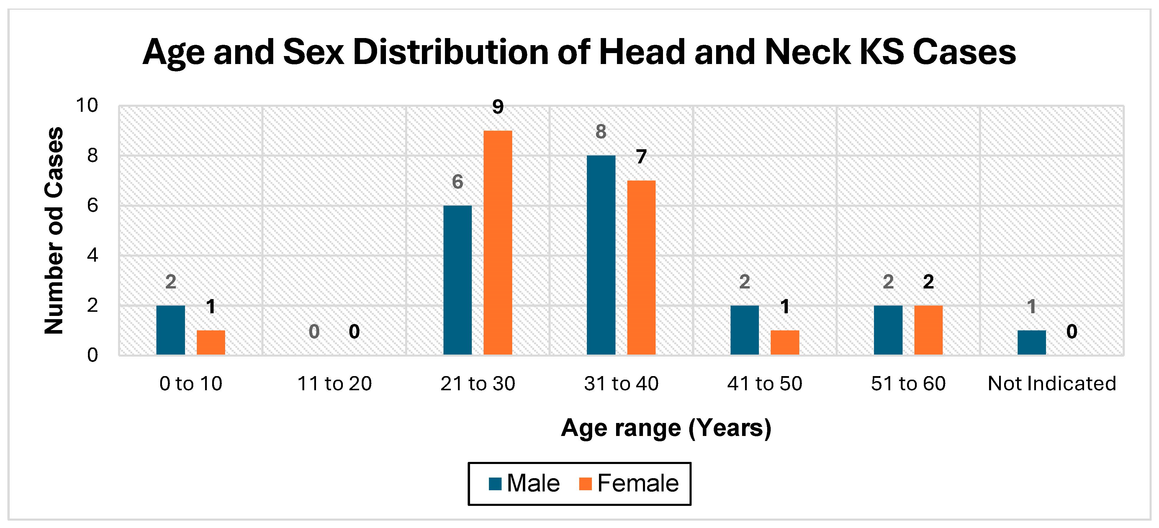
A total of 41 HNKS cases were analysed over the 21-year study period. A slight male predominance was observed, with 21 male (51.2%) and 20 female (48.8%) cases, and a male: female ratio of 1.05:1 (Figure 1). The mean age at diagnosis was 33.4 ± 11.6 years (range 3–59 years). The mean age for females was 32.8 ± 10.2 and for males it was 33.9 ± 12.8. The 21–30 year and 31–40-year age groups were the most frequently affected, each accounting for 17.1% of cases. Gender-specific analysis revealed that females predominated in the 21–30-year age group, while males were more prevalent in the 31–40-year age group (Figure 1).
Figure 1.
The age and sex distribution of head and neck Kaposi sarcoma.
The majority of patients 35 (85.4%) were HIV-positive at the time of HNKS diagnosis, with a mean age of 31.9 ± 5.8 years. No HIV-negative cases were documented in this cohort. HIV status was not indicated in six (14.6%), with a mean age of 41.5 ± 11.7 years. Regarding ART status at the time of diagnosis, 41.5% of patients (n = 17) were on ART, 6 (15%) were ART-naïve, and ART status was unknown in 18 (45%), as presented in Table 2.
Table 2.
HIV characteristics of head and neck Kaposi sarcoma.
3.1. Clinical Presentation and Anatomical Distribution
The most common clinical manifestation was lesions (macules, papules, plaques, and violaceous nodules) in 34% of cases, followed by tumour masses in 27%, swelling in 19%, and ulceration in 12%. Cutaneous KS was the most common involvement (23 cases, 56.1%). The facial skin (17.1%) and neck skin (17.1%) were the most frequent cutaneous sites; the hard palate (12.2%) was the most common mucosal site (Table 3). Multifocal involvement was documented in eight patients (19.5%).
Table 3.
Anatomical site distribution of head and neck Kaposi sarcoma cases.
3.2. Histopathological Stage and Variant Distribution
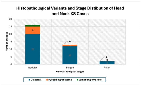
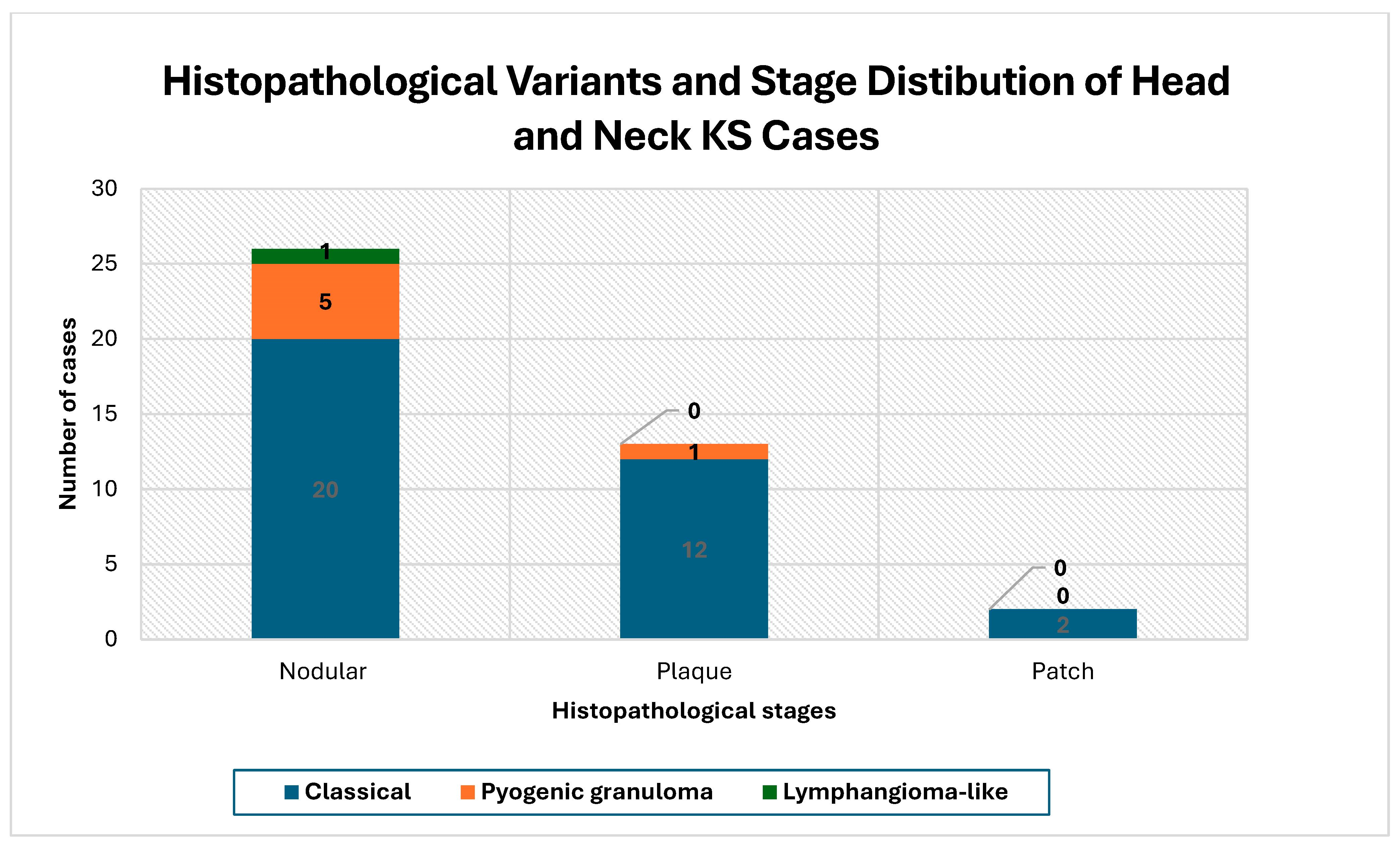
Histopathological classification and staging were done on all 41 cases; one case had insufficient tissue for further IHC analysis. Nodular stage predominated (26 cases, 63.4%), Classical KS was the most common variant (34 cases, 82.9%), followed by pyogenic granuloma-like KS (6 cases, 15.0%) and lymphangioma-like KS (1 case, 2.5%) (Figure 2). All non-classical variants were confined exclusively to the nodular stage. There was no statistically significant association between histological variant and histopathological stage (p = 0.837).
Figure 2.
The histopathological stages and variants distribution of HNKS.
3.3. Immunohistochemical Findings
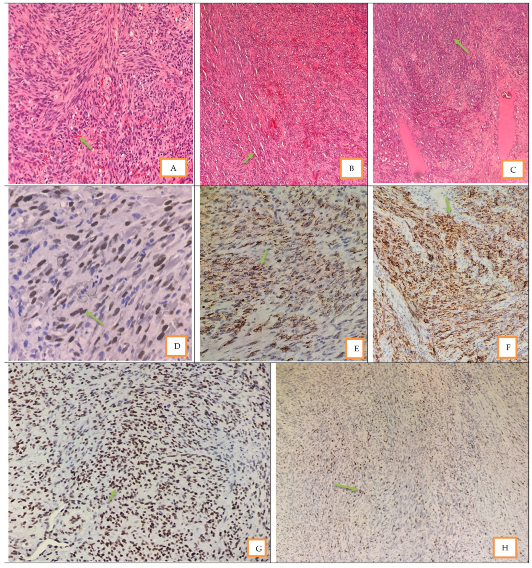
A total of 41 cases were included for immunohistochemistry analysis; one case was excluded from analysis of other biomarkers due to insufficient tissue. Figure 3 represent the haematoxylin and eosin and immunohistochemistry staining patterns for HNKS09, the figures (3D-H) show clear nuclear or cytoplasmic staining patterns of the diagnostic and prognostic markers. All 41 cases were CD34-positive (100.0%), confirming vascular and endothelial lineage across the entire cohort, irrespective of histopathological stage, histological variant, HIV status, or ART status.
Figure 3.
H&E and immunohistochemistry micrographs of a HNKS09 case. Micrographs (A–D) are haematoxylin and eosin stains of a conventional KS case. Subfigure (A) shows classical KS extravasation of red blood cells indicated by the green arrow at 40× magnification, with (B) showing slit-like spaces as indicated by green arrows at 4× magnification. Subfigure (C) shows proliferation of spindle cells as indicated by the green arrows at 4× magnification. Subfigure (D) shows positive HHV8 at 40× magnification; (E) positive CD34 at 40× magnification; (F) positive D2-40 at 10× magnification; (G) positive P53 at 10× magnification; and (H) positive KI67 at 4× magnification, with arrows pointing at positive cells.
3.4. Diagnostic Markers
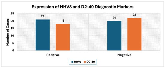
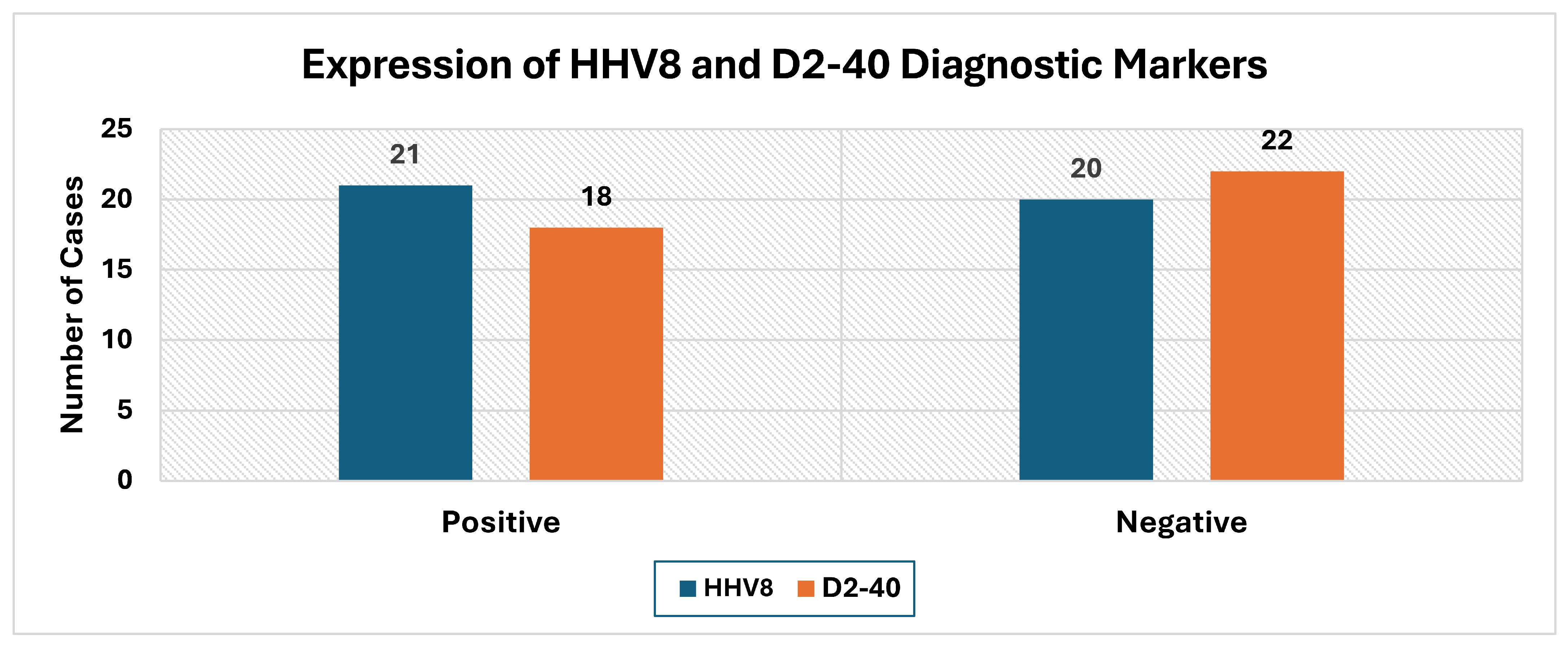
The staining patterns for the IHC makers are displayed on Figure 3. HHV-8 (LANA-1) expression was detected in 21 cases (51.2%). Of the 21 HHV-8-positive cases, the majority were in the nodular stage (17, 65.4%). The D2-40 (podoplanin) was positive in 18 cases (45.0%), consistent with lymphatic endothelial differentiation (Figure 4). HHV-8 and D2-40 were co-expressed in 10 cases (25.0%); at least one of the two diagnostic markers was positive in 28 cases (70.0%). All the HHV-8-positive cases were also HIV-positive.
Figure 4.
The diagnostic immunohistochemistry antibodies for HNKS.
3.5. Prognostic Markers
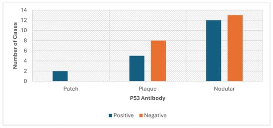
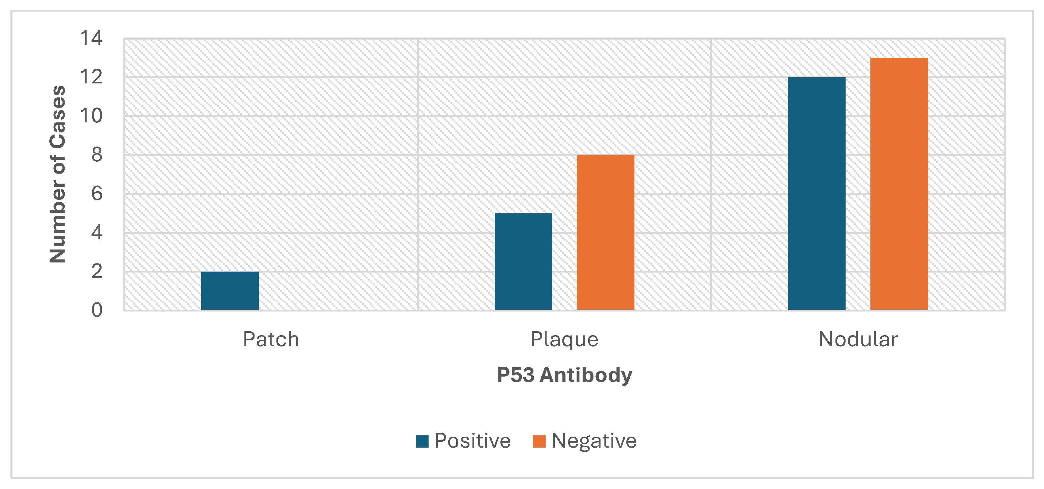
Table 4 illustrate the full IHC profiles of the HNKS cases. The P53 antibody was positive in 19 cases (47.5%); 63.2% of p53-positive cases were in the nodular stage (n = 12), 26.3% in the plaque stage (n = 5), and 10.5% in the patch stage (n = 2) (Figure 5). The Ki-67 proliferative index was low (≤15%) in 30 of 40 cases (75.0%), with a mean proliferative index of 12.6%. High Ki-67 expression (>15%) was observed in 10 cases (25%), predominantly in nodular-stage lesions (n = 9). A summary of all IHC marker results is presented in Table 4; the Ki-67 distribution by histopathological stage is shown in Table 5.
Figure 5.
P53 expression and histopathological stage for HNKS cases.
Table 4.
Immunohistochemical marker expression in head and neck Kaposi sarcoma.
Table 5.
Ki-67 proliferative index by histopathological stage in head and neck Kaposi sarcoma.
3.6. Associations Between Histopathological Stage and Clinicopathological Characteristics
Fisher’s exact test demonstrated no statistically significant associations between histopathological stage and most of the demographic, clinicopathological, and immunohistochemical variables analysed (all p > 0.05). HHV-8 showed a statistically significant association with histopathological stage at p = 0.040, with nodular stage being most common, as presented in Table 6.
Table 6.
Association between histopathological stage and demographic, clinicopathological, and immunohistochemical variables in head and neck Kaposi sarcoma.
4. Discussion
The findings of this study provide a comprehensive clinicopathological and immunohistochemical characterisation of HNKS within a high HIV-prevalence, resource-limited setting over a 21-year period. HNKS predominantly affected young HIV-positive adults in the fourth decade of life, with a near-equal sex distribution reflecting the feminisation of the HIV epidemic in sub-Saharan Africa [9,10,19,20]. Nodular-stage and classical-variant disease predominated, consistent with delayed clinical presentation and advanced disease at the time of diagnosis, a pattern reported across multiple African and Asian cohort studies [9,17,18,20,21,22,23,24]. This is the first South African study that integrated clinicopathological and IHC characterisation from this institution, contributing novel data to the limited literature on HNKS tumour microenvironment studies within the global population.
The study observed a slight male predominance (male: female ratio 1.05:1), with peak incidence in the third and fourth decade of life and the majority of cases being HIV-positive. The near-equal sex distribution is notably less pronounced than that reported in European and North American series, where male: female ratios of up to 10:1 have been documented [9,11]. This narrower sex ratio likely reflects the high HIV prevalence among South African women, as sub-Saharan Africa carries a disproportionate burden of HIV among females globally [24,25,26].
The strong association between HNKS and HIV infection (85.4% HIV-positive, with no confirmed HIV-negative cases) is consistent with the published literature, where HIV-positive prevalence in HNKS ranges from 38.1% to 82% [10,11]. Zeinaty et al. (2023) postulated that the higher HIV prevalence in African females, driven by biological, socio-cultural, and socioeconomic vulnerabilities, is directly associated with the higher rates of AIDS-related KS in women in this region, a pattern consistent with the predominantly epidemic KS subtype observed in this cohort [27].
The high proportion of unknown ART status (43.9%) underscores persistent challenges in early HIV diagnosis, documentation and treatment adherence in this resource-limited setting. Consequently, although the role of HIV in HNKS tumourigenesis is clearly evident in this cohort, the contribution of immune reconstitution through ART or combined therapy to disease suppression or progression could not be fully evaluated.
The predominance of nodular stage disease (63.4%) is consistent with findings from Nigerian (48.1%) [17], Tanzanian (60.5%) [22], and Pakistani (74.5%) [23] case series studies and likely reflects delayed healthcare presentation. Furthermore, the classical KS histopathological variant has been reported as the most common subtype at 82.9%, consistent with reports in the literature [3,10,11,15,16,17,18]. All non-classical histological variants were confined to nodular-stage lesions, consistent with the greater architectural complexity that emerges with disease progression and disease aggressiveness. The pyogenic granuloma-like KS rate (15.0%) is a clinically important finding, as this variant closely resembles lobular capillary haemangioma and requires IHC confirmation to avoid misdiagnosis [28].
The 51.2% HHV-8 positivity rate is substantially lower than the near-universal positivity (approaching 100%) reported in most published case series [10,15,16]; this finding indicates the usefulness of histopathological characterisation of cases especially within the ART and other treatment modalities that sometimes gives false-negative HHV-8 IHC results due to viral suppression or low viral count. The findings of the current study are closely related to those reported by Xu et al. (2026), where a substantial number of KS cases were negative for HHV-8 IHC; however, the study associated HHV-8 intensity with prognosis and did not really discuss the fate of the HHV-8-negative cases [29]. Platform-related variability in HHV-8 detection has also been documented [25,30,31,32]; prolonged archival FFPE storage (up to 20 years in this cohort) may also compromise antigen preservation [30]; and ART-induced suppression of HHV-8 viral replication may also reduce intranuclear LANA-1 availability [30]. The positive and negative controls used were recently processed tissue; the degradation linked to prolonged sample storage could not be determined as cases that were initially reported as HHV-8 were still HHV-8-positive and if fading was suspected, the repeated stain was positive.
Various studies in the literature have reported negative IHC HHV-8 results even upon repeated assay; however, most of the cases were positive when repeated using a molecular technique [25,31,32]. Interestingly, Speicher et al., 2015, in Kenya reported a diagnostic conundrum of HHV-8 PCR as it amplified HHV-8 on both KS samples and KS mimics, although the authors cited technical incompetencies and overamplification associated with the PCR technique [33]. These findings have direct diagnostic implications: when HHV-8 IHC yields a negative result despite strong histopathological suspicion; complementary markers (D2-40 and CD34) and PCR confirmation/in situ hybridisation should be considered. Although there is growing evidence in the literature that suggests that negative HHV-8 KS cases might represent a special entity, the current study repeated the negative IHC HHV-8 cases manually but could not perform molecular techniques due to a lack of resources. More evidence is required to regard HHV-8-negative IHC cases as a distinct clinical subtype, although some authors have made such reports [34].
The CD34 universal positivity (100%) in this cohort confirms vascular endothelial differentiation across all stages and variants, irrespective of HHV-8 status, supporting its utility as an ancillary confirmatory marker in resource-limited settings. The p53 and Ki-67 results, while not indicating statistical significance, demonstrated numerically higher expression in nodular-stage lesions (63.2% of p53-positive cases and 90% of high Ki-67 cases). This pattern is consistent with HHV-8 LANA-1-mediated p53 suppression in early-stage disease with progressive accumulation in advanced lesions [35,36], and with increasing proliferative activity with tumour progression. Larger prospective studies are needed to establish the prognostic utility of these markers in HNKS.
The finding that all HHV-8-positive cases in this cohort were also HIV-positive underscores the critical oncogenic synergy between these two viruses in HNKS development. The latency-associated nuclear antigen 1 (LANA-1) of HHV-8 further contributes to oncogenesis by inhibiting p53-mediated apoptosis and dysregulating the Rb/E2F cell cycle pathway, thereby promoting unrestricted tumour cell survival [35,36,37]. HIV, through progressive CD4 + T-lymphocyte depletion and chronic immune activation, disrupts the immunosurveillance mechanisms that normally suppress HHV-8 lytic replication and tumour cell expansion [9,38,39]. The resulting state of combined viral oncogenesis and HHV-8-driven endothelial transformation in the context of HIV-mediated immunosuppression creates a permissive tumour microenvironment that facilitates HNKS development, progression to advanced nodular-stage disease, and multifocal involvement, as observed in the current cohort [10,17,22,40]. This synergy further explains the late-stage predominance in this HIV-endemic setting, where immune reconstitution through ART initiation and adherence remains inconsistent [13,30,38,39,41].
The combined IHC panel comprising CD34, D2-40, and HHV-8 (LANA-1) enhanced diagnostic accuracy; CD34 universal positivity (100%) was particularly valuable in confirming endothelial lineage in cases where HHV-8 and D2-40 yielded negative results, underscoring its role as a critical first-line diagnostic marker in resource-limited settings [17]. The prognostic markers p53 and Ki-67 demonstrated biologically relevant trends in nodular-stage disease, consistent with LANA-1-mediated p53 suppression and escalating proliferative activity with tumour progression [3,22], warranting further investigation in larger prospective cohorts. These findings collectively highlight the diagnostic challenges inherent to HNKS in resource-limited settings, advocate for the adoption of a reliable, confirmed IHC panel to improve diagnostic accuracy [1,10,16,40]. Furthermore, there is a need for routine implementation of molecular techniques like in situ hybridisation and polymerase chain reaction for HHV-8 confirmation in IHC-negative cases.
5. Conclusions
Head and neck KS predominantly affected young, HIV-positive adults in this high HIV-prevalent, resource-limited setting, with nodular-stage and classical-variant disease predominating. The CD34 universal positivity confirms vascular endothelial lineage in all cases and supports its use as a complementary diagnostic marker. The suboptimal HHV-8 and D2-40 detection rates observed with archival FFPE material underscore the importance of combined diagnostic IHC panels and PCR/molecular-based HHV-8 confirmation when IHC results are equivocal. The trends observed for p53 and Ki-67 in nodular-stage disease suggest biological relevance as a potential prognostic/predictive biomarker for advanced disease; larger prospective studies are needed to establish their clinical utility. Strengthened HIV surveillance, early ART initiation, and improved treatment adherence remain essential to reducing the burden of HNKS in sub-Saharan Africa.
Author Contributions
A.O.M.—Protocol development, ethics application, data collection and cleaning, data analysis and manuscript editing. L.N.M.—Conceptualisation, supervision, study design, methodology, data analysis and manuscript editing. E.K.M.—Supervision, study design, methodology, data analysis, original manuscript draft and manuscript editing. N.E.M.—Data collection, data analysis and manuscript editing. P.K.S.—Data collection and cleaning, data analysis and manuscript editing. All authors have read and agreed to the published version of the manuscript.
Funding
This study was funded by a Thuthuka Grant (National Research Foundation, South Africa; Reference No: TTK240426216124).
Institutional Review Board Statement
The study received ethics approval from the Sefako Makgatho Health Sciences University Research Ethics Committee (SMUREC) under reference number SMUREC (Reference No.: SMUREC/M/258/2025: PG; Approval Date: 26 June 2025). The confidentiality of the cases was strictly adhered to throughout the study period. Permission to utilise data was granted by the NHLS AARMS with reference: PR2559401.
Informed Consent Statement
Patient consent was not applicable, as the study used archival formalin-fixed–paraffin-embedded tissue samples, for which permission was granted by the NHLS AARMS management and the Head of Department.
Data Availability Statement
The data presented in this study are available on request from the NHLS AARMS. The data is not publicly available due to patient privacy and ethical restrictions associated with the use of clinical and pathological patient information.
Acknowledgments
The authors are grateful to the Department of Anatomical Pathology at SMU; special thanks go to Bukiwe Mboba and Ephraim Moabi for their technical assistance. The authors also acknowledge the HOD of Anatomical Pathology at SMU for support, and MO Keetse for assistance with histopathological and immunohistochemistry analysis. This article is partially based on author AOM’s Honours Anatomical Pathology dissertation completed at Sefako Makgatho Health Sciences University, South Africa, in December 2025. During the preparation of this manuscript, the authors used ChatGPT version 4 to create the graphical abstract and Grammarly to improve overall readability and language.
Conflicts of Interest
The authors declare that this is their own work; all the sources used in this article have been duly acknowledged, and there are no conflicts of interest.
Abbreviations
The following abbreviations are used in this manuscript:
| AARMS | Academic affairs and research management system |
| AIDS | Acquired immunodeficiency syndrome |
| ART | Antiretroviral therapy |
| CDW | Central Data Warehouse |
| DGMAL | Dr George Mukhari Academic Laboratory |
| FFPE | Formalin-fixed, paraffin-embedded |
| GLOBOCAN | Global Cancer Observatory |
| HHV8 | Human herpesvirus 8 |
| HIV | Human immunodeficiency virus |
| HNKS | Head and neck Kaposi sarcoma |
| IHC | Immunohistochemical |
| KS | Kaposi sarcoma |
| KSHV | Kaposi sarcoma-associated herpesvirus |
| MSM | Men who have sex with men |
| NHLS | National Health Laboratory Service |
| LANA-1 | Latency-associated nuclear antigen 1 |
| PLWHIV | People living with HIV |
| SSA | Sub-Saharan Africa |
| SNOMED | Systematized Nomenclature of Medicine |
References
- Lebbe, C.; Garbe, C.; Stratigos, A.J.; Harwood, C.; Peris, K.; del Marmol, V.; Malvehy, J.; Zalaudek, I.; Hoeller, C.; Dummer; et al. Diagnosis and treatment of Kaposi’s sarcoma: European consensus-based interdisciplinary guideline (EDF/EADO/EORTC). Eur. J. Cancer 2019, 114, 117–127. [Google Scholar] [CrossRef] [Scilit] [PubMed]
- Ulloa-Padilla, J.P.; Ghassibi, M.P.; Dubovy, S.R.; Kerr, D.A. Clinicopathologic correlation of Kaposi sarcoma involving the ocular adnexa: Immunophenotyping of diagnostic and therapeutic targets. Ophthalmic Plast. Reconstr. Surg. 2020, 36, 185–190. [Google Scholar] [CrossRef] [Scilit] [PubMed]
- Bunn, B.K.; Carvalho, M.V.; Louw, M.; Vargas, P.A.; van Heerden, W.F. Microscopic diversity in oral Kaposi sarcoma. Oral Surg. Oral Med. Oral Pathol. Oral Radiol. 2013, 115, 241–248. [Google Scholar] [CrossRef] [Scilit] [PubMed]
- Denis, D.; Seta, V.; Regnier-Rosencher, E.; Kramkimel, N.; Chanal, J.; Avril, M.F.; Dupin, N. A fifth subtype of Kaposi’s sarcoma, classic Kaposi’s sarcoma in men who have sex with men: A cohort study in Paris. J. Eur. Acad. Dermatol. Venereol. 2018, 32, 1377–1384. [Google Scholar] [CrossRef] [Scilit] [PubMed]
- Morales, A.E.; Benson, G.; Glavan, S.; Giuliano, R.; Dickson, M.A. Fifth subtype of Kaposi sarcoma in HIV-negative MSM: A retrospective single-arm cohort study from a tertiary care center in NYC from 2000 to 2022. Oncologist 2025, 30, oyaf024. [Google Scholar] [CrossRef] [Scilit] [PubMed]
- Tourlaki, A.; Nazzaro, G.; Wei, Y.; Pontini, P.; Brambilla, L. Clinical, dermoscopic, ultrasonographic, and histopathologic correlations in Kaposi’s sarcoma lesions and their differential diagnoses: A single-center prospective study. J. Clin. Med. 2023, 12, 278. [Google Scholar] [CrossRef] [Scilit] [PubMed]
- Ferlay, J.; Ervik, M.; Lam, F.; Laversanne, M.; Colombet, M.; Mery, L.; Marion, P.; Ariana, Z.; Isabelle, S.; Freddie, B. Global Cancer Observatory: Cancer Today; International Agency for Research on Cancer: Lyon, France, 2024; Available online: https://gco.iarc.who.int/today (accessed on 4 May 2025).
- Sung, H.; Filho, A.M.; Laversanne, M.; Ferlay, J.; Siegel, R.L.; Soerjomataram, I.; Jemal, A.; Bray, F. Global cancer statistics 2024: GLOBOCAN estimates of incidence and mortality worldwide for 34 cancers in 186 countries. CA Cancer J. Clin. 2026, 76, e70090. [Google Scholar] [CrossRef] [Scilit] [PubMed]
- Russo, I.; Marino, D.; Cozzolino, C.; Piaserico, S.; Alaibac, M. Kaposi’s sarcoma: Evaluation of clinical features, treatment outcomes, and prognosis in a single-center retrospective case series. Cancers 2024, 16, 691. [Google Scholar] [CrossRef] [Scilit] [PubMed]
- Agaimy, A.; Mueller, S.K.; Harrer, T.; Bauer, S.; Thompson, L.D.R. Head and neck Kaposi sarcoma: Clinicopathological analysis of 11 cases. Head Neck Pathol. 2018, 12, 511–516. [Google Scholar] [CrossRef] [Scilit] [PubMed]
- Marcoval, J.; Bonfill-Ortí, M.; Martínez-Molina, L.; Valentí-Medina, F.; Penín, R.M.; Moreno, A. Evolution of Kaposi sarcoma in the past 30 years in a tertiary hospital of the European Mediterranean basin. Clin. Exp. Dermatol. 2019, 44, 32–39. [Google Scholar] [CrossRef] [Scilit] [PubMed]
- Huang, A.Y.Y.; Lin, C.L.; Chen, G.S.; Hu, S.C.S. Clinical features of Kaposi’s sarcoma: Experience from a Taiwanese medical center. Int. J. Dermatol. 2019, 58, 1388–1397. [Google Scholar] [CrossRef] [Scilit] [PubMed]
- Moreira, D.G.L.; da Silva, L.P.; de Morais, E.F.; Queiroz, S.I.M.L.; de Moura Santos, E.; de Souza, L.B.; de Almeida Freitas, R. The occurrence and pattern of head and neck sarcomas. Eur. Arch. Otorhinolaryngol. 2020, 277, 1473–1480. [Google Scholar] [CrossRef] [Scilit] [PubMed]
- Radu, O.; Pantanowitz, L. Kaposi sarcoma. Arch. Pathol. Lab. Med. 2013, 137, 289–294. [Google Scholar] [CrossRef] [Scilit] [PubMed]
- Lee, K.B.; Lee, H.S.; Lee, H.E.; Kim, M.A.; Kim, W.H. Immunohistochemical characteristics of Kaposi sarcoma and its mimicries. Korean J. Pathol. 2006, 40, 361–367. [Google Scholar]
- El-Ashmawy, A.A.; El-Ayat, G.A. Kaposi sarcoma: Immunohistochemical study of some diagnostic and prognostic markers. J. Egypt. Women’s Dermatol. Soc. 2012, 9, 77–85. [Google Scholar]
- Forae, G.D.; Obaseki, D.E. Trends and histopathological patterns of Kaposi sarcoma at the University of Benin Teaching Hospital, Nigeria. Oman Med. J. 2018, 33, 337–341. [Google Scholar] [CrossRef] [Scilit] [PubMed]
- Genedy, R.M.; Hamza, A.M.; Abdel Latef, A.A.; El-Beity, A.A. Sensitivity and specificity of D2-40 in differentiating Kaposi sarcoma from its mimickers. J. Egypt. Women’s Dermatol. Soc. 2021, 18, 67–74. [Google Scholar] [CrossRef] [Scilit] [PubMed]
- Parra-Fariñas, R.; Insua, C.; Buznego, L.; Runza, P.; Armiñanzas, C.; Gutierrez-Cuadra, M.; Marcos-González, S.; de Las Revillas, F.A. Conjunctival and cutaneous Kaposi’s Sarcoma as the first manifestation of HIV infection. Rev. Esp. Quimioter. 2025, 38, 352–354. [Google Scholar] [CrossRef] [Scilit] [PubMed]
- Masele, A.; Sohal, K.S.; Kalyanyama, B.M.; Owibingire, S.S.; Simon, E.N. Head and neck lesions among HIV/AIDS patients on highly active antiretroviral therapy attending the Muhimbili National Hospital in Dar es Salaam, Tanzania. Front. Oral Maxillofac. Med. 2020, 3, 2. [Google Scholar] [CrossRef] [Scilit]
- Zwane, N.B.; Mohangi, G.U.; Shangase, S.L. Head and neck cancers among HIV-positive patients: A five year retrospective study from a Johannesburg hospital, South Africa. S. Afr. Dent. J. 2018, 7, 121–126. Available online: http://www.scielo.org.za/scielo.php?script=sci_arttext&pid=S0011-85162018000300002&lng=en (accessed on 7 March 2025).
- Gervas, R.; Mgaya, E. Histopathological patterns and topographical distribution of Kaposi sarcoma at Muhimbili National Hospital, Tanzania. Afr. Health Sci. 2021, 21, 1733–1738. [Google Scholar] [CrossRef] [Scilit] [PubMed]
- Anique, M.; Akhlaq, H.; Azhar, S.; Saher, R.G.; Khan, H.U.; Arshad, M. Determining the stage of Kaposi sarcoma through histopathological analysis. Pak. J. Health Sci. 2024, 5, 36–40. [Google Scholar] [CrossRef] [Scilit]
- Motlhale, M.; Sitas, F.; Bradshaw, D.; Chen, W.C.; Singini, M.G.; de Villiers, C.B.; Lewis, C.M.; Muchengeti, M.; Waterboer, T.; Mathew, C.G.; et al. Epidemiology of Kaposi’s sarcoma in sub-Saharan Africa. Cancer Epidemiol. 2022, 78, 102167. [Google Scholar] [CrossRef] [Scilit] [PubMed]
- Buhle, A.C.; Phillips, M.A.; Hijazi, Y.M.; Wiid, M.; Grider, D.J. Kaposi sarcoma: What to do with a negative human herpesvirus 8 immunohistochemical stain? J. Cutan. Pathol. 2022, 49, 460–462. [Google Scholar] [CrossRef] [Scilit] [PubMed]
- UNAIDS Global AIDS Update 2022: In Danger; Joint United Nations Programme on HIV/AIDS: Geneva, Switzerland, 2022; Available online: https://www.unaids.org/sites/default/files/media_asset/2022-global-aids-update_en.pdf (accessed on 21 November 2025).
- Zeinaty, P.E.; Lebbé, C.; Delyon, J. Endemic Kaposi’s Sarcoma. Cancers 2023, 15, 872. [Google Scholar] [CrossRef] [Scilit] [PubMed]
- Ho, B.; Rinaldi, G.; Khan, I.; Szakacs, S. Pyogenic granuloma-like Kaposi sarcoma presenting in an HIV-negative man. BMJ Case Rep. 2020, 13, e237420. [Google Scholar] [CrossRef] [Scilit] [PubMed]
- Xu, J.; Zhou, J.; Huang, X.; Wu, M.; Guo, W.; Liu, K.; Yang, Y.; Yang, G.; Song, S. Comparative study of clinical features, pathology, and immunophenotype between HIV-related cutaneous and visceral Kaposi’s sarcoma. BMC Cancer 2026, 26, 576. [Google Scholar] [CrossRef] [Scilit] [PubMed]
- Manuraj, S.; Steve, M.; Heinz, K.; Alistair, R. Negative HHV-8 immunoreactivity in HIV-associated Kaposi’s sarcoma. J. Cutan. Pathol. 2016, 43, 626–628. [Google Scholar] [CrossRef] [Scilit] [PubMed]
- Anderlini, C.; Gaggino, M.P.; Zanitti, S.; Mazzotta, M.M.; Kurpis, M.; Valente, E. Negative Herpesvirus-8 Immunoreactivity in Kaposi’s Sarcoma after Liver Transplantation. Indian J. Dermatol. 2023, 68, 355. [Google Scholar] [CrossRef] [Scilit] [PubMed]
- Elias, E.M.; Mwakigonja, A.R. Evaluation of Clinical and Histopathological Diagnosis of Kaposi’s Sarcoma at Muhimbili National Hospital, Dar es Salaam, Tanzania. East Afr. Health Res. J. 2024, 8, 394–401. [Google Scholar] [CrossRef] [Scilit] [PubMed]
- Speicher, D.J.; Wanzala, P.; D’Lima, M.; Njiru, A.; Chindia, M.; Dimba, E.; Johnson, N.W. Diagnostic challenges of oral and cutaneous Kaposi’s sarcoma in resource-constrained settings. J. Oral Pathol. Med. 2015, 44, 842–849. [Google Scholar] [CrossRef] [Scilit] [PubMed]
- Kumar, P. Human herpes virus-8-negative kaposiform sarcoma in a HIV-positive male: Novel variant. J. Cancer Res. Ther. 2020, 16, 177–179. [Google Scholar] [CrossRef] [Scilit] [PubMed]
- Friborg, J., Jr.; Kong, W.P.; Hottiger, M.O.; Nabel, G.J. p53 inhibition by the LANA protein of KSHV protects against cell death. Nature 1999, 402, 889–894. [Google Scholar] [CrossRef] [Scilit] [PubMed]
- Katano, H.; Sato, Y.; Sata, T. Expression of p53 and HHV-8-encoded LANA with inhibition of apoptosis in HHV-8-associated malignancies. Cancer 2001, 92, 3076–3084. [Google Scholar] [CrossRef] [Scilit]
- Radkov, S.A.; Kellam, P.; Boshoff, C. The latent nuclear antigen of Kaposi sarcoma-associated herpesvirus targets the retinoblastoma-E2F pathway and with the oncogene Hras transforms primary rat cells. Nat. Med. 2000, 6, 1121–1127. [Google Scholar] [CrossRef] [Scilit] [PubMed]
- Ensoli, B.; Sgadari, C.; Barillari, G.; Sirianni, M.C.; Stürzl, M.; Monini, P. Biology of Kaposi’s sarcoma. Eur. J. Cancer 2001, 37, 1251–1269. [Google Scholar] [CrossRef] [Scilit] [PubMed]
- Majaya, E.; Girdler-Brown, B.V.; Muchengeti, M.; Singh, E. The impact of the South African ART programme on the age-standardised incidence rate of Kaposi sarcoma, 1999–2016. Int. J. Infect. Dis. 2021, 102, 20–27. [Google Scholar] [CrossRef] [Scilit] [PubMed]
- Cesarman, E.; Damania, B.; Krown, S.E.; Martin, J.; Bower, M.; Whitby, D. Kaposi sarcoma. Nat. Rev. Dis. Primers 2019, 5, 9. [Google Scholar] [CrossRef] [Scilit] [PubMed]
- Khammissa, R.A.; Pantanowitz, L.; Feller, L. Oral HIV-associated Kaposi sarcoma: A clinical study from the Ga-Rankuwa area, South Africa. AIDS Res. Treat. 2012, 2012, 873171. [Google Scholar] [CrossRef] [Scilit] [PubMed]

Disclaimer/Publisher’s Note: The statements, opinions and data contained in all publications are solely those of the individual author(s) and contributor(s) and not of MDPI and/or the editor(s). MDPI and/or the editor(s) disclaim responsibility for any injury to people or property resulting from any ideas, methods, instructions or products referred to in the content. |
© 2026 by the authors. Licensee MDPI, Basel, Switzerland. This article is an open access article distributed under the terms and conditions of the Creative Commons Attribution (CC BY) license.