


Abstract
Metabolic dysfunction-associated steatotic liver disease (MASLD) affects 32% of the world’s population. Its pathogenesis is closely linked to metabolic dysfunction and lipotoxicity. The main pathogenic processes in MASLD are lipotoxicity, oxidative stress, and activation of inflammasome assembly. These processes sustain chronic inflammation, recruit immune cells to the liver, activate Kupffer cells, and subsequently drive the differentiation of hepatic stellate cells into myofibroblasts and the development of fibrosis. Oxidized low- and high-density lipoproteins (oxLDL and oxHDL) are involved in the progression of MASLD to metabolic dysfunction-associated steatohepatitis (MASH). OxLDL and oxHDL are formed through enzymatic oxidation (involving NOX, MPO, and 15-LOX) as well as non-enzymatic oxidation of native lipoproteins and acquire an affinity for scavenger receptors. Scavenger receptors are often associated with Toll-like receptors (TLRs) 2/4 and are involved in the pathogenesis of the transition from MASLD to MASH and fibrosis, in which activation of the LOX-1/NF κB/NLRP3 axis plays a central role. This review examines drugs in clinical trials that act on downstream signaling pathways. Studies of inhibitors targeting components of this signaling cascade have shown promising results. Analysis of therapeutic strategies for MASH suggests that a combination of lowering lipoprotein levels and blocking inflammatory signaling may be most effective, as it targets both the metabolic and inflammatory components of the disease.
1. Introduction
Based on a 2022 meta-analysis of 72 publications that included a sample of 1 million people from 17 countries, the estimated global prevalence of metabolic dysfunction-associated steatotic liver disease (MASLD) was 32.4% [1]. The prevalence of MASLD increased significantly from 25.5% in 2005 to 37.8% in 2016. The disease is significantly more common in men (39.7%) than in women (25.6%) [2].
The earliest hypothesis to explain the pathogenesis of metabolic dysfunction-associated steatohepatitis (MASH) is the “double hit” theory, which proposes that steatosis is present along with a “second hit” from other factors, such as oxidative stress [3]. The “first hit” is the development of hepatic steatosis due to disruption of normal lipid metabolism, leading to increased lipid intake and decreased lipid elimination. This is accompanied by increased consumption of polyunsaturated fatty acids (PUFAs), increased influx of free fatty acids (FFAs) into the liver, and enhanced de novo lipogenesis [4]. The “second hit” involves additional pathogenic factors, such as oxidative stress, mitochondrial dysfunction, lipotoxicity, insulin resistance (IR), and cytokine and adipokine imbalance, which, in addition to the contributors mentioned above, lead to severe liver damage [5].
However, this concept has since expanded and evolved into the “multiple parallel hits” hypothesis [6]. According to this hypothesis, pathogenic factors affect the liver, where the development of MASH may be primary rather than secondary to MASLD [6]. These factors include IR, characterized by dysfunctional adipose tissue with altered secretion of inflammatory cytokines and adipokines, increased lipogenesis, and impaired suppression of lipolysis in adipocytes, all of which together increase the flux of fatty acids into the liver [3].
Recently, metabolic syndrome (MS) has been added to these factors [7]. MS is a cluster of interconnected metabolic disorders, including abdominal obesity, IR, dyslipidemia, arterial hypertension, and other related factors [8]. MS increases the risk of developing MASLD, which is one of its most common complications [9]. The prevalence of MASLD among patients with MS is as high as 80% [10]. According to the World Health Organization (WHO) recommendations, metabolic health criteria are based on cardiometabolic risk (CMR) assessment [10], the classification of Metabolically Healthy Obesity (MHO), and the criteria for Harmonized Metabolic Syndrome (HMS) [11,12,13].
The criteria include the following lipoproteins and their fractions: very-low-density lipoproteins (VLDL), small dense low-density lipoprotein (sdLDL), reduced levels of high-density lipoproteins (HDL), and associated protein markers such as apolipoprotein B (ApoB) and lipoprotein(a) (Lp(a)) [14,15].
In the context of CMR, these lipoproteins form a specific cluster of abnormalities known as atherogenic dyslipidemia [16]. Lipid metabolism disorders, including altered levels of triglycerides (TGs), remnant lipoprotein particles, apolipoproteins, and FFAs, are associated with liver dysfunction [16]. The accumulation of TGs in the liver is accompanied by increased lipotoxicity caused by high levels of FFAs and other lipid metabolites, leading to mitochondrial dysfunction, oxidative stress, and increased production of reactive oxygen species (ROS) [16].
The gut-liver axis also plays a role in MASLD pathogenesis: disruption of the intestinal microflora increases small intestinal permeability, which leads to increased absorption of fatty acids and activation of inflammatory processes with the release of pro-inflammatory cytokines [17]. In addition, individual bacterial metabolites can influence bile acid metabolism and tissue insulin sensitivity [17]. Hepatocyte damage may also result from chronic inflammation caused by genetic or epigenetic factors [18].
This narrative review examines the pathogenetic role of oxidized low- and high-density lipoproteins (oxLDL and oxHDL) in the initiation and progression of MASLD/MASH, analyzes the therapeutic potential of key metabolic targets, and synthesizes the available information thematically.
2. Metabolic Syndrome as a Pathogenetic Basis of MASLD: The Role of Lipotoxicity, Adipokine Dysregulation, and Oxidative Stress
MASLD is closely associated with MS and may be a hepatic manifestation of MS, whose main pathogenetic mechanism is IR [19]. MASLD and MS are characterized by similar patterns of adipokines, cytokines, and other blood metabolites. For example, plasminogen activator inhibitor-1 (PAI-1) levels increase in the tissues of patients with progressive fibrosis, leading to inhibition of tissue proteolytic activity and increased fibrogenesis [20,21]. Elevated PAI-1 levels are associated with obesity, IR, and type 2 diabetes (T2D) [22]. Additionally, dysregulated adipokines in abdominal obesity (characterized by decreased adiponectin levels and increased pro-inflammatory mediators) increase systemic inflammation and IR, promoting TG accumulation in hepatocytes and activating liver macrophages [5].
Lipotoxicity is another mechanism of liver damage. Excessive influx of non-esterified fatty acids (NEFAs) from visceral adipose tissue and increased de novo lipogenesis during hyperinsulinemia lead to the accumulation of FFAs and ceramides, membrane damage, and lipoapoptosis in hepatocytes [23]. In adipose tissue, lipogenesis decreases and lipolysis increases during IR, resulting in increased amounts of saturated fatty acids (SFAs) entering the bloodstream. In MS, the primary flow of FFAs enters the liver through the portal vein [24]. Paradoxically, lipogenesis is preserved, including the synthesis of fatty acids from glucose and fructose and the activities of the enzymes acetyl-CoA carboxylase (ACC), fatty acid synthase (FASN), and stearoyl-CoA desaturase-1 (SCD1), which are associated with sterol regulatory element-binding protein 1 (SREBP-1c) and carbohydrate-responsive element-binding protein (ChREBP) [25]. In addition, β-oxidation of fatty acids increases compensatorily in the early stages of the disease. As fatty infiltration progresses, mitochondrial membrane potential is depleted, and fatty acids are oxidized in peroxisomes [26].
In the initial stages, TGs are stored in hepatocytes. As the disease progresses, levels of intermediate lipid metabolites with cytotoxic properties increase, including diacylglycerols, ceramides, acylcarnitines, and free cholesterol [27]. Lipotoxicity involves diacylglycerol, which activates protein kinase Cε (PKCε) and contributes to the development of IR [27]. Ceramides alter mitochondrial membrane permeability, induce apoptosis, and inhibit the phosphatidylinositol 3-kinase (PI3K)/protein kinase B (AKT) signaling pathway [27,28]. Free cholesterol can accumulate in the membranes of mitochondria and the endoplasmic reticulum, altering their properties and disrupting the function of membrane proteins [28]. Gradually, this damage is accompanied by the development of oxidative stress [28]. Thus, mitochondrial dysfunction develops, and within the electron transport chain, some electrons react with oxygen to form superoxide anions [29].
The oxidation of fatty acids in peroxisomes is accompanied by the formation of hydrogen peroxide and is referred to as peroxisomal β-oxidation, not lipid peroxidation [30]. PUFAs in membranes undergo oxidation, and lipid peroxidation generates malondialdehyde (MDA) and 4-hydroxynonenal (4-HNE) [30]. These products damage proteins, lipids, and DNA [30]. As a result, damaged cells release damage-associated molecular patterns (DAMPs), which are recognized by innate immune receptors, including those on Kupffer cells [31]. This activates pro-inflammatory signaling pathways, including nuclear factor kappa B (NF-κB), and the production of associated pro-inflammatory cytokines [5]. Afterward, monocytes, neutrophils, and T cells migrate to the liver, helping sustain the chronic inflammatory process [32].
Taken together, impaired lipid efflux in MASLD completes a vicious cycle of pathogenesis: lipid export from the liver is reduced because of a lack of structural phospholipids in the VLDL membrane, lipotoxicity-induced ApoB deficiency, and decreased microsomal triglyceride transfer protein (MTP) activity [33]. Together, these factors create an imbalance among lipid intake, synthesis, oxidation, and elimination. MASLD not only accompanies MS but often represents its hepatic manifestation: IR and abdominal obesity trigger systemic inflammation, adipokine dysregulation, and lipotoxicity, leading to progression to MASH and fibrosis.
3. Lipoprotein Transport in Metabolic Syndrome
Lipids are a diverse group of substances that are insoluble in water but soluble in organic solvents [34]. Lipids involved in the pathogenesis of MS can be classified into several groups. Glycerolipids consist of glycerol and three fatty acids. Phospholipids contain glycerol, two fatty acids, and a polar phosphate group. Sphingolipids are based on sphingosine, and sterols include cholesterol and its derivatives. FFAs serve as a source of energy and as substrates for the synthesis of other lipids [34].
Cholesterol and TGs are nonpolar lipid molecules, so they are transported through the aqueous plasma by lipoproteins [35]. The main function of lipoproteins is to transport hydrophobic lipids in an aqueous environment [35]. Lipoproteins include chylomicrons, VLDL, intermediate-density lipoproteins (IDL), low-density lipoproteins (LDL), and HDL [35]. Chylomicrons produced by enterocytes in the small intestine transport dietary TGs and cholesterol to peripheral tissues and the liver [35]. ApoB is involved in chylomicron assembly and plays a key role in the absorption of dietary fats. VLDL delivers endogenous TGs to muscle and adipose tissue, where it releases TGs and forms remnants. As these remnants lose TGs and become enriched with cholesterol, they are converted into LDL [36]. LDL delivers cholesterol to tissues, while HDL transports cholesterol back to the liver [35]. Blood lipid profile parameters typically include levels of total cholesterol, LDL, HDL, TGs, and ApoB. Because each particle contains only one ApoB molecule, its concentration is a reliable indicator of the number of atherogenic lipoprotein particles.
Thus, lipids form a complex, functionally significant system that is transported in the blood as part of lipoproteins, and analyzing their fractions and apolipoproteins allows an objective assessment of lipid metabolism in MS.
4. The Influence of Lipid Parameters on MASLD Progression
Lipid assessment criteria are used to evaluate metabolic health. The widely recognized classifications of Metabolically Healthy Obesity (MHO), Harmonized Metabolic Syndrome (HMS), and CMR are applied to assess parameters of metabolic disorders [37,38,39]. These parameters include several criteria: the absence of MS, a limited number of its components, indicators of IR, or a combination of these factors [37]. One advantage of the CMR criteria [37] is that they use more sensitive threshold values for lipid metabolism indicators, such as TGs, compared with traditional criteria for defining MS [37]. The TG level according to the CMR criteria is thought to more accurately reflect the formation of sdLDL, which have more pronounced atherogenic properties than larger LDL particles [37,38,39].
The use of CMR parameters is also relevant for individuals who are not obese. Increasing attention in modern scientific literature is being paid to a phenotype referred to as Metabolically Unhealthy Normal Weight (MUNW) [40]. This phenomenon describes a condition in which individuals with a normal body mass index (BMI) exhibit metabolic disturbances characteristic of patients with MS [40]. Metabolic disorders in non-obese individuals are driven by the same fundamental mechanisms as in obesity, including IR, adipose tissue dysfunction, and chronic inflammation [41]. Thus, patients with MASLD who are not obese have lipid metabolism disorders at levels comparable to those of patients with obesity [42]. This has been demonstrated in studies showing that the frequency of abnormal TG and HDL levels does not differ between these groups [43,44]. This confirms that key metabolic abnormalities can be present independently of BMI.
Thus, using CMR parameters is justified for all patients, regardless of BMI. Assessing factors such as medical history, glucose levels, and, most importantly, lipid metabolism parameters makes it possible to identify individuals at increased risk for metabolic liver complications.
5. Mechanism of Oxidative Modification of LDL
Oxidized lipids are formed through both enzymatic and non-enzymatic reactions from PUFAs, cholesterol, and phospholipids [45]. Oxidation products undergo further modification and include oxidized phospholipids, oxysterols, oxylipins, lipid peroxides, and reactive aldehydes [45]. Oxidation products can be present in free form or bound to ApoB, with several classes of oxidized lipids localized within the lipoprotein [46]. OxLDL promotes cellular stress, inflammatory signaling, and immune dysregulation [47].
OxLDL forms when native LDL is modified in the subendothelial space of hepatic blood vessels [46], in blood plasma, and at sites of inflammation. Native LDL penetrates the endothelium and binds to extracellular matrix proteoglycans. This process involves lipid peroxidation of the lipophilic core and ApoB-100, generating cytotoxic products [48,49,50,51,52,53].
Native LDL oxidation can occur via enzymatic or non-enzymatic pathways. The former involves nicotinamide adenine dinucleotide phosphate (NADPH) oxidases (NOX, generating superoxide anions and hydrogen peroxide), myeloperoxidase (MPO, producing hypochlorous acid and tyrosyl radicals), and lipoxygenases (LOX, forming hydroperoxides). In the latter case, oxidation occurs through free radicals of other origins, transition metal ions, or chain autoxidation [50,51]. Oxidants produced by these enzymes and metal ions can oxidize LDL to varying degrees. Two oxidation stages are distinguished: minimally modified LDL (mmLDL) and extensively oxidized LDL (oxLDL) (Figure 1). mmLDL represents the initial stage of LDL oxidation, and because the ApoB-100 structure remains intact, mmLDL is still recognized by the native LDL receptor (LDLR) [45]. OxLDL represents the final stage of oxidation, during which the ApoB-100 protein is also modified, leading to conformational changes in ApoB-100 and the acquisition of high affinity for scavenger receptors [45]. The term “oxLDL” is used as a broad category for all oxidized forms of LDL. To distinguish between mmLDL and oxLDL, a method is used to measure the level of substances that react with thiobarbituric acid, which reflects the degree of lipid peroxidation [49,50]. This method does not always allow clear differentiation between mmLDL and oxLDL. Specific monoclonal antibodies to modified ApoB epitopes are required for more accurate discrimination [48]. NOX and lipoxygenases predominantly generate mmLDL, which is characterized by the presence of oxidized lipids and preservation of the native ApoB structure [52]. mmLDL has low affinity for macrophage scavenger receptors (SR), but it induces the recruitment of inflammatory cells and the production of chemokines and cytokines through activation of Toll-like receptors 4 (TLR4). MPO and NOX, in combination with NO synthase, oxidize LDL extensively through the formation of peroxynitrite (ONOO−), affecting both the lipid and protein components of ApoB [53]. Highly oxidized oxLDL is readily taken up by macrophages (including liver Kupffer cells) through SR [54,55] (Figure 1).
Figure 1.
Formation of oxidized lipoproteins in the liver. Native LDL is oxidized enzymatically (NOX, MPO, 15-LOX) or non-enzymatically to form mmLDL and oxLDL. Oxidation is accompanied by the formation of MDA/4-HNE adducts with ApoB and a decrease in affinity for LDLR. During oxidation, native HDL is transformed into oxHDL, losing its antioxidant properties and impairing reverse cholesterol transport.
The enzymatic cascade leading to the formation of oxLDL involves the coordinated actions of NOX, MPO, and LOX [50,51]. During the initiation phase, membrane-bound NOX complexes generate superoxide anions, which subsequently form hydrogen peroxide [50,53,55]. Hydrogen peroxide is a substrate for MPO [50,51]. Localized on the surface of LDL particles, MPO catalyzes the chlorination and oxidation of ApoB amino acid residues through the generation of hypochlorous acid and tyrosyl radicals [50,53]. 15-LOX catalyzes the oxygenation of PUFAs in LDL cholesteryl esters and phospholipids, triggering a lipid peroxidation cascade that releases the aldehydes MDA and 4-HNE. MDA and 4-HNE contain electrophilic carbonyl groups that react with the nucleophilic side chains of amino acids in the ApoB protein [50,53]. As a result of nucleophilic addition reactions that form Schiff bases or Michael adducts, covalent bonds are created between the lipid and the protein [56,57,58]. Modification of lysine residues reduces the positive charge of ApoB-100, resulting in conformational changes and loss of its native tertiary structure [50,53].
Thus, oxidation can be enzymatic, involving myeloperoxidase, NADPH oxidase, and lipoxygenase, or non-enzymatic, involving transition metal ions and free radicals. Oxidative modification of lipoproteins in MASLD, however, cannot be fully explained by the classical model of lipid peroxidation. These pathways are not interchangeable and, when activated, generate qualitatively different oxidized species with distinct biological properties. Moderate oxidation mediated by NOX and 15-LOX produces minimally modified LDL (mmLDL) that retains native ApoB, whereas extensive oxidation by MPO and NO synthase generates fully modified oxLDL with MDA/4-HNE adducts.
The sources of oxidative modification in the liver are poorly understood. Myeloperoxidase and NADPH oxidases of endothelial cells and macrophages play a key role in the vascular wall. In the liver, lipotoxicity-induced mitochondrial dysfunction can serve as an additional source of ROS, potentially triggering local oxidation of lipoproteins. This is supported by studies showing the presence of oxLDL in hepatocytes and Kupffer cells in MASH [56].
HDL normally mediates the reverse transport of lipid hydroperoxides to the liver for subsequent degradation. OxLDL loses its affinity for the classical LDL receptor, and the liver loses its ability to take up and utilize cholesterol (Figure 1).
One of the key mechanisms linking these processes is the LOX-1/NF-κB/NLRP3 signaling axis. The pathogenetic effects of oxLDL are mediated by hepatocytes, Kupffer cells, and hepatic stellate cells (HSCs). The negative charge of OxLDL facilitates binding to SRs: LOX-1 on the endothelium and hepatocytes, as well as CD36 and SR-A on liver Kupffer cells [59]. Kupffer cells increase oxLDL uptake via CD36 [60]. Hepatocytes receive signals via LOX-1 that activate the NLR family pyrin domain containing 3 (NLRP3) inflammasome. SR-A mediates oxLDL uptake in liver macrophages and promotes MASLD progression. The combined effects of hepatocytes (lipotoxicity, DAMPs), Kupffer cells (interleukin (IL)-1β and tumor necrosis factor (TNF)-α production), and stellate cells (fibrogenesis), acting through activation of the LOX-1/NF-κB/NLRP3 axis, mediate the transition from MASLD to MASH.
Despite compelling evidence linking oxidized lipoproteins to the severity of MASLD, a number of unresolved questions remain. The relative contribution of enzymatic and non-enzymatic oxidation pathways in the liver is still uncertain. OxLDL is an important pathogenetic factor in the development and progression of MASLD to MASH.
6. Oxidized LDL in the Pathogenesis of MASLD
Quantitative and qualitative changes in oxidized lipoproteins in MASLD are characterized by several key features. First, oxHDL levels show a stepwise increase as liver fibrosis progresses, without correlating with total HDL levels. This indicates that the qualitative composition of HDL (the proportion of oxidized forms), rather than its absolute quantity, is a more significant parameter. Second, serum oxLDL concentration is positively correlated with the severity of MASLD, which allows it to be considered a potential biomarker of disease progression. Third, hepatic oxLDL content and the expression of scavenger receptors (CD36, LOX-1) increase as the disease progresses, reaching a maximum in cirrhosis.
The association of circulating and hepatic oxidized lipoproteins with the severity of MASLD is supported by extensive clinical data. The level of oxHDL increases stepwise from steatosis to steatohepatitis and then to fibrosis, which makes it a promising biomarker for assessing the stage of the disease. A similar pattern is seen for oxLDL: its concentration correlates with histological activity and the stage of fibrosis. It is important to emphasize that this relationship apparently reflects not just oxidative stress as such, but the active pathogenetic role of oxidized lipoproteins in the activation of the NLRP3 inflammasome and fibrogenesis. OxLDL plays a key pathogenic role in the development of MASLD and its progression to MASH. Research shows that oxLDL contributes to steatosis, oxidative stress, and liver cell death, including pyroptosis and ferroptosis [61]. Endothelial cells and macrophages generate ROS, and the resulting free radicals attack fatty acids, triggering a chain reaction of LDL peroxidation. The interaction of OxLDL with receptors triggers inflammatory signaling pathways, such as NF-κB, which promote cytokine production [62]. In hepatocytes, OxLDL exacerbates mitochondrial dysfunction and ROS generation, induces lipotoxicity and hepatocyte apoptosis, and leads to the accumulation of free cholesterol and fatty acids in hepatocytes, causing endoplasmic reticulum stress and hepatocyte death (ballooning) [62,63].
Some oxLDL signaling pathways are associated with HSC activation, which induces a transition to a fibroblast-like phenotype. Activated HSCs produce collagen and contribute to the development of fibrosis [63]. Two pathways are involved: a direct pathway, in which oxLDL binds to TLR4 on HSCs and activates them, and an indirect pathway, in which oxLDL binds to Kupffer cells, which then secrete transforming growth factor β (TGF-β), activating HSCs [64].
OxLDL components can directly or indirectly activate TLR4 and TLR2 on Kupffer cells and HSCs, leading to activation of the classical MyD88-dependent pathway. This stimulates the expression of C-C motif chemokine ligand 2 (CCL2)/Monocyte chemoattractant protein-1 (MCP-1) chemokines, which recruit new circulating monocytes into the liver and exacerbate lobular inflammation. Another activator, in addition to NF-κB and LOX-1, is proprotein convertase subtilisin/kexin type 9 (PCSK9), which is induced by oxLDL and NF-κB. It also increases the expression of LOX-1 and TLR4 via the TLR4/NF-κB pathway, creating a vicious cycle in MASH pathogenesis [65,66].
One of the key signaling pathways is the LOX-1/NF-κB/NLRP3 pathway [66,67]. To date, it has been linked to the progression of MASLD to MASH [68]. OxLDL binds to LOX-1, which is expressed on the membranes of sinusoidal endothelial cells (LSECs), Kupffer cells, and hepatocytes [59,69]. OxLDL is considered an endogenous DAMP and activates intracellular NOX. This interaction leads to excessive ROS production and causes mitochondrial dysfunction, mitochondrial DNA (mtDNA) damage, and endoplasmic reticulum stress [68,69]. The next step involves activation of NF-κB and initiation of NLRP3 inflammasome assembly. In the nucleus, NF-κB activates the transcription of genes encoding inflammasome components and other pro-inflammatory cytokines [68,69]. This creates a vicious cycle, as the LOX-1 gene promoter also contains an NF-κB binding site. As a result, NF-κB activation increases LOX-1 expression, amplifying the pathogenic effects of oxLDL. HSCs are sensitive to damage and interact with the NLRP3 complex [67]. In addition to the oxLDL/LOX-1/NF-κB/NLRP3 axis, oxLDL activates pathogenic pathways in the liver through scavenger receptors on Kupffer cells, monocyte-derived macrophages (MDMs), and hepatocytes [70]. The scavenger receptor CD36 is the primary mediator of oxLDL uptake [70].
Oxidized phospholipids within oxLDL interact with peroxisome proliferator-activated receptor gamma (PPARγ), a nuclear receptor. Cells with enhanced internalization take up more oxLDL, accumulate cholesterol in lysosomes, and undergo transformation. Furthermore, when oxLDL binds to CD36, cells become immobilized and anchored at sites of liver inflammation, maintaining chronic infiltration and continuous secretion of pro-inflammatory cytokines (TNF-α, IL-6) [71]. SR-A1 recognizes oxidation-specific epitopes on ApoB-100, which overloads the lysosomal system and leads to leakage of cathepsins into the cytosol, mitochondrial dysfunction, induction of hepatocyte apoptosis, and subsequent necrosis [71].
In the pathogenesis of MASH, the oxLDL/LOX-1/NF-κB/NLRP3 signaling axis acts as an integrated two-component trigger: oxLDL binding to the LOX-1 induces NF-κB-dependent inflammasome priming, thereby increasing NLRP3 and IL1B transcription, while the accumulation of free cholesterol in macrophages and Kupffer cells activates NLRP3 inflammasome. Thus, oxLDL contributes to the pathogenesis of MASLD and MASH, enhancing lipotoxicity, oxidative stress, Kupffer cell activation, and fibrogenesis through specific receptors.
7. Oxidized HDL in the Pathogenesis of MASLD
OxHDL, like oxLDL, has been well studied in the context of atherosclerosis, where it exhibits pronounced atherogenic potential [72]. Few studies have examined the involvement of oxHDL in the pathogenesis of MASLD/MASH, but its role in dyslipidemia is well established [73]. Clinical studies in patients with MASLD have shown that serum oxHDL concentrations increase as liver fibrosis worsens [74]. Moreover, oxHDL levels are not associated with HDL levels [74].
Oxidative modification disrupts the physiological functions of HDL, including its role in cholesterol efflux [75] and in neutralizing lipid peroxidation [76].
In addition to losing its native properties, oxidized HDL activates the LOX-1/NOX2/ROS axis [77,78] and can induce oxidative stress in liver sinusoidal cells, thereby exacerbating hepatocyte damage [76,79]. Studies of HUECs, animal models, and patients with atherosclerotic lesions have provided evidence that oxHDL may contribute to endothelial fibrosis by inducing endothelial-to-mesenchymal transition via the LOX-1/NOX-2/ROS/NF-κB pathway, increasing TGF-β secretion, and activating ALK-5/Smad signaling [76,79,80].
Some studies report that HDL, when oxidized, can stimulate pathological processes and increase cardiovascular risk, a phenomenon termed “Paradox-Idation.” The structure of ApoA-I is damaged, and oxidized HDL loses the ability to bind to its receptors, thereby blocking cholesterol efflux. Oxidized HDL also loses affinity for its receptor, scavenger receptor class B type I (SR-BI), and activates the scavenger receptors LOX-1, CD36, and SR-A [80].
Native HDL normally carries the enzyme paraoxonase-1 (PON1), which protects LDL from damage. Oxidation inactivates PON1, triggering a cascade of lipid oxidation in the blood. Oxidation of HDL leads to the replacement of proteins and enzymes with the acute-phase protein serum amyloid A (SAA), as well as the accumulation of lipid hydroperoxides and oxidized cholesteryl esters, which can then be transported into tissues [81,82,83,84,85].
Some authors dispute the pathogenic role of oxHDL; earlier animal studies showed that the liver rapidly takes up and reduces lipid hydroperoxides associated with HDL [82]. Oxidized HDL can prevent the oxidation of LDL by metal ions, although the mechanism underlying this effect remains unknown [82,83]. Specific methionine residues in ApoA-I are proposed to enhance the reduction of lipid hydroperoxides to alcohols, which challenges the idea that HDL loses its physiological properties upon oxidation [86].
Despite the above, the pathogenetic role of oxHDL in metabolic disorders, particularly MASLD, remains controversial and requires further research, especially regarding its contribution to MASH progression.
8. Scavenger Receptor Signaling Pathways in the Liver
CD36 in hepatocytes and liver sinusoidal cells activates lipogenesis via the enzymes FASN, ACC, and SCD1 and induces a transcriptional cascade mediated by the factors PPAR-α, PPAR-γ, and SREBP-1c [87]. Oxidative stress and lipotoxicity lead to the activation of c-Jun N-terminal kinase 1 (JNK1) and IκB kinase β (IKKβ) and the inhibition of insulin receptor substrate 1 (IRS-1), resulting in insulin resistance (IR). In Kupffer cells (liver-resident macrophages) and HSCs, CD36 promotes pro-inflammatory and profibrotic responses to metabolic stress. In addition, CD36 in macrophages activates the Src–NF-κB axis [87]. In parallel, intracellular oxidative stress induces inflammasome assembly and pro-inflammatory cytokine secretion, driving HSC activation and their differentiation into myofibroblasts, thereby promoting liver fibrosis [87] (Figure 2).
Figure 2.
OxLDL and oxHDL activate scavenger receptors and trigger signaling cascades involved in MASLD/MASH progression: inflammation (NF-κB/NLRP3), lipotoxicity (JNK/PPARγ), oxidative stress (NOX, ROS), and fibrogenesis (TGF-β).
CD36 and SR-A are the main receptors for oxLDL uptake by Kupffer cells, which triggers a pro-inflammatory and profibrotic cascade. The SR-A receptor signaling pathway is associated with regulating the inflammatory immune response, endocytosis, and cell survival in macrophages [87]. Receptor activation causes phosphorylation of intracellular tyrosine residues, which activates the mitogen-activated protein kinases extracellular signal-regulated kinase (ERK), p38, and JNK, as well as the PI3K and Akt pathways. SR-A crosstalks with TLRs, such as TLR4, activating the IKK complex and promoting nuclear translocation of the transcription factor NF-κB, triggering the expression of pro-inflammatory cytokines. Interestingly, the receptor plays a multidirectional role that depends on the microenvironment: SR-A can act as either an activator or a suppressor of the immune response in macrophages [88] by competing with SR-A and TLR4 for LPS binding and inhibiting TLR4 (Figure 2). However, SR-A deletion leads to increased production of pro-inflammatory cytokines and enhanced activation of NF-κB.
CD36 is a receptor with a wide range of ligands, including long-chain fatty acids, thrombospondins, and molecular patterns associated with injury or pathogens [60]. Notably, when binding different ligands, CD36 interacts with different membrane partners—neurokinin receptor A, TLRs, integrins, and tetraspanins—forming distinct signaling complexes [46,47]. Consequently, activation of the same receptor can trigger intracellular cascades that differ in direction, depending on the ligand environment. LOX-1 activates a cascade of inflammatory, pro-oxidant, and apoptotic intracellular pathways [89]. LOX-1 activation is not solely due to oxLDL-mediated signaling. Assembly of NOX2/NOX4 initiates oxidative stress, which activates the mitogen-activated protein kinases p38 (MAPK) and ERK1/2 kinases. This leads to the degradation of the IκB inhibitor and, at the same time, to the activation of the mitochondrial apoptotic pathway through phosphorylation of the p66Shc (the 66-kDa isoform of the growth factor adapter Shc) [89]. In addition, LOX-1 can initiate endothelial dysfunction and tumorigenesis via RhoA/ROCK signaling by suppressing basal endothelial nitric oxide synthase (eNOS) activity and reducing nitric oxide levels [55,90]. In tumorigenesis, LOX-1 stabilizes hypoxia-inducible factor 1α (HIF-1α) and vascular endothelial growth factor (VEGF), activating angiogenesis and the epithelial–mesenchymal transition (EMT) program [91]. In patients with MASLD, fibrosis may develop through alternative non-LOX-1 pathways, suggesting that the receptor is functionally redundant. SREC-I (scavenger receptor expressed by endothelial cells I) initiates actin-dependent pinocytosis and endocytosis [92]. Forms heteromeric plasmalemmal complexes with TLR4, activating the MyD88/TNF receptor-associated factor 6 (TRAF6)/TGF-β-activated kinase 1 (TAK1) pathway and p38 MAPK and JNK1/2 kinases, which in turn activate NF-κB and trigger the pro-inflammatory response [93]. SREC-I activates actin-dependent pinocytosis through the small guanosine triphosphatases (GTPases) cell division control protein 42 homolog (Cdc42) and Ras-related C3 botulinum toxin substrate (Rac1) [94]. SREC-I may have anti-inflammatory properties through integrin αvβ3 and trigger an anti-inflammatory cascade via the PI3K/Akt axis [95]. Its increased expression on the endothelium promotes the recruitment of pro-inflammatory T cells, exacerbating tissue damage [93]. At the same time, its ability to induce M2 polarization of Kupffer cells through PI3K/Akt/STAT3 signaling is an important protective mechanism [92] that helps resolve inflammation and restore homeostasis.
Stabilin-1 (STAB1) is activated via its cytoplasmic tail containing an NPXY-like motif [96]. Upon binding oxLDL, this motif recruits the PTB domain-containing adaptor protein GULP1 (GULP PTB domain-containing engulfment adapter protein 1) and the vesicular transport regulator GGA2 (Golgi Associated, Gamma Adaptin Ear Containing, ARF Binding Protein 2) [96]. The STAB1/GULP1 complex activates the small GTPase Rac1 in clathrin-independent endocytosis [96]. oxLDL undergoes endosomal-lysosomal degradation. In parallel, Stabilin-1 activates the PI3K/Akt/mTOR cell survival pathway, involving other PI3K/Akt pathways, signal transducer and activator of transcription 6 (STAT6), and PPARγ [55,95]. Activation of this pathway blocks the expression of pro-inflammatory cytokines and stimulates the secretion of anti-inflammatory factors (IL-10, TGF-β), thereby suppressing the immune response.
Signaling from scavenger receptors is often context-dependent and cross-regulated. Future studies should assess the contribution of each receptor at different stages of MASLD and functionally characterize their signaling cascades in tissues.
9. Pharmacologically Targeted Drugs to Control MASLD/MASH Progression
Blocking components of the LOX-1/NF-κB/NLRP3 signaling cascade is considered one of the most promising therapeutic strategies for controlling the development of MASH and preventing its progression to cirrhosis [97]. These molecular targets are the focus of contemporary preclinical and clinical research [97].
The first approach is to block LOX-1, preventing oxLDL formation at the earliest stage and the subsequent pathological processes [98]. Several research groups are developing targeted approaches to blocking, such as monoclonal antibodies and antisense oligonucleotides [99,100]. The study results demonstrated that suppression of LOX-1 in the liver reduces oxLDL uptake by Kupffer cells and prevents excessive NF-κB activation. This process can be indirectly influenced by existing drugs, such as statins and PCSK9 inhibitors [101].
Another approach is to use molecules that target mitochondrial dysfunction. MitoQ and SkQ1 can scavenge ROS that accumulate on the inner mitochondrial membrane [102,103]. NADPH oxidase inhibitors, such as setanaxib, can block the enzymes NOX1 and NOX4, which are activated when oxLDL binds to LOX-1 [104,105]. Setanaxib is one of the drugs targeting the LOX-1/NF-κB/NLRP3 axis that have undergone clinical trials. It has demonstrated a significant antifibrotic effect in liver fibrosis by protecting HSCs from activation. Another drug that underwent a Phase 2a clinical trial was HU6, 2,4-dinitrophenol (DNP), a mild mitochondrial uncoupler of oxidative phosphorylation [105,106] (Table 1).
Direct NF-κB inhibition poses significant safety concerns, given its essential role in immune regulation. Therefore, indirect inhibition, such as that mediated by PPAR-γ and PPAR-α, is preferred. PPAR-γ agonists and dual PPAR-α/γ agonists, thiazolidinediones, and newer glitazars exhibit potent anti-inflammatory effects in the liver. However, this approach is associated with a high incidence of severe side effects. Despite this, pioglitazone is currently included in many clinical guidelines as a drug capable of reducing necroinflammation in MASH [107,108,109,110,111,112,113] (Table 1).
Blocking the NLRP3 inflammasome reduces inflammation induced by oxLDL. The inhibitor MCC950 prevented inflammasome assembly, halted hepatocyte pyroptosis, reduced IL-1β levels, and halted fibrogenesis, but exhibited high hepatotoxicity. Insomelide was one of the first drugs to selectively inhibit NLRP3. Newer inhibitors include MCC950 and NT-0796, which are in earlier stages of development and are undergoing trials to assess their impact on systemic inflammation [114,115,116] (Table 1).
In addition, inhibitors of hepatocyte cell death are being studied. For example, disulfiram blocks the ability of cleaved gasdermin to form membrane pores and trigger pyroptosis [117]. Emricasan blocks caspase-1 and apoptotic caspases-3 and -8. In patients with MASH, it effectively reduced transaminase (ALT, AST) levels and cell death markers.
There is a group of drugs that act as fibroblast growth factor (FGF) and farnesoid X receptor (FXR) agonists. The hormone FGF21, by interacting with the FGFR1c receptor and the β-Klotho (KLB) coreceptor, is involved in regulating energy homeostasis and has anti-inflammatory properties [118]. FGF21 agonists activate AMP-activated protein kinase (AMPK), increase fatty acid oxidation, reduce lipogenesis, and increase LDL receptor expression. Efruxifermin [119], pegosafermin [120], and pegbelfermin [121] are FGF21 agonists (Table 1).
The FXR regulates the inflammatory response through NF-κB, bile acid synthesis, and lipid and carbohydrate metabolism. FXR activates PPARα, suppressing lipogenesis. Tropifexor and vonafexor reduced steatosis and liver fibrosis but caused side effects such as pruritus and a worsening lipid profile (NCT04328077, NCT03812029). The FXR agonist EDP-305 had similar effects on MASLD without significantly affecting lipid metabolism (NCT03421431). Obeticholic acid is an FXR agonist. Despite promising results with obeticholic acid in liver fibrosis, a new randomized controlled trial showed that obeticholic acid worsened blood lipoprotein profiles, leading to the suspension of drug development [120,122]. Using metabolic agents (e.g., FGF21 or farnesoid X receptor agonists) that reduce LDL levels and overall steatosis, in combination with targeted inflammatory response inhibitors (LOX-1, NOX, and NLRP3 inhibitors, as well as PPAR-γ and PPAR-α/γ agonists), will simultaneously limit the cause (excess modified lipids) and the destructive consequence (cell death and fibrosis) (Table 1).
Table 1.
Potential drugs in clinical trials for the treatment of MASH.
10. Conclusions
Metabolic disorders lead to disturbances in the lipid profile and its modifications. Lipoproteins can be partially oxidized—affecting only the lipid portion (minimally modified LDL [mmLDL])—or oxidized along with the protein portion of ApoB-100 (extensively oxidized LDL [ext-oxLDL]). The dysfunctional particles oxLDL and oxHDL lose their physiological properties. oxLDL induces lipotoxicity and an inflammatory response and sustains oxidative stress.
Oxidative modification of HDL disrupts its role in reverse cholesterol transport [50,81]. oxHDL loses the ability to effectively stimulate cholesterol efflux from peripheral cells via the transporters ABCA1/ABCG1 (ATP-binding cassette transporter subfamily A member 1/ATP-binding cassette transporter subfamily G member 1), which promotes lipid accumulation in macrophages and their transformation into foam cells [75,80]. In addition, oxHDL loses affinity for the SR-BI receptor, which normally mediates the selective uptake of cholesteryl esters from HDL by hepatocytes for subsequent excretion into bile [78,80]. Disruption of this stage leads to reduced cholesterol clearance and worsening dyslipidemia, which is an important link in the pathogenesis of MASLD [74,76]. Both modified lipoproteins interact with SRs. In the liver, oxLDL and oxHDL interact with LOX-1, CD36, and SR-A, while oxHDL also has affinity for SREC-I and STAB1.
The key molecular mechanism linking oxidative stress to MASH progression is oxLDL/LOX-1/NF-κB/NLRP3 signaling. This pathway regulates the main pathogenetic processes of MASH—lipotoxicity, oxidative stress, and initiation of inflammasome assembly. The associated signaling pathways downstream of SRs and TLR2/4 form a complex network of interconnected mechanisms that sustain chronic inflammation and immune cell recruitment to the liver, followed by activation of Kupffer cells, transformation of stellate cells, and development of fibrosis. Studies of inhibitors targeting components of the signaling cascade involved in MASLD/MASH progression have shown promising results in both preclinical and clinical studies. Analysis of therapeutic effectiveness indicates that a combination approach aimed at reducing lipoprotein levels and blocking components of inflammatory signaling pathways, thereby allowing simultaneous intervention in the metabolic and inflammatory components of pathogenesis, appears most promising.
Oxidized lipoproteins trigger inflammatory and profibrotic reactions in the liver. Further research in this area offers prospects for developing personalized approaches to MASLD/MASH treatment based on targeted modulation of key signaling axes. Limitations are related to the difficulty of validating and standardizing methods for measuring oxLDL and oxHDL concentrations as biomarkers of MASLD/MASH progression. For example, oxHDL can be considered an independent marker of worsening liver fibrosis [74]; however, the lack of a standardized method for its determination in blood does not yet allow a threshold level to be established. Regarding the role of oxHDL, uncertainty remains as to whether it has proatherogenic and profibrotic effects [77,78] or retains antioxidant properties [83,84,85,86], which would contradict the loss of function of native HDL during oxidative modification.
Information about the role of oxLDL and oxHDL is mainly derived from cross-sectional studies, which makes it difficult to draw conclusions about causal relationships between oxidized lipoprotein levels and disease progression. Prospective multicenter studies with long-term patient follow-up would make it possible to more effectively assess the role of oxLDL and oxHDL as biomarkers of fibrosis stage.
It is important to remember the heterogeneity of patients with steatosis, for whom several stratification criteria exist, necessitating a differentiated approach to the management of patients with MASLD/MASH.
One informative approach may be to characterize the LOX-1/NF-κB/NLRP3 signaling axis, which would help identify patients with the most pronounced inflammatory component, in whom targeted therapy may be most effective.
Analysis of existing therapeutic approaches has shown limited effectiveness of treatments targeting any single component of pathogenesis. For example, reducing lipoprotein levels without controlling inflammatory signaling does not prevent the damaging effects of oxLDL and oxHDL on hepatocytes and stellate cells, and blockade of inflammatory mediators without correcting the lipid profile does not eliminate the source of oxidative stress. The most reasonable strategy appears to be a combination approach involving FGF21 agonists (efruxifermin, pegozafermin, and pegbelfermin) [117,118,119,120] with inhibitors of LOX-1, NOX, or NLRP3 [107,108,109,110,111,112,113]. The optimal therapeutic doses, drug administration regimens, assessment of drug interactions, and the need to conduct lengthy and expensive clinical trials remain unresolved. In addition, blocking inflammatory signaling pathways carries several serious risks. Direct inhibition of NF-κB is associated with severe side effects due to its fundamental role in immunoregulation. NLRP3 inhibitors (MCC950) are characterized by high hepatotoxicity [114,115,116]. FXR agonists cause undesirable changes in the lipid profile, which led to the suspension of their development [119,121].
Achieving clinical impact by targeting molecular mechanisms requires solving a set of problems, including standardizing methods for determining oxidized lipoproteins, developing reproducible criteria for patient stratification, creating safe combined therapeutic regimens, and conducting large-scale prospective studies. Resolving these issues will be a focus of future research in this area.
Author Contributions
Conceptualization—D.A.S. and L.S.L., Original Draft—N.M.T., D.A.S. and A.A.K., Visualization—E.D.G. and M.A.V., Review and Editing—R.T., M.M.B. and S.S.V., Supervision—M.A.V. and K.A.Y. All authors have read and agreed to the published version of the manuscript.
Funding
The study was carried out with financial support from the Russian Science Foundation (Project No. 23-15-00061-P).
Institutional Review Board Statement
Not applicable.
Informed Consent Statement
Not applicable.
Data Availability Statement
The original contributions presented in this study are included in the article. Further inquiries can be directed to the corresponding authors.
Acknowledgments
During the preparation of this manuscript, the authors used InstaText (Slovenia) to check the grammar of the text.
Conflicts of Interest
The authors declare no conflicts of interest.
Abbreviations
The following abbreviations are used in this manuscript:
| 4-HNE | 4-hydroxynonenal |
| ACC | acetyl-CoA carboxylase |
| ABCA1 | ATP-Binding Cassette transporter subfamily A member 1 |
| ABCG1 | ATP-Binding Cassette transporter subfamily G member 1 |
| ApoA-I | apolipoprotein A-I |
| ApoB | apolipoprotein B |
| ApoB-100 | apolipoprotein B-100 |
| BMI | body mass index |
| CCL2 | C-C motif chemokine ligand 2 |
| CD36 | cluster of differentiation 36 |
| ChREBP | carbohydrate-responsive element-binding protein |
| CMR | cardiometabolic risk |
| DAMP | damage-associated molecular pattern |
| DNP | 2,4-dinitrophenol |
| EDP-305 | FXR agonist (non-bile acid) |
| eNOS | endothelial nitric oxide synthase |
| ERK | extracellular signal-regulated kinase |
| FASN | fatty acid synthase |
| FFA | free fatty acid |
| FGF21 | fibroblast growth factor 21 |
| FGFR | fibroblast growth factor receptor |
| FGFR1c | fibroblast growth factor receptor 1c |
| FXR | farnesoid X receptor |
| GSDMD | gasdermin D |
| GULP1 | GULP PTB domain-containing engulfment adapter protein 1 |
| GGA2 | golgi Associated, Gamma Adaptin Ear Containing, ARF Binding Protein 2 |
| HDL | high-density lipoprotein |
| HIF-1α | hypoxia-inducible factor 1-alpha |
| HMS | Harmonized Metabolic Syndrome |
| HSC | hepatic stellate cell |
| IDL | intermediate-density lipoprotein |
| IKKβ | inhibitor of nuclear factor kappa-B kinase subunit beta |
| IL-1β | interleukin-1 beta |
| IL-6 | interleukin-6 |
| IL-10 | interleukin-10 |
| IR | insulin resistance |
| IRS-1 | insulin receptor substrate 1 |
| IκB | inhibitor of nuclear factor kappa-B |
| JNK | c-Jun N-terminal kinase |
| KC | Kupffer cell |
| KLB | β-Klotho |
| LDL | low-density lipoprotein |
| LDLR | low-density lipoprotein receptor |
| LOX | lipoxygenase |
| LOX-1 | lectin-like oxidized low-density lipoprotein receptor-1 |
| Lp(a) | lipoprotein(a) |
| LSEC | liver sinusoidal endothelial cell |
| MAPK | mitogen-activated protein kinase |
| MASH | metabolic dysfunction-associated steatohepatitis |
| MASLD | metabolic dysfunction-associated steatotic liver disease |
| MCP-1 | monocyte chemoattractant protein-1 |
| MDA | malondialdehyde |
| MDM | monocyte-derived macrophage |
| MHO | metabolically healthy obesity |
| mmLDL | minimally oxidized low-density lipoprotein |
| MPO | myeloperoxidase |
| MS | metabolic syndrome |
| MTP | microsomal triglyceride transfer protein |
| mtDNA | mitochondrial DNA |
| MUNW | metabolically unhealthy normal weight |
| MyD88 | myeloid differentiation primary response 88 |
| NEFA | non-esterified fatty acid |
| NF-κB | nuclear factor kappa-B |
| NLRP3 | NLR family pyrin domain-containing 3 |
| NO | nitric oxide |
| NOX | NADPH oxidase |
| oxHDL | oxidized high-density lipoprotein |
| oxLDL | oxidized low-density lipoprotein |
| PAI-1 | plasminogen activator inhibitor-1 |
| PCSK9 | proprotein convertase subtilisin/kexin type 9 |
| PI3K | phosphoinositide 3-kinase |
| PKCε | protein kinase C epsilon |
| PON1 | paraoxonase-1 |
| PPAR | peroxisome proliferator-activated receptor |
| PPAR-α | peroxisome proliferator-activated receptor alpha |
| PPAR-γ | peroxisome proliferator-activated receptor gamma |
| PTB | phosphotyrosine-binding |
| PUFA | polyunsaturated fatty acid |
| Rac1 | Ras-related C3 botulinum toxin substrate 1 |
| RhoA | Ras homolog family member A |
| ROCK | Rho-associated coiled-coil containing protein kinase |
| ROS | reactive oxygen species |
| SCD1 | stearoyl-CoA desaturase-1 |
| sdLDL | small dense low-density lipoprotein |
| SFA | saturated fatty acid |
| SHP | small heterodimer partner |
| SR-A | scavenger receptor class A |
| SR-A1 | scavenger receptor class A type 1 |
| SR-BI | scavenger receptor class B type I |
| SREC-I | scavenger receptor expressed by endothelial cells I |
| SREBP-1c | sterol regulatory element-binding protein 1c |
| STAB1 | stabilin-1 |
| STAT6 | signal transducer and activator of transcription 6 |
| T2D | type 2 diabetes |
| TAK1 | transforming growth factor beta-activated kinase 1 |
| TGF-β | transforming growth factor beta |
| TG | triglyceride |
| TLR2 | Toll-like receptor 2 |
| TLR4 | Toll-like receptor 4 |
| TNF-α | tumor necrosis factor alpha |
| TRAF6 | TNF receptor-associated factor 6 |
| VLDL | very-low-density lipoprotein |
| WHO | World Health Organization |
References
- Riazi, K.; Azhari, H.; Charette, J.H.; Underwood, F.E.; King, J.A.; Afshar, E.E.; Swain, M.G.; Congly, S.E.; Kaplan, G.G.; Shaheen, A.-A. The Prevalence and Incidence of NAFLD Worldwide: A Systematic Review and Meta-Analysis. Lancet Gastroenterol. Hepatol. 2022, 7, 851–861. [Google Scholar] [CrossRef] [Scilit] [PubMed]
- Han, S.K.; Baik, S.K.; Kim, M.Y. Non-Alcoholic Fatty Liver Disease: Definition and Subtypes. Clin. Mol. Hepatol. 2023, 29, S5–S16. [Google Scholar] [CrossRef] [Scilit] [PubMed]
- Buzzetti, E.; Pinzani, M.; Tsochatzis, E.A. The Multiple-Hit Pathogenesis of Non-Alcoholic Fatty Liver Disease (NAFLD). Metabolism 2016, 65, 1038–1048. [Google Scholar] [CrossRef] [Scilit] [PubMed]
- Ferramosca, A.; Zara, V. Modulation of Hepatic Steatosis by Dietary Fatty Acids. World J. Gastroenterol. 2014, 20, 1746–1755. [Google Scholar] [CrossRef] [Scilit] [PubMed]
- Vahrenbrink, M.; Coleman, C.D.; Kuipers, S.; Lurje, I.; Hammerich, L.; Kunkel, D.; Keye, J.; Dittrich, S.; Schjeide, B.M.; Hiß, R.; et al. Dynamic changes in macrophage populations and resulting alterations in Prostaglandin E2 sensitivity in mice with diet-induced MASH. Cell Commun. Signal. 2025, 23, 227. [Google Scholar] [CrossRef] [Scilit] [PubMed]
- Gou, C.; Zhang, W.; Xu, H.; Zhang, H.; Ding, R.; Zhang, X. Pathogenesis of Metabolic Dysfunction-Associated Steatotic Liver Disease and Donor Liver Damage. iLIVER 2025, 4, 100195. [Google Scholar] [CrossRef] [Scilit] [PubMed]
- Tilg, H.; Petta, S.; Stefan, N.; Targher, G. Metabolic Dysfunction-Associated Steatotic Liver Disease in Adults: A Review. JAMA 2026, 335, 163–174. [Google Scholar] [CrossRef] [Scilit] [PubMed]
- Al Maimani, B.; Farhin, F.; Saha, S.; Mahbub, F.; Supty, D.; Paul, A.K.; Tina, N.M.; Maksud, F.B.; Sharmin, N.; Islam, S. The Global Prevalence of Metabolic Syndrome in Adults and Its Association with Lifestyle Factors: A Systematic Review and Meta-Analysis. Cureus 2026, 18, e105453. [Google Scholar] [CrossRef] [Scilit] [PubMed]
- Radu, F.; Potcovaru, C.-G.; Salmen, T.; Filip, P.V.; Pop, C.; Fierbințeanu-Braticievici, C. The Link between NAFLD and Metabolic Syndrome. Diagnostics 2023, 13, 614. [Google Scholar] [CrossRef] [Scilit] [PubMed]
- Almeda-Valdés, P.; Cuevas-Ramos, D.; Aguilar-Salinas, C.A. Metabolic Syndrome and Non-Alcoholic Fatty Liver Disease. Ann. Hepatol. 2009, 8, S18–S24. [Google Scholar] [CrossRef] [Scilit]
- Blüher, M. Metabolically Healthy Obesity. Endocr. Rev. 2020, 41, bnaa004. [Google Scholar] [CrossRef] [Scilit] [PubMed]
- Čermáková, E.; Forejt, M. Metabolically Healthy Obesity and Health Risks—A Review of Meta-Analyses. Cent. Eur. J. Public Health 2024, 32, 3–8. [Google Scholar] [CrossRef] [Scilit] [PubMed]
- Onesi, S.O.; Ignatius, U.E. Metabolic Syndrome: Performance of Five Different Diagnostic Criterias. Indian. J. Endocrinol. Metab. 2014, 18, 496–501. [Google Scholar] [CrossRef] [Scilit] [PubMed]
- Duell, P.B.; Welty, F.K.; Miller, M.; Chait, A.; Hammond, G.; Ahmad, Z.; Cohen, D.E.; Horton, J.D.; Pressman, G.S.; Toth, P.P.; et al. Nonalcoholic Fatty Liver Disease and Cardiovascular Risk: A Scientific Statement from the American Heart Association. Arterioscler. Thromb. Vasc. Biol. 2022, 42, e168–e185. [Google Scholar] [CrossRef] [Scilit] [PubMed]
- Luque-Linero, P.; Castilla-Guerra, L. Management of Cardiovascular Risk in the Non-Alcoholic Fatty Liver Disease Setting. Eur. Cardiol. 2024, 19, e02. [Google Scholar] [CrossRef] [Scilit] [PubMed]
- Fresneda, S.; Abbate, M.; Busquets-Cortés, C.; López-González, A.; Fuster-Parra, P.; Bennasar-Veny, M.; Yáñez, A.M. Sex and Age Differences in the Association of Fatty Liver Index-Defined Non-Alcoholic Fatty Liver Disease with Cardiometabolic Risk Factors: A Cross-Sectional Study. Biol. Sex. Differ. 2022, 13, 64. [Google Scholar] [CrossRef] [Scilit] [PubMed]
- Fousekis, F.S.; Mpakogiannis, K.; Lianos, G.D.; Antonelli, E.; Bassotti, G.; Katsanos, K.H. Gut-Liver Axis, Microbiota, Bile Acids, and Immune Response in Pathogenesis of Primary Sclerosing Cholangitis: An Overview. J. Clin. Med. 2025, 14, 7817. [Google Scholar] [CrossRef] [Scilit] [PubMed]
- Al Qassab, M.; Chaarani, N.; Hamou, A.; Harb, R.; Jradi, A.; Zeineddine, M.; Ghadieh, H.E.; Khattar, Z.A.; Azar, S.; Kanaan, A.; et al. The Gut Microbiota–Insulin Resistance Axis: Mechanisms, Clinical Implications, and Therapeutic Potential. FASEB Bioadv. 2026, 8, e70080. [Google Scholar] [CrossRef] [Scilit] [PubMed]
- Jin, X.; Yip, T.C.-F.; Wong, G.L.-H.; Wong, V.W.-S.; Lai, J.C.-T. The New Definition of Metabolic Dysfunction-Associated Steatotic Liver Disease: The Role of Ultrasound and Elastography. Ultrasonography 2025, 44, 189–201. [Google Scholar] [CrossRef] [Scilit] [PubMed]
- Pan, Z.; Örmeci, N.; Chen, J.; Ghazinian, H.; Payawal, D.; Eslam, M. What Is New in the 2025 APASL Guidelines for Metabolic Dysfunction-Associated Fatty Liver Disease? Hepatobiliary Surg. Nutr. 2026, 15, 16. [Google Scholar] [CrossRef] [Scilit] [PubMed]
- Zhao, Y.; Yakufu, M.; Ma, C.; Wang, B.; Yang, J.; Hu, J. Transcriptomics Reveal a Molecular Signature in the Progression of Nonalcoholic Steatohepatitis and Identifies PAI-1 and MMP-9 as Biomarkers in in Vivo and in Vitro Studies. Mol. Med. Rep. 2024, 29, 15. [Google Scholar] [CrossRef] [Scilit] [PubMed]
- Altalhi, R.; Pechlivani, N.; Ajjan, R.A. PAI-1 in Diabetes: Pathophysiology and Role as a Therapeutic Target. Int. J. Mol. Sci. 2021, 22, 3170. [Google Scholar] [CrossRef] [Scilit] [PubMed]
- Ibrahim, S.H.; Kohli, R.; Gores, G.J. Mechanisms of Lipotoxicity in NAFLD and Clinical Implications. J. Pediatr. Gastroenterol. Nutr. 2011, 53, 131–140. [Google Scholar] [CrossRef] [Scilit] [PubMed]
- Item, F.; Konrad, D. Visceral Fat and Metabolic Inflammation: The Portal Theory Revisited. Obes. Rev. 2012, 13, 30–39. [Google Scholar] [CrossRef] [Scilit] [PubMed]
- Ferré, P.; Foufelle, F. Hepatic Steatosis: A Role for de Novo Lipogenesis and the Transcription Factor SREBP-1c. Diabetes Obes. Metab. 2010, 12, 83–92. [Google Scholar] [CrossRef] [Scilit] [PubMed]
- Gusdon, A.M.; Song, K.; Qu, S. Nonalcoholic Fatty Liver Disease: Pathogenesis and Therapeutics from a Mitochondria-Centric Perspective. Oxid. Med. Cell Longev. 2014, 2014, 637027. [Google Scholar] [CrossRef] [Scilit] [PubMed]
- Caligiuri, A.; Gentilini, A.; Marra, F. Molecular Pathogenesis of NASH. Int. J. Mol. Sci. 2016, 17, 1575. [Google Scholar] [CrossRef] [Scilit] [PubMed]
- Bikman, B.T.; Summers, S.A. Ceramides as Modulators of Cellular and Whole-Body Metabolism. J. Clin. Investig. 2011, 121, 4222–4230. [Google Scholar] [CrossRef] [Scilit] [PubMed]
- Arumugam, M.K.; Gopal, T.; Kalari Kandy, R.R.; Boopathy, L.K.; Perumal, S.K.; Ganesan, M.; Rasineni, K.; Donohue, T.M.; Osna, N.A.; Kharbanda, K.K. Mitochondrial Dysfunction-Associated Mechanisms in the Development of Chronic Liver Diseases. Biology 2023, 12, 1311. [Google Scholar] [CrossRef] [Scilit] [PubMed]
- Ayala, A.; Muñoz, M.F.; Argüelles, S. Lipid Peroxidation: Production, Metabolism, and Signaling Mechanisms of Malondialdehyde and 4-Hydroxy-2-Nonenal. Oxid. Med. Cell Longev. 2014, 2014, 360438. [Google Scholar] [CrossRef] [Scilit] [PubMed]
- Xu, G.-X.; Wei, S.; Yu, C.; Zhao, S.-Q.; Yang, W.-J.; Feng, Y.-H.; Pan, C.; Yang, K.-X.; Ma, Y. Activation of Kupffer Cells in NAFLD and NASH: Mechanisms and Therapeutic Interventions. Front. Cell Dev. Biol. 2023, 11, 1199519. [Google Scholar] [CrossRef] [Scilit] [PubMed]
- Zhang, F.; Li, W. The Role of Neutrophils in Non-Alcoholic Fatty Liver Disease: Mechanisms and Clinical Significance. J. Inflamm. Res. 2026, 19, 585447. [Google Scholar] [CrossRef] [Scilit] [PubMed]
- Chen, A.; Zhang, C.; Ding, W.-X.; Ni, H.-M. Mechanisms of Scramblases in Regulating Hepatic Lipoprotein Secretion and Autophagy. J. Lipid Res. 2026, 67, 100979. [Google Scholar] [CrossRef] [Scilit] [PubMed]
- Ahmed, S.; Shah, P.; Ahmed, O. Biochemistry, Lipid Metabolism. In StatPearls; StatPearls Publishing: Treasure Island, FL, USA, 2026. [Google Scholar]
- Feingold, K.R. Introduction to Lipids and Lipoproteins. In Endotext; Feingold, K.R., Adler, R.A., Ahmed, S.F., Anawalt, B., Blackman, M.R., Chrousos, G., Corpas, E., de Herder, W.W., Dhatariya, K., Dungan, K., et al., Eds.; MDText.com, Inc.: South Dartmouth, MA, USA, 2000. [Google Scholar]
- Glavinovic, T.; Thanassoulis, G.; de Graaf, J.; Couture, P.; Hegele, R.A.; Sniderman, A.D. Physiological Bases for the Superiority of Apolipoprotein B over Low-Density Lipoprotein Cholesterol and Non-High-Density Lipoprotein Cholesterol as a Marker of Cardiovascular Risk. J. Am. Heart Assoc. 2022, 11, e025858. [Google Scholar] [CrossRef] [Scilit] [PubMed]
- Writing Committee Members; Blumenthal, R.S.; Morris, P.B.; Gaudino, M.; Johnson, H.M.; Anderson, T.S.; Bittner, V.A.; Blankstein, R.; Brewer, L.C.; Cho, L.; et al. 2026 ACC/AHA/AACVPR/ABC/ACPM/ADA/AGS/APhA/ASPC/NLA/PCNA Guideline on the Management of Dyslipidemia: A Report of the American College of Cardiology/American Heart Association Joint Committee on Clinical Practice Guidelines. Circulation 2026, 153, e1154–e1276, Correction in Circulation 2026, 154, 10. https://doi.org/10.1161/CIR.0000000000001475. [Google Scholar] [CrossRef] [Scilit] [PubMed]
- Gómez-Ambrosi, J.; Catalán, V.; Ramírez, B.; Salmón-Gómez, L.; Marugán-Pinos, R.; Rodríguez, A.; Becerril, S.; Aguas-Ayesa, M.; Yárnoz-Esquíroz, P.; Olazarán, L.; et al. Cardiometabolic Risk Stratification Using a Novel Obesity Phenotyping System Based on Body Adiposity and Waist Circumference. Eur. J. Intern. Med. 2024, 124, 54–60. [Google Scholar] [CrossRef] [Scilit] [PubMed]
- Kuk, J.L.; Ardern, C.I.; Church, T.S.; Sharma, A.M.; Padwal, R.; Sui, X.; Blair, S.N. Edmonton Obesity Staging System: Association with weight history and all-cause and cardiovascular mortality. Appl. Physiol. Nutr. Metab. 2011, 36, 570–576. [Google Scholar] [CrossRef] [Scilit] [PubMed]
- Pál, S.; Sepsey, A. Undiagnosed Diabetes in Metabolically Unhealthy Normal Weight Adults: A Cross-Sectional Analysis of National Health and Nutrition Examination Survey Cycle 2017–2020 in the United States. J. Clin. Med. 2026, 15, 1385. [Google Scholar] [CrossRef] [Scilit] [PubMed]
- Gómez-Zorita, S.; Queralt, M.; Vicente, M.A.; González, M.; Portillo, M.P. Metabolically Healthy Obesity and Metabolically Obese Normal Weight: A Review. J. Physiol. Biochem. 2021, 77, 175–189. [Google Scholar] [CrossRef] [Scilit] [PubMed]
- Kuchay, M.S.; Martínez-Montoro, J.I.; Choudhary, N.S.; Fernández-García, J.C.; Ramos-Molina, B. Non-Alcoholic Fatty Liver Disease in Lean and Non-Obese Individuals: Current and Future Challenges. Biomedicines 2021, 9, 1346. [Google Scholar] [CrossRef] [Scilit] [PubMed]
- Wang, A.Y.; Dhaliwal, J.; Mouzaki, M. Lean Non-Alcoholic Fatty Liver Disease. Clin. Nutr. 2019, 38, 975–981. [Google Scholar] [CrossRef] [Scilit] [PubMed]
- Kumar, R.; Mohan, S. Non-Alcoholic Fatty Liver Disease in Lean Subjects: Characteristics and Implications. J. Clin. Transl. Hepatol. 2017, 5, 216–223. [Google Scholar] [CrossRef] [Scilit] [PubMed]
- Poznyak, A.V.; Nikiforov, N.G.; Markin, A.M.; Kashirskikh, D.A.; Myasoedova, V.A.; Gerasimova, E.V.; Orekhov, A.N. Overview of OxLDL and Its Impact on Cardiovascular Health: Focus on Atherosclerosis. Front. Pharmacol. 2021, 11, 613780. [Google Scholar] [CrossRef] [Scilit] [PubMed]
- Khatana, C.; Saini, N.K.; Chakrabarti, S.; Saini, V.; Sharma, A.; Saini, R.V.; Saini, A.K. Mechanistic Insights into the Oxidized Low-Density Lipoprotein-Induced Atherosclerosis. Oxid. Med. Cell Longev. 2020, 2020, 5245308. [Google Scholar] [CrossRef] [Scilit] [PubMed]
- Srivastava, N.; Wan, X. From Damage Signals to Immune Modulators: Oxidized Lipids in Immunometabolic Inflammation. J. Lipid Res. 2026, 67, 100990. [Google Scholar] [CrossRef] [Scilit] [PubMed]
- Taleb, A.; Witztum, J.L.; Tsimikas, S. Oxidized Phospholipids on apoB-100-Containing Lipoproteins: A Biomarker Predicting Cardiovascular Disease and Cardiovascular Events. Biomark. Med. 2011, 5, 673–694. [Google Scholar] [CrossRef] [Scilit] [PubMed]
- Kaplan, M.; Aviram, M. Oxidized Low Density Lipoprotein: Atherogenic and Proinflammatory Characteristics during Macrophage Foam Cell Formation. An Inhibitory Role for Nutritional Antioxidants and Serum Paraoxonase. Clin. Chem. Lab. Med. 1999, 37, 777–787. [Google Scholar] [CrossRef] [Scilit] [PubMed]
- Brites, F.; Martin, M.; Guillas, I.; Kontush, A. Antioxidative Activity of High-Density Lipoprotein (HDL): Mechanistic Insights into Potential Clinical Benefit. BBA Clin. 2017, 8, 66–77. [Google Scholar] [CrossRef] [Scilit] [PubMed]
- Bonilha, I.; Hajduch, E.; Luchiari, B.; Nadruz, W.; Le Goff, W.; Sposito, A.C. The Reciprocal Relationship between LDL Metabolism and Type 2 Diabetes Mellitus. Metabolites 2021, 11, 807. [Google Scholar] [CrossRef] [Scilit] [PubMed]
- Giles, D.A.; Moreno-Fernandez, M.E.; Divanovic, S. IL-17 Axis Driven Inflammation in Non-Alcoholic Fatty Liver Disease Progression. Curr. Drug Targets 2015, 16, 1315–1323. [Google Scholar] [CrossRef] [Scilit] [PubMed]
- Gillotte, K.L.; Hörkkö, S.; Witztum, J.L.; Steinberg, D. Oxidized Phospholipids, Linked to Apolipoprotein B of Oxidized LDL, Are Ligands for Macrophage Scavenger Receptors. J. Lipid Res. 2000, 41, 824–833. [Google Scholar] [CrossRef] [Scilit]
- Babakr, A.T. Oxidized Low-Density Lipoproteins and Their Contribution to Atherosclerosis. Explor. Cardiol. 2025, 3, 101246. [Google Scholar] [CrossRef] [Scilit]
- Zhao, W.; Ma, G.; Chen, X. Lipopolysaccharide Induced LOX-1 Expression via TLR4/MyD88/ROS Activated p38MAPK-NF-κB Pathway. Vasc. Pharmacol. 2014, 63, 162–172. [Google Scholar] [CrossRef] [Scilit] [PubMed]
- Liu, Y.; Zhang, M.; Huang, Y.; Zhang, Y.; Zhu, C.; Guan, J.; Xia, S.; Liao, J. Involvement of Ferroptosis in Metabolic Dysfunction-Associated Steatohepatitis-Related Liver Diseases. J. Mol. Med. 2025, 103, 1299–1315. [Google Scholar] [CrossRef] [Scilit] [PubMed]
- Yang, C.; Wang, J.; Krutchinsky, A.N.; Chait, B.T.; Morrisett, J.D.; Smith, C.V. Selective Oxidation in Vitro by Myeloperoxidase of the N-Terminal Amine in Apolipoprotein B-100. J. Lipid Res. 2001, 42, 1891–1896. [Google Scholar] [CrossRef] [Scilit]
- He, A.; Ji, R.; Shao, J.; He, C.; Jin, M.; Xu, Y. TLR4-MyD88-TRAF6-TAK1 Complex-Mediated NF-κB Activation Contribute to the Anti-Inflammatory Effect of V8 in LPS-Induced Human Cervical Cancer SiHa Cells. Inflammation 2016, 39, 172–181. [Google Scholar] [CrossRef] [Scilit] [PubMed]
- Catar, R.; Chen, L.; Zhao, H.; Wu, D.; Kamhieh-Milz, J.; Lücht, C.; Zickler, D.; Krug, A.W.; Ziegler, C.G.; Morawietz, H.; et al. Native and Oxidized Low-Density Lipoproteins Increase the Expression of the LDL Receptor and the LOX-1 Receptor, Respectively, in Arterial Endothelial Cells. Cells 2022, 11, 204. [Google Scholar] [CrossRef] [Scilit] [PubMed]
- Li, Y.; Zhang, L.; Jiao, J.; Ding, Q.; Li, Y.; Li, Z. Hepatocyte CD36 protects mice from NASH diet-induced liver injury and fibrosis via blocking N1ICD production. Biochim. Biophys. Acta (BBA)—Mol. Basis Dis. 2023, 1869, 166800. [Google Scholar] [CrossRef] [Scilit] [PubMed]
- Liu, S.; Wu, Y.; Chen, L.; Hu, L.; Yin, J. OxLDL-Induced Ferroptosis and Pyroptosis in Atherosclerosis: A Mini Review. Front. Immunol. 2026, 17, 1831457. [Google Scholar] [CrossRef] [Scilit] [PubMed]
- Zingg, J.-M.; Vlad, A.; Ricciarelli, R. Oxidized LDLs as Signaling Molecules. Antioxidants 2021, 10, 1184. [Google Scholar] [CrossRef] [Scilit] [PubMed]
- Kragh Petersen, S.; Bilkei-Gorzo, O.; Govaere, O.; Härtlova, A. Macrophages and Scavenger Receptors in Obesity-associated Non-alcoholic Liver Fatty Disease (NAFLD). Scand. J. Immunol. 2020, 92, e12971. [Google Scholar] [CrossRef] [Scilit] [PubMed]
- Butera, E.; Termite, F.; Esposto, G.; Galasso, L.; Mignini, I.; Borriello, R.; Ainora, M.E.; Miele, L.; Gasbarrini, A.; Zocco, M.A. Exploring the Role of Bempedoic Acid in Metabolic Dysfunction Associated Steatotic Liver Disease: Actual Evidence and Future Perspectives. Int. J. Mol. Sci. 2024, 25, 6938. [Google Scholar] [CrossRef] [Scilit] [PubMed]
- Castellano-Castillo, D.; Núñez-Sánchez, M.Á.; Balaguer-Román, A.; Martínez-Sánchez, M.A.; Fernández-Ruiz, V.E.; Ferrer-Gómez, M.; Martínez, C.M.; Frutos, M.D.; Queipo-Ortuño, M.I.; Cardona, F.; et al. The Role of PCSK9 in Metabolic Dysfunction-Associated Steatotic Liver Disease and Its Impact on Bariatric Surgery Outcomes. Surg. Obes. Relat. Dis. 2024, 20, 652–659. [Google Scholar] [CrossRef] [Scilit] [PubMed]
- Mijiti, T.; Chen, X.; Ma, X.; Ma, Y.; Ma, X.; Chen, B. Inhibition of Hepatic PCSK9 as a Novel Therapeutic Target Ameliorates Metabolic Steatohepatitis in Mice. Int. Immunopharmacol. 2024, 143, 113621. [Google Scholar] [CrossRef] [Scilit] [PubMed]
- Kong, P.; Cui, Z.-Y.; Huang, X.-F.; Zhang, D.-D.; Guo, R.-J.; Han, M. Inflammation and Atherosclerosis: Signaling Pathways and Therapeutic Intervention. Sig Transduct. Target. Ther. 2022, 7, 131. [Google Scholar] [CrossRef] [Scilit] [PubMed]
- Huang, R.; Yang, Y.; Zhou, S.; Zhu, X.; Hu, C. LOX—1 Gene Knockout Improves Metabolic Dysfunction-Associated Steatohepatitis in Mice. Zhong Nan Da Xue Xue Bao Yi Xue Ban 2025, 50, 2038–2050. [Google Scholar] [CrossRef] [Scilit] [PubMed]
- Ho, C.-M.; Ho, S.-L.; Jeng, Y.-M.; Lai, Y.-S.; Chen, Y.-H.; Lu, S.-C.; Chen, H.-L.; Chang, P.-Y.; Hu, R.-H.; Lee, P.-H. Accumulation of Free Cholesterol and Oxidized Low-Density Lipoprotein Is Associated with Portal Inflammation and Fibrosis in Nonalcoholic Fatty Liver Disease. J. Inflamm. 2019, 16, 7. [Google Scholar] [CrossRef] [Scilit] [PubMed]
- Dai, Z.; Liu, X.; Liang, Y.; Zhou, G.; Chen, F.; Xie, W.; Lei, S.-Y.; Lian, L.-Y.; Chen, S.-D.; Muthiah, M.D.; et al. CD36-PPARγ-SPP1 Axis Mediates Hepatocyte-Macrophage Coordination to Drive MASLD-Related Liver Fibrosis. JHEP Rep. 2026, 8, 101745. [Google Scholar] [CrossRef] [Scilit] [PubMed]
- Sheng, W.; Ji, G.; Zhang, L. Role of Macrophage Scavenger Receptor MSR1 in the Progression of Non-Alcoholic Steatohepatitis. Front. Immunol. 2022, 13, 1050984. [Google Scholar] [CrossRef] [Scilit] [PubMed]
- Nakajima, T.; Origuchi, N.; Matsunaga, T.; Kawai, S.; Hokari, S.; Nakamura, H.; Inoue, I.; Katayama, S.; Nagata, A.; Komoda, T. Localization of Oxidized HDL in Atheromatous Plaques and Oxidized HDL Binding Sites on Human Aortic Endothelial Cells. Ann. Clin. Biochem. 2000, 37, 179–186. [Google Scholar] [CrossRef] [Scilit] [PubMed]
- Kotani, K.; Sakane, N.; Ueda, M.; Mashiba, S.; Hayase, Y.; Tsuzaki, K.; Yamada, T.; Remaley, A.T. Oxidized High-Density Lipoprotein Is Associated with Increased Plasma Glucose in Non-Diabetic Dyslipidemic Subjects. Clin. Chim. Acta 2012, 414, 125–129. [Google Scholar] [CrossRef] [Scilit] [PubMed]
- Miura, K.; Arai, N.; Goka, R.; Morimoto, N.; Watanabe, S.; Isoda, N.; Yamamoto, H.; Kotani, K. Oxidized High-Density Lipoprotein Shows a Stepwise Increase as Fibrosis Progresses in Patients with Nonalcoholic Fatty Liver Disease. Antioxidants 2021, 10, 239. [Google Scholar] [CrossRef] [Scilit] [PubMed]
- Laguna-Maldonado, K.D.; Uribe-Ramírez, D.; Vázquez-Carrada, M.; Matuz-Mares, D.; Vilchis-Landeros, M.M. Dysfunction of the ABCA1 and ABCG1 Transporters and Their Impact on HDL Metabolism. Antioxidants 2025, 14, 1362. [Google Scholar] [CrossRef] [Scilit] [PubMed]
- Damba, T.; Bourgonje, A.R.; Abdulle, A.E.; Pasch, A.; Sydor, S.; van den Berg, E.H.; Gansevoort, R.T.; Bakker, S.J.L.; Blokzijl, H.; Dullaart, R.P.F.; et al. Oxidative Stress Is Associated with Suspected Non-alcoholic Fatty Liver Disease and All-cause Mortality in the General Population. Liver Int. 2020, 40, 2148–2159. [Google Scholar] [CrossRef] [Scilit] [PubMed]
- Rojas, M.; Prado, Y.; Tapia, P.; Carreño, L.J.; Cabello-Verrugio, C.; Simon, F. Oxidized High-Density Lipoprotein Induces Endothelial Fibrosis Promoting Hyperpermeability, Hypotension, and Increased Mortality. Antioxidants 2022, 11, 2469. [Google Scholar] [CrossRef] [Scilit] [PubMed]
- Itabe, H.; Sawada, N.; Makiyama, T.; Obama, T. Structure and Dynamics of Oxidized Lipoproteins In Vivo: Roles of High-Density Lipoprotein. Biomedicines 2021, 9, 655. [Google Scholar] [CrossRef] [Scilit] [PubMed]
- Santoro, N.; Caprio, S.; Giannini, C.; Kim, G.; Kursawe, R.; Pierpont, B.; Shaw, M.M.; Feldstein, A.E. Oxidized fatty acids: A potential pathogenic link between fatty liver and type 2 diabetes in obese adolescents? Antioxid. Redox Signal. 2014, 20, 383–389. [Google Scholar] [CrossRef] [Scilit] [PubMed]
- Napolitano, G.; Fasciolo, G.; Tomajoli, M.T.M.; Carlucci, A.; Ascione, E.; Salvatore, A. Effects of Superoxide Anion Attack on the Lipoprotein HDL. Mol. Cell Biochem. 2023, 478, 1059–1066. [Google Scholar] [CrossRef] [Scilit] [PubMed]
- Bergt, C.; Oram, J.F.; Heinecke, J.W. Oxidized HDL: The Paradox-Idation of Lipoproteins. Arterioscler. Thromb. Vasc. Biol. 2003, 23, 1488–1490. [Google Scholar] [CrossRef] [Scilit] [PubMed]
- Christison, J.; Karjalainen, A.; Brauman, J.; Bygrave, F.; Stocker, R. Rapid Reduction and Removal of HDL- but Not LDL-Associated Cholesteryl Ester Hydroperoxides by Rat Liver Perfused in Situ. Biochem. J. 1996, 314, 739–742. [Google Scholar] [CrossRef] [Scilit] [PubMed]
- Parthasarathy, S.; Barnett, J.; Fong, L.G. High-Density Lipoprotein Inhibits the Oxidative Modification of Low-Density Lipoprotein. Biochim. Biophys. Acta 1990, 1044, 275–283. [Google Scholar] [CrossRef] [Scilit] [PubMed]
- Van Lenten, B.J.; Navab, M.; Shih, D.; Fogelman, A.M.; Lusis, A.J. The Role of High-Density Lipoproteins in Oxidation and Inflammation. Trends Cardiovasc. Med. 2001, 11, 155–161. [Google Scholar] [CrossRef] [Scilit] [PubMed]
- Leeuwenburgh, C.; Rasmussen, J.E.; Hsu, F.F.; Mueller, D.M.; Pennathur, S.; Heinecke, J.W. Mass Spectrometric Quantification of Markers for Protein Oxidation by Tyrosyl Radical, Copper, and Hydroxyl Radical in Low Density Lipoprotein Isolated from Human Atherosclerotic Plaques. J. Biol. Chem. 1997, 272, 3520–3526. [Google Scholar] [CrossRef] [Scilit] [PubMed]
- Garner, B.; Waldeck, A.R.; Witting, P.K.; Rye, K.A.; Stocker, R. Oxidation of High Density Lipoproteins. II. Evidence for Direct Reduction of Lipid Hydroperoxides by Methionine Residues of Apolipoproteins AI and AII. J. Biol. Chem. 1998, 273, 6088–6095. [Google Scholar] [CrossRef] [Scilit] [PubMed]
- Alquraini, A.; Khoury, J.E. Scavenger Receptors. Curr. Biol. 2020, 30, R790–R795. [Google Scholar] [CrossRef] [Scilit] [PubMed]
- Linares-Alcántara, E.; Mendlovic, F. Scavenger Receptor A1 Signaling Pathways Affecting Macrophage Functions in Innate and Adaptive Immunity. Immunol. Investig. 2022, 51, 1725–1755. [Google Scholar] [CrossRef] [Scilit] [PubMed]
- Thakkar, S.; Wang, X.; Khaidakov, M.; Dai, Y.; Gokulan, K.; Mehta, J.L.; Varughese, K.I. Structure-Based Design Targeted at LOX-1, a Receptor for Oxidized Low-Density Lipoprotein. Sci. Rep. 2015, 5, 16740. [Google Scholar] [CrossRef] [Scilit] [PubMed]
- Kattoor, A.J.; Goel, A.; Mehta, J.L. LOX-1: Regulation, Signaling and Its Role in Atherosclerosis. Antioxidants 2019, 8, 218. [Google Scholar] [CrossRef] [Scilit] [PubMed]
- Chan, H.-C.; Chan, H.-C.; Wang, L.-F.; Chan, M.-L.; Huang, W.-C.; Bender, D.; Ko, Y.-M.; Yu, M.-L.; Ke, G.-M.; Chou, M.-C.; et al. The Lysophosphatidylcholine-HIF-1α Axis Enhances Apolipoprotein E Sialylation and Promotes Electronegative LDL Accumulation. iScience 2026, 29, 116167. [Google Scholar] [CrossRef] [Scilit] [PubMed]
- Patten, D.A. SCARF1: A Multifaceted, yet Largely Understudied, Scavenger Receptor. Inflamm. Res. 2018, 67, 627–632. [Google Scholar] [CrossRef] [Scilit] [PubMed]
- Murshid, A.; Gong, J.; Prince, T.; Borges, T.J.; Calderwood, S.K. Scavenger Receptor SREC-I Mediated Entry of TLR4 into Lipid Microdomains and Triggered Inflammatory Cytokine Release in RAW 264.7 Cells upon LPS Activation. PLoS ONE 2015, 10, e0122529. [Google Scholar] [CrossRef] [Scilit] [PubMed]
- Azim, A.C.; Barkalow, K.; Chou, J.; Hartwig, J.H. Activation of the Small GTPases, Rac and Cdc42, after Ligation of the Platelet PAR-1 Receptor. Blood 2000, 95, 959–964. [Google Scholar] [CrossRef] [Scilit] [PubMed]
- Ramirez-Ortiz, Z.G.; Pendergraft, W.F.; Prasad, A.; Byrne, M.H.; Iram, T.; Blanchette, C.J.; Luster, A.D.; Hacohen, N.; El Khoury, J.; Means, T.K. The Scavenger Receptor SCARF1 Mediates the Clearance of Apoptotic Cells and Prevents Autoimmunity. Nat. Immunol. 2013, 14, 917–926. [Google Scholar] [CrossRef] [Scilit] [PubMed]
- Prevo, R.; Banerji, S.; Ni, J.; Jackson, D.G. Rapid Plasma Membrane-Endosomal Trafficking of the Lymph Node Sinus and High Endothelial Venule Scavenger Receptor/Homing Receptor Stabilin-1 (Feel-1/Clever-1)*. J. Biol. Chem. 2004, 279, 52580–52592. [Google Scholar] [CrossRef] [Scilit] [PubMed]
- Ma, W.; Wang, Y.; Liu, J. NLRP3 Inflammasome Activation in Liver Disorders: From Molecular Pathways to Therapeutic Strategies. J. Inflamm. Res. 2025, 18, 8277–8294. [Google Scholar] [CrossRef] [Scilit] [PubMed]
- Reyes, C.; Nova-Lamperti, E.; Duran-Sandoval, D.; Rojas, D.; Gajardo, J.; Guzman-Gutierrez, E.; Bustos-Ruiz, C.; Ormazábal, V.; Zúñiga, F.A.; Escudero, C.; et al. Loxin Reduced the Inflammatory Response in the Liver and the Aortic Fatty Streak Formation in Mice Fed with a High-Fat Diet. Int. J. Mol. Sci. 2022, 23, 7329. [Google Scholar] [CrossRef] [Scilit] [PubMed]
- Twigg, M.W.; Freestone, K.; Homer-Vanniasinkam, S.; Ponnambalam, S. The LOX-1 Scavenger Receptor and Its Implications in the Treatment of Vascular Disease. Cardiol. Res. Pract. 2012, 2012, 632408. [Google Scholar] [CrossRef] [Scilit] [PubMed]
- Liu, S.; Ding, Z. LOX-1 Biology and Targeting LOX-1 in Cardiovascular Diseases. Cardiovasc. Disord. Med. 2016, 1, 29–32. [Google Scholar] [CrossRef] [Scilit]
- Ding, Z.; Liu, S.; Wang, X.; Theus, S.; Deng, X.; Fan, Y.; Zhou, S.; Mehta, J.L. PCSK9 Regulates Expression of Scavenger Receptors and Ox-LDL Uptake in Macrophages. Cardiovasc. Res. 2018, 114, 1145–1153, Correction in Cardiovasc. Res. 2022, 118, 1849. https://doi.org/10.1093/cvr/cvab354. [Google Scholar] [CrossRef] [Scilit] [PubMed]
- Zaparina, O.; Kovner, A.; Petrova, V.; Kolosova, N.; Mordvinov, V.; Pakharukova, M. Plastoquinone-Derivative SkQ1 Improved the Biliary Intraepithelial Neoplasia during Liver Fluke Infection. Curr. Issues Mol. Biol. 2024, 46, 1593–1606. [Google Scholar] [CrossRef] [Scilit] [PubMed]
- Radosavljevic, T.; Brankovic, M.; Samardzic, J.; Djuretić, J.; Vukicevic, D.; Vucevic, D.; Jakovljevic, V. Altered Mitochondrial Function in MASLD: Key Features and Promising Therapeutic Approaches. Antioxidants 2024, 13, 906. [Google Scholar] [CrossRef] [Scilit] [PubMed]
- Cammisotto, V.; Valeriani, E.; Pignatelli, P.; Violi, F. Nicotinamide Adenine Dinucleotide Phosphate Oxidases and Metabolic Dysfunction-Associated Steatotic Liver Disease. Antioxidants 2025, 14, 83. [Google Scholar] [CrossRef] [Scilit] [PubMed]
- Thannickal, V.J.; Jandeleit-Dahm, K.; Szyndralewiez, C.; Török, N.J. Pre-clinical Evidence of a Dual NADPH Oxidase 1/4 Inhibitor (Setanaxib) in Liver, Kidney and Lung Fibrosis. J. Cell Mol. Med. 2023, 27, 471–481. [Google Scholar] [CrossRef] [Scilit] [PubMed]
- Taylor, S.I.; Yazdi, Z.S.; Raufman, J.-P. Uncoupler Therapy for NAFLD: Is Flushing a Possible Harbinger of a Safety Concern? Lancet Gastroenterol. Hepatol. 2024, 9, 105. [Google Scholar] [CrossRef] [Scilit] [PubMed]
- Pereira, I.V.A.; de Oliveira, A.B.S.; Zitelli, P.M.Y.; Barbieri, L.A.; Cardoso, A.C.; Monteiro, M.J.d.S.D.; de Oliveira, J.S.; Stefano, J.T.; Altikes, R.; Reis, A.L.G.; et al. Long-Term Pioglitazone Use in MASLD Patients: Insights from a Multicentric Preliminary Study. Clinics 2025, 80, 100737. [Google Scholar] [CrossRef] [Scilit] [PubMed]
- Poxel SA. A 36-Week, Randomized, Double-Blind, Placebo-Controlled, Parallel Group Trial to Assess the Efficacy and Safety of PXL065 Versus Placebo in Noncirrhotic Biopsy-Proven NonAlcoholic SteatoHepatitis (NASH) Patients; Poxel SA: Lyon, France, 2023. [Google Scholar]
- Gupta, M.; Lachhwani, H.; Chandra, K.P.; Awasthi, R.; Kumar, D. Effectiveness of Saroglitazar in MASLD Patients: A Prospective, Real-World Assessment of Liver and Metabolic Health. Endocrinol. Diabetes Metab. 2025, 9, e70144. [Google Scholar] [CrossRef] [Scilit] [PubMed]
- Zydus Therapeutics Inc. A Phase 2, Prospective, Randomized, Double-Blind, Placebo-Controlled Study to Evaluate the Safety and Efficacy of Saroglitazar Magnesium 2 Mg and 4 Mg in Patients with Non-Alcoholic Steatohepatitis (NASH); Zydus Therapeutics Inc.: Pennington, NJ, USA, 2024. [Google Scholar]
- Cooreman, M.P.; Vonghia, L.; Francque, S.M. MASLD/MASH and Type 2 Diabetes: Two Sides of the Same Coin? From Single PPAR to Pan-PPAR Agonists. Diabetes Res. Clin. Pract. 2024, 212, 111688. [Google Scholar] [CrossRef] [Scilit]
- Sugihara, T. Selective PPARα Modulator (SPPARMα) in the Era of the MASLD Pandemic: Current Insights and Future Prospects. Yonago Acta Med. 2025, 68, 91–105. [Google Scholar] [CrossRef] [Scilit] [PubMed]
- Inventiva Pharma. A Randomized, Double-Blind, Placebo-Controlled, Multicenter, Dose-Range, Proof-of-Concept, 24-Week Treatment Study of IVA337 in Adult Subjects with Nonalcoholic Steatohepatitis (NASH); Inventiva Pharma: Daix, France, 2023. [Google Scholar]
- Niu, C.; Gao, W.; Tan, M.; Chen, Y.; Shi, Y.; Geng, Y.; Tao, H.; Xu, Z.; Zhang, T.; Yang, J. Galactose-Decorated Lipid Nanoparticle-Mediated Delivery of a Selective NLRP3 Inhibitor Attenuates Hepatic Inflammation in Metabolic Dysfunction-Associated Steatotic Liver Disease. J. Mater. Chem. B 2026, 14, 4173–4182. [Google Scholar] [CrossRef] [Scilit] [PubMed]
- Kaufmann, B.; Kui, L.; Reca, A.; Leszczynska, A.; Kim, A.D.; Booshehri, L.M.; Wree, A.; Friess, H.; Hartmann, D.; Broderick, L.; et al. Cell-specific Deletion of NLRP3 Inflammasome Identifies Myeloid Cells as Key Drivers of Liver Inflammation and Fibrosis in Murine Steatohepatitis. J. Hepatol. 2022, 77, 1037–1048, Correction in J. Hepatol. 2024, 17, 319. https://doi.org/10.1016/j.jcmgh.2023.11.004. [Google Scholar] [CrossRef] [Scilit] [PubMed]
- Wei, S.; He, Y.; Luo, C.; He, X. The NLRP3 Inflammasome in Metabolic Dysfunction-Associated Steatotic Liver Disease: Role and Advances in Therapeutic Targeting. Front. Immunol. 2026, 17, 1924392. [Google Scholar] [CrossRef] [Scilit] [PubMed]
- Lei, Y.; Tang, L.; Chen, Q.; Wu, L.; He, W.; Tu, D.; Wang, S.; Chen, Y.; Liu, S.; Xie, Z.; et al. Disulfiram Ameliorates Nonalcoholic Steatohepatitis by Modulating the Gut Microbiota and Bile Acid Metabolism. Nat. Commun. 2022, 13, 6862. [Google Scholar] [CrossRef] [Scilit] [PubMed]
- Bhatt, D.L.; Bays, H.E.; Miller, M.; Cain, J.E.; Wasilewska, K.; Andrawis, N.S.; Parli, T.; Feng, S.; Sterling, L.; Tseng, L.; et al. The FGF21 Analog Pegozafermin in Severe Hypertriglyceridemia: A Randomized Phase 2 Trial. Nat. Med. 2023, 29, 1782–1792, Correction in Nat. Med. 2024, 30, 1788–1789. https://doi.org/10.1038/s41591-024-02890-2. [Google Scholar] [CrossRef] [Scilit] [PubMed]
- Akero Therapeutics, Inc. A Phase 2a, Randomized, Double-Blind, Placebo-Controlled Study Evaluating the Safety and Efficacy of Efruxifermin in Subjects with Nonalcoholic Steatohepatitis (NASH); Akero Therapeutics, Inc.: San Francisco, CA, USA, 2022. [Google Scholar]
- Zhang, X.; Lau, H.C.-H.; Yu, J. Pharmacological Treatment for Metabolic Dysfunction–Associated Steatotic Liver Disease and Related Disorders: Current and Emerging Therapeutic Options. Pharmacol. Rev. 2025, 77, 100018. [Google Scholar] [CrossRef] [Scilit] [PubMed]
- Bristol-Myers Squibb. A Randomized, Double-Blind, Placebo-Controlled, Parallel Group, Multiple Dose Study to Evaluate the Safety, Pharmacokinetics and Pharmacodynamic Effects of BMS-986036 in Adults with Non-Alcoholic Steatohepatitis; Bristol-Myers Squibb: New York, NY, USA, 2021. [Google Scholar]
- Siddiqui, M.S.; Van Natta, M.L.; Connelly, M.A.; Vuppalanchi, R.; Neuschwander-Tetri, B.A.; Tonascia, J.; Guy, C.; Loomba, R.; Dasarathy, S.; Wattacheril, J.; et al. Impact of Obeticholic Acid on the Lipoprotein Profile in Patients with Non-Alcoholic Steatohepatitis. J. Hepatol. 2020, 72, 25–33. [Google Scholar] [CrossRef] [Scilit] [PubMed]
- Calliditas Therapeutics AB. Comparison of the Relative Oral Bioavailability of GKT137831 Formulated in Capsules or in Tablets; Calliditas Therapeutics AB: Stockholm, Sweden, 2022. [Google Scholar]
- Calliditas Therapeutics AB. Study to Assess Safety & Efficacy of GKT137831 in Patients with Primary Biliary Cholangitis Receiving Ursodiol; Calliditas Therapeutics AB: Stockholm, Sweden, 2022. [Google Scholar]
- Antipodean Pharmaceuticals, Inc. A Study to Compare MitoQ and Placebo to Treat Non-alcoholic Fatty Liver Disease (NAFLD); Antipodean Pharmaceuticals, Inc.: Menlo Park, CA, USA, 2011. [Google Scholar]
- Rivus Pharmaceuticals, Inc. A Trial to Investigate Safety, Exposure, and Efficacy of HU6 Compared with Placebo in Adult Participants with Metabolic Dysfunction-associated Steatohepatitis (MASH); Rivus Pharmaceuticals, Inc.: Charlottesville, VA, USA, 2026. [Google Scholar]
- University of Florida. Low-Dose Pioglitazone in Patients with NASH (AIM 2); University of Florida: Gainesville, FL, USA, 2025. [Google Scholar]
- The University of Texas Medical Branch, Galveston. Fatty Liver Study in Patients with Type II Diabetes; The University of Texas Medical Branch: Galveston, TX, USA, 2018. [Google Scholar]
- Poxel SA. Study of PXL065 in Patients with Nonalcoholic Steatohepatitis (NASH); Poxel SA: Lyon, France, 2023. [Google Scholar]
- Zydus Therapeutics Inc. Saroglitazar Magnesium for the Treatment of Nonalcoholic Steatohepatitis with Fibrosis; Zydus Therapeutics Inc.: Pennington, NJ, USA, 2021. [Google Scholar]
- Gilead Sciences. A Study to Evaluate Seladelpar in Subjects with Nonalcoholic Steatohepatitis (NASH); Gilead Sciences: Foster City, CA, USA, 2022. [Google Scholar]
- Gilead Sciences. Study to Evaluate the Pharmacokinetics, Safety, and Tolerability of Seladelpar in Subjects with Hepatic Impairment and Healthy Subjects; Gilead Sciences: Foster City, CA, USA, 2018. [Google Scholar]
- Inventiva Pharma. A Phase 3 Study Evaluating Efficacy and Safety of Lanifibranor Followed by an Active Treatment Extension in Adult Patients with (NASH) and Fibrosis Stages F2 and F3 (NATiV3); Inventiva Pharma: Daix, France, 2026. [Google Scholar]
- Novartis Pharmaceuticals. Study of Safety and Efficacy of Tropifexor (LJN452) in Patients with Non-alcoholic Steatohepatitis (NASH); Novartis Pharmaceuticals: Basel, Switzerland, 2021. [Google Scholar]
- Novartis Pharmaceuticals. Study of Safety, Tolerability, and Efficacy of a Combination Treatment of LJN452 and CVC in Adult Patients with NASH and Liver Fibrosis; Novartis Pharmaceuticals: Basel, Switzerland, 2022. [Google Scholar]
- Enyo Pharma. Safety, Tolerability, Pharmacokinetics and Efficacy of EYP001a in Patients with Nonalcoholic Steatohepatitis (NASH); Enyo Pharma: Lyon, France, 2023. [Google Scholar]
- Enanta Pharmaceuticals, Inc. A Study to Assess the Safety, Tolerability, Pharmacokinetics and Efficacy of EDP-305 in Subjects with Non-Alcoholic Steatohepatitis; Enanta Pharmaceuticals, Inc.: Watertown, MA, USA, 2021. [Google Scholar]
- National Institute of Diabetes and Digestive and Kidney Diseases (NIDDK). The Farnesoid X Receptor (FXR) Ligand Obeticholic Acid in NASH Treatment Trial (FLINT); National Institute of Diabetes and Digestive and Kidney Diseases: Bethesda, DC, USA, 2018.
- Inflazome UK Ltd. Safety and Tolerability, Pharmacokinetic and Pharmacodynamic Study with Inzomelid; Inflazome UK Ltd.: Welwyn Garden City, UK, 2020. [Google Scholar]
- Rezera (formerly NodThera Limited). Evaluate the Efficacy and Safety of NT-0796 in Participants with Obesity With or Without Type 2 Diabetes Mellitus; Rezera: Boston, MA, USA, 2026. [Google Scholar]
- Akero Therapeutics, Inc. A Study of Efruxifermin in Non-Cirrhotic Subjects with Histologically Confirmed Nonalcoholic Steatohepatitis (NASH); Akero Therapeutics, Inc.: San Francisco, CA, USA, 2025. [Google Scholar]
- Akero Therapeutics, Inc. A Study Evaluating Efruxifermin in Subjects with Compensated Cirrhosis Due to NASH/MASH; Akero Therapeutics, Inc.: San Francisco, CA, USA, 2026. [Google Scholar]
- 89bio, Inc. A Study Evaluating the Efficacy and Safety of Pegozafermin in Participants with MASH and Fibrosis (ENLIGHTEN-Fibrosis); 89bio, Inc.: San Francisco, CA, USA, 2026. [Google Scholar]
- 89bio, Inc. A Study to Evaluate the Efficacy and Safety of Pegozafermin in Participants with Compensated Cirrhosis Due to MASH; 89bio, Inc.: San Francisco, CA, USA, 2026. [Google Scholar]
- Bristol-Myers Squibb. A Study of Experimental Medication BMS-986036 in Adults with Nonalcoholic Steatohepatitis (NASH) and Liver Cirrhosis; Bristol-Myers Squibb: Tokyo, Japan, 2022. [Google Scholar]
- Bristol-Myers Squibb. A Study of Experimental Medication BMS-986036 in Adults with Nonalcoholic Steatohepatitis (NASH) and Stage 3 Liver Fibrosis; Bristol-Myers Squibb: Tokyo, Japan, 2022. [Google Scholar]
- Conatus Pharmaceuticals Inc. Emricasan, a Caspase Inhibitor, for Evaluation in Subjects with Non-Alcoholic Steatohepatitis (NASH) Fibrosis; Conatus Pharmaceuticals Inc.: San Diego, CA, USA, 2019. [Google Scholar]
- Histogen. Emricasan, an Oral Caspase Inhibitor, in Subjects with NASH Cirrhosis and Severe Portal Hypertension; Histogen: San Diego, CA, USA, 2019. [Google Scholar]
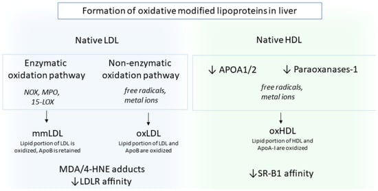
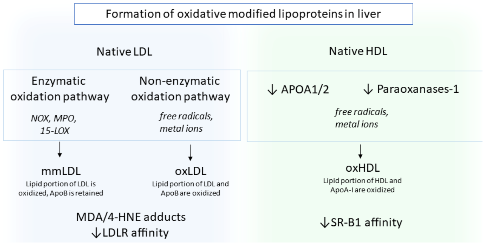
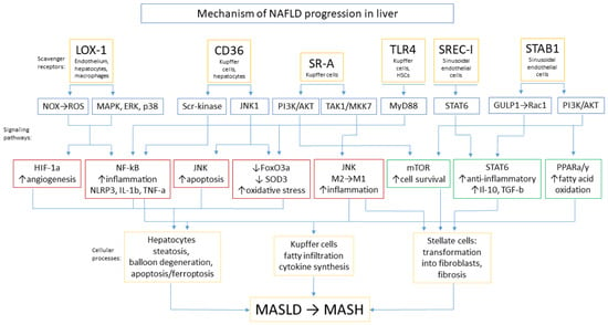
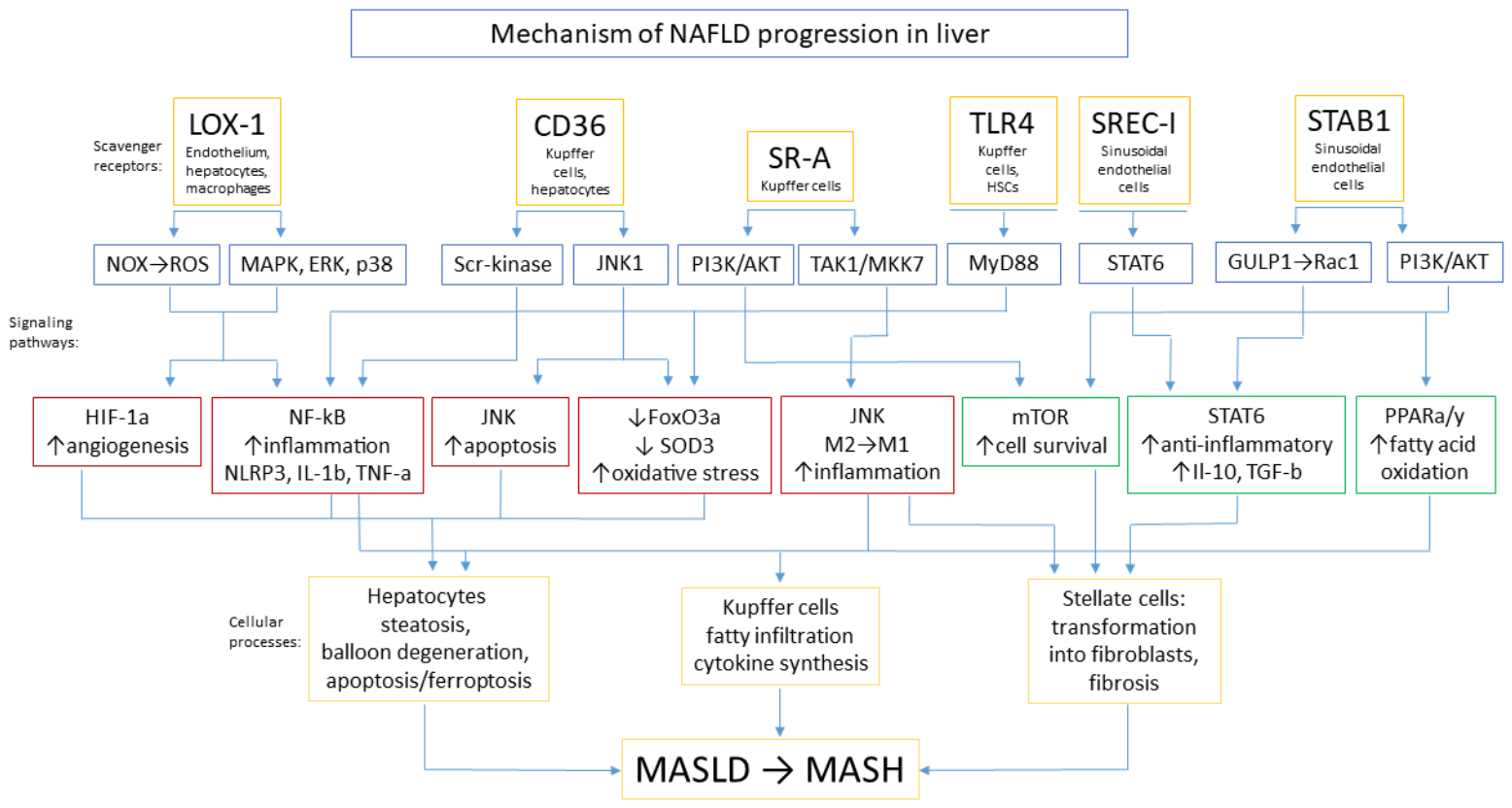
Disclaimer/Publisher’s Note: The statements, opinions and data contained in all publications are solely those of the individual author(s) and contributor(s) and not of MDPI and/or the editor(s). MDPI and/or the editor(s) disclaim responsibility for any injury to people or property resulting from any ideas, methods, instructions or products referred to in the content. |
© 2026 by the authors. Licensee MDPI, Basel, Switzerland. This article is an open access article distributed under the terms and conditions of the Creative Commons Attribution (CC BY) license.