


Abstract
Crepidotus represents a group of broadly distributed saprophytic fungi distinguished by remarkable species diversity. Herein, two undescribed species from China are introduced, supported by integrative evidence such as molecular phylogenetics, morphological features, host associations, and biogeographic distribution. Crepidotus hainanensis C.G. Song & Z.F. Jia, sp. nov., is characterized by its tomentose, plano-convex, white to pale mouse grey or straw yellow pileus, clamped hyphae, and hyaline, ellipsoid, thin-walled, smooth basidiospores. Phylogenetically, it forms an isolated terminal lineage without an immediate sister species. Crepidotus subasiaticus C.G. Song & Z.F. Jia, sp. nov., is characterized by its stipitate basidiomata, pinkish buff, cinnamon buff, clay-pink to yellowish-brown pileus, clamped hyphae, and olivaceous buff to yellowish-brown, ellipsoid, thick-walled, finely verrucose basidiospores, and phylogenetically it is closely allied to C. asiaticus. Morphological illustrations and taxonomic descriptions of the new proposed species are provided.
1. Introduction
Crepidotus (Fr.) Staude (Crepidotaceae, Agaricales) was established by Staude in 1857 and typified by Crepidotus mollis (Schaeff.) Staude [1]. Crepidotus is a genus of saprophytic fungi that primarily colonize and derive nutrients from decaying wood and branches [2]. As saprotrophs, Crepidotus species grow less commonly on other plant debris or soil, and rarely on bryophyte thalli or fungal fruiting bodies [3,4,5]. This genus has a wide geographical distribution across tropical to subpolar zones and is considered taxonomically prevalent in natural environments [6]. Crepidotus species are characterized by small pleurotoid basidiomata, lamellate hymenophore and dark-colored basidiospores [2,7].
Regarding the classification of Crepidotus, this differs among the various authors. Singer [8] classified Crepidotus into two sections based solely on spore ornamentation: sect. Levisporae, characterized by smooth basidiospores, and sect. Echinospori, characterized by ornamented basidiospores. Subsequently, Singer [9,10] reclassified the former section as sect. Crepidotus. Hesler and Smith [11] divided the genus into three subgeneric groups: Subg. Crepidotus, Subg. Dochmiopus, and Subg. Sphaerula, based on the presence or absence of clamp connections. Each subgenus was subsequently subdivided into sections and subsections according to the morphological characteristics of the basidiospores. Senn-Irlet [7,12] established a new classification system. Building on the original framework of Hesler and Smith [11], Senn-Irlet incorporated the calyptrate structure as an additional criterion for subgeneric delimitation, and accordingly divided the genus into two subgenera: Subg. Crepidotus and Subg. Dochmiopus. Consiglio and Setti [4] centered their work primarily on European collections, revised the classification by segregating Crepidotus into two subgenera and aggregated globose-spored taxa within C. subg. Dochmiopus as sect. Sphaerula Hesler & A.H. Sm. Accordingly, different taxonomic treatments recognize either two or three subgenera within Crepidotus, i.e., Subg. Crepidotus, Subg. Dochmiopus, and Subg. Sphaerula. Species classified in Subg. Crepidotus are characterized by smooth, almond-shaped basidiospores and clamp connections [2,7]. In contrast, members of the other two subgenera typically produce ornamented basidiospores and hyphae with clamps. Furthermore, with the exception of some species (e.g., C. epibryus (Fr.) Quél., which lack clamp connections and produce smooth basidiospores), most species can be classified based on this [7,13].
A total of 587 taxa have been recorded for Crepidotus according to Index Fungorum (17 November 2025). The majority of these species have been documented across European and American regions [2,4,7,11,14]. In recent years, research on this genus has increased in Asia, with more than 80 species being discovered and described in China [2,15,16,17,18,19,20,21,22].
In the 20th century, Crepidotus species were studied exclusively using morphology [3,7,11]. At present, molecular phylogenetic approaches have been employed to delimit several species, and numerous new species have been described. Ge and Bau [23] described six new species of Crepidotus from China, integrating morphological observations supported by phylogenetic analyses based on nuclear ITS sequences. Jančovičová et al. [6] identified a novel species, C. pini Jančovič, hosted by Pinus sylvestris, through phylogenetic and morphological analyses. Manoj Kumar et al. [18] recognized three new species from India based on nrLSU sequences. Han et al. [2] carried out phylogenetic and morphological analyses on Crepidotus subg. Dochmiopus and recognized four new species. Yang et al. [22], based on morphological and phylogenetic analyzes described a new species and a new record from China.
To date, the taxonomic inventory of Crepidotus in tropical southern China remains incomplete, with only a limited number of species formally documented from this region. Few investigations have focused on this genus in tropical Chinese habitats. During our fieldwork in southern China, two new collections were found after a taxonomic and phylogenetic study (using a combination of ITS + nLSU sequences), which were concluded to correspond to two undescribed species proposed in this work. The two novel species reported in the present study therefore add to our knowledge of Crepidotus diversity in tropical southern China.
2. Materials and Methods
2.1. Sampling and Morphological Studies
Field sampling and specimen preservation follow the procedure described by Song et al. [24]. Sample collection for the present study was conducted in the Yanuoda Tropical Rainforest, Hainan Province, China, in the month of January during the rainy season. Detailed specimen information, host substrates, ecological characteristics, location, altitude, collector, and date were recorded, and the field photographs of fungal basidiomata and their growing habitats were captured following Rathnayaka et al. [25]. The specimens were dried in situ with a portable drying oven to avoid decay and microbial contamination. The drying process was conducted under a stable temperature range of 40–45 °C. After drying, the specimens were promptly packed and stored for long-term preservation. All fungal materials collected and investigated in this work have been formally preserved in the Fungarium of Liaocheng University’s College of Life Sciences (LCUF). Macromorphological descriptions were based on the detailed field observations and records and measurements of herbarium specimens. Microscopic observations, measurements, and image documentation were performed on slide preparations stained with Cotton Blue and Melzer’s reagent. All microscopic examinations were conducted under a light microscope at a maximum magnification of ×100, following the protocols described by Song et al. [24]. Basidiospores were measured from sections cut from the lamellae. Abbreviations used in this study are defined as follows: IKI = Melzer’s reagent; IKI– = neither amyloid nor dextrinoid; KOH = 5% potassium hydroxide; CB = Cotton Blue; CB– = acyanophilous; L = mean spore length (arithmetic average of all spores); W = mean spore width (arithmetic average of all spores); Q = variation in the L/W ratios between the specimens studied; n (a/b) = the number of spores (a) measured from a given number (b) of specimens. Author abbreviations of fungal taxa are obtained from Index Fungorum (www.indexfungorum.org; accessed on 23 March 2026).
2.2. Molecular Study and Phylogenetic Analysis
Genomic DNA was extracted by the CTAB plant genome rapid extraction Hi-DNA-secure Plant Kit (Tiangen, Beijing, China). Samples used for DNA extraction were obtained from dried specimens. The extracted genomic DNA was utilized for polymerase chain reaction (PCR) amplification, following the manufacturer’s standard protocols with minor procedural modifications [26]. ITS and nLSU gene regions were amplified with the primer pairs ITS5/ITS4 and LR0R/LR7, respectively [27]. PCR amplification of the ITS region was performed under the following conditions: initial denaturation at 95 °C for 3 min, 35 cycles of denaturation at 94 °C for 40 s, annealing at 56 °C for 45 s, elongation at 72 °C for 1 min, and a final extension step at 72 °C for 10 min. PCR amplification of the nLSU region was performed under the following conditions: initial denaturation at 94 °C for 1 min, 35 cycles of denaturation at 94 °C for 30 s, annealing at 50 °C for 1 min, elongation at 72 °C for 1.5 min, and a final extension step at 72 °C for 10 min. Amplified PCR products were purified and bidirectionally sequenced at the Beijing Genomics Institute (China) using the same primer sets. All novel sequences obtained in this study were submitted to GenBank (Table 1).
Table 1.
List of species and their information used in the molecular phylogenetic analysis (new sequences are shown in bold; sequences with asterisks (T) are type specimens).
Sequences generated in this study, together with reference sequences retrieved, were aligned via MAFFT 7 [28] and manually adjusted in BioEdit [29]. The sequences of Neopaxillus dominicanus Angelini & Vizzini and N. echinospermus (Speg.) Singer from GenBank were selected as the outgroups, according to Vizzini et al. [30]. Sequence alignments were assembled using Mesquite v. 3.2. The optimal nucleotide substitution model for the datasets was determined via the Akaike Information Criterion (AIC) in MrModeltest 2.3. [31].
Maximum Likelihood (ML) analysis was performed in RAxML v.7.2.8 under the GTR + G + I model [32]. All model parameters were automatically calculated by the software, and only the optimal ML tree screened from all search replicates was retained for subsequent analysis. The optimal evolution model corresponding to individual datasets for Bayesian inference (BI) was identified using MrModeltest 2.3 [31,33]. Bayesian inference (BI) phylogenetic analysis was implemented in MrBayes 3.2.6 via the Abe resource on the Cipres Science Gateway (www.phylo.org; accessed on 23 March 2026) [34]. The analysis was implemented with two independent runs initiated from random starting trees, utilizing four simultaneous Markov chain Monte Carlo (MCMC) chains. A total of 5 million generations were executed, with one tree sampled at every 100-generation interval.
The initial 25% of the sampled trees were discarded as burn-in, and a majority-rule consensus tree was subsequently constructed from the remaining retained trees. Branches with ML bootstrap values ≥50 and BI posterior probabilities ≥0.95 were regarded as significantly supported. The phylogenetic tree was visualized by FigTree version 1.4.4 [35].
3. Results
3.1. Phylogenetic Analyses
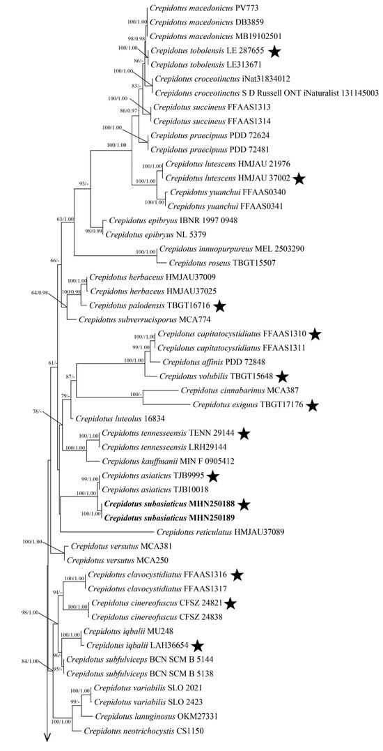
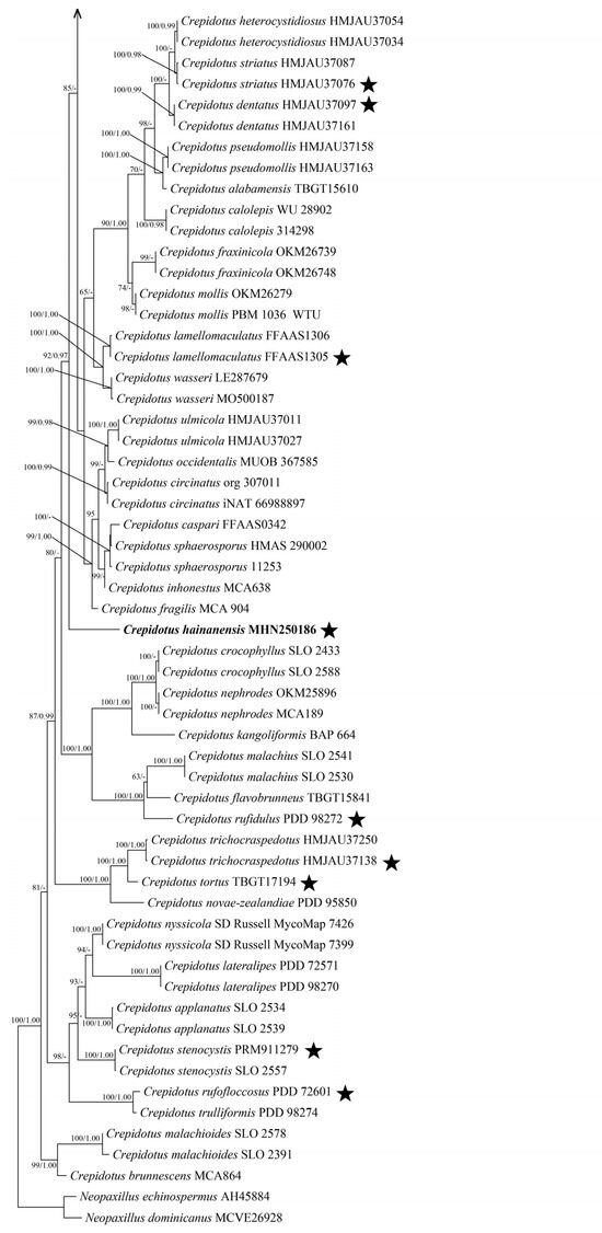
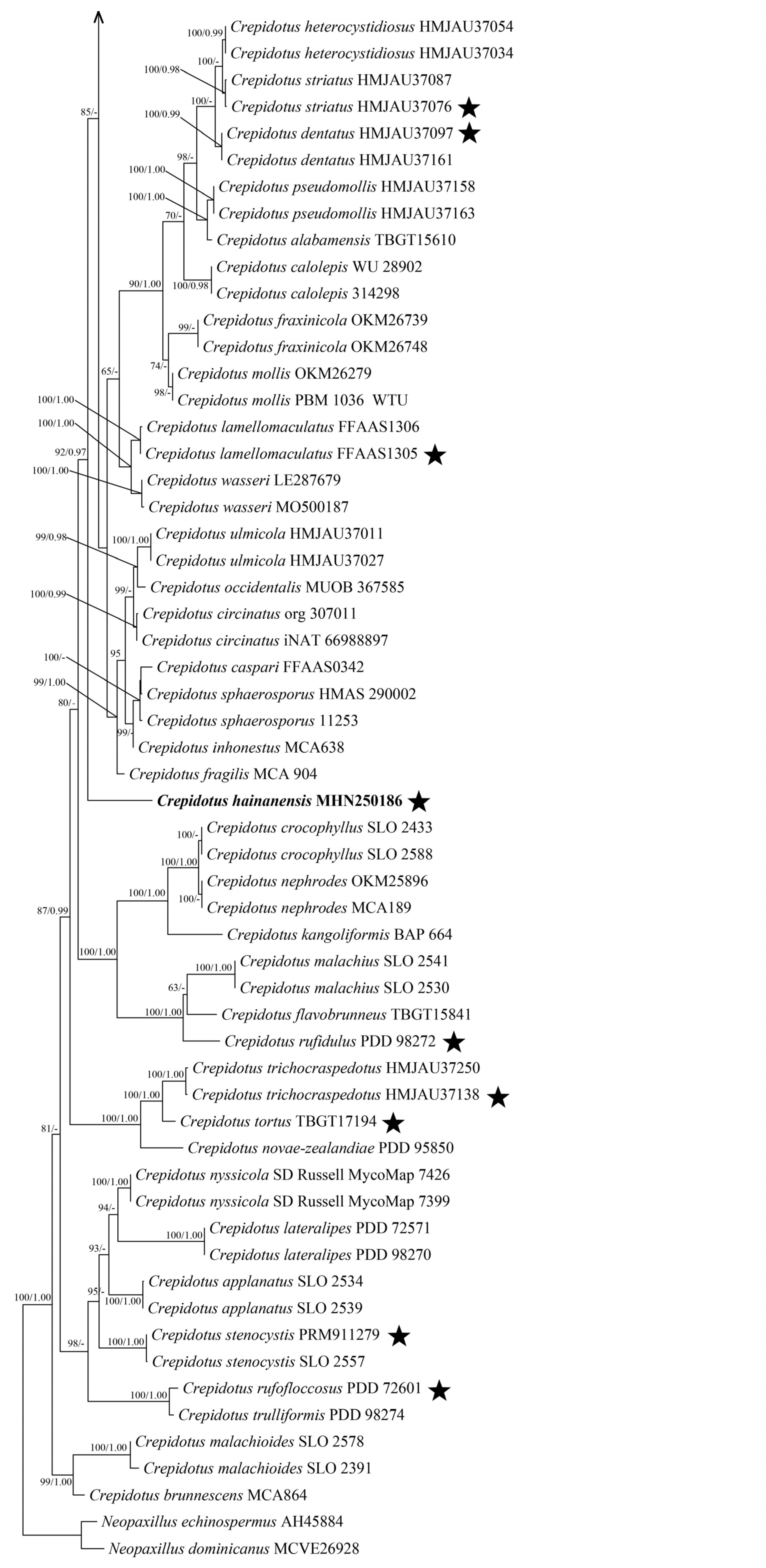
The combined ITS + nLSU dataset comprised sequences from 110 fungal samples corresponding to 69 taxa. The best-fit model, selected on the basis of AIC, was TVM + F + I + G4 for ITS and TIM3e + R3 for nLSU. BI yielded a phylogenetic topology highly consistent with the ML tree, with an average standard deviation of split frequencies of 0.009999. Node support values on the ML topology include ML bootstrap values (≥50%) and Bayesian posterior probabilities (BPP ≥ 0.95), as illustrated in Figure 1. Phylogenetic datasets generated in this work are available in Zenodo (DOI: 10.5281/zenodo.21915411).
Figure 1.
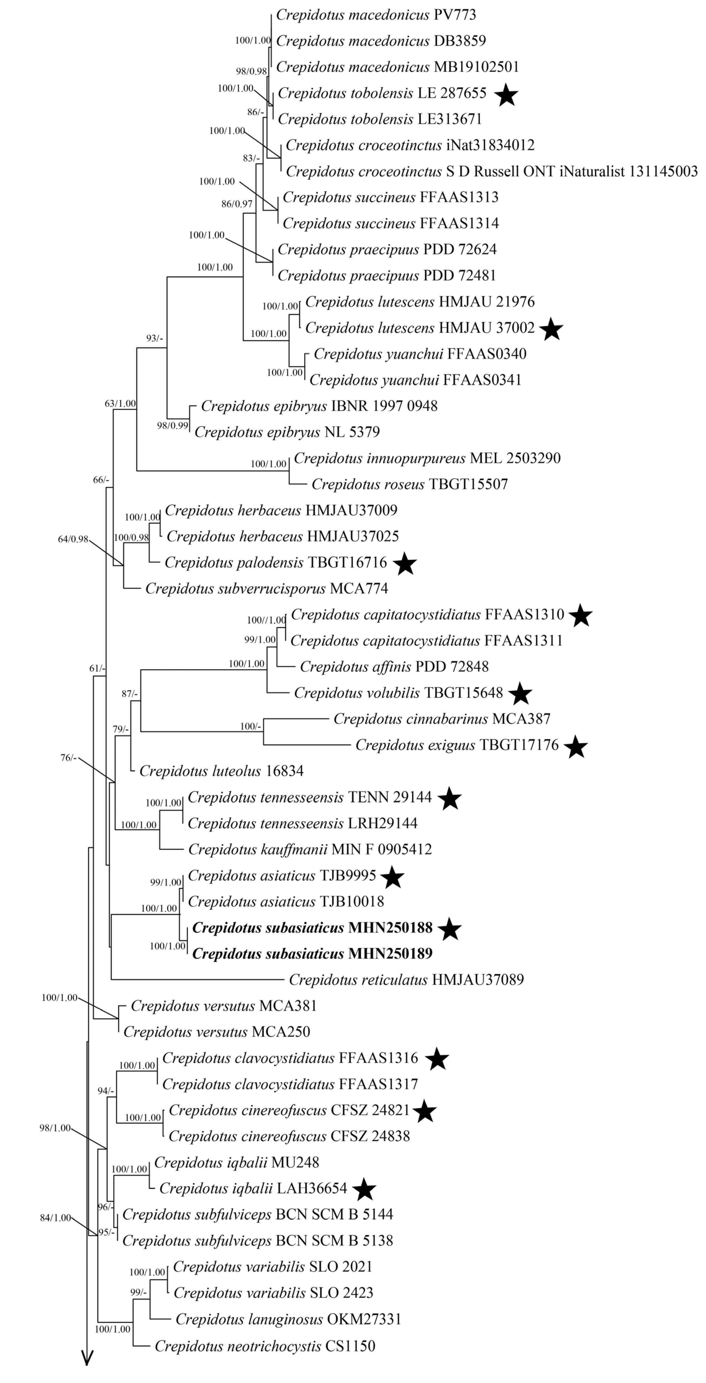
Crepidotus phylogeny based on the combined ITS + nLSU sequence dataset. Branches in the ML tree are labeled with Maximum Likelihood bootstrap higher than ML ≥ 50% and BPP ≥ 0.95, respectively. New species are indicated in bold. Black stars = type sequences.
In this study, our ITS + nLSU-based phylogeny is largely congruent with the topology reported by Han et al. [2], which divided Crepidotus into twelve major clades corresponding to morphologically defined subgenera and sections. Several morphology-based infrageneric taxa are not monophyletic in our tree. For instance, subg. Dochmiopus sect. Autochthoni falls into phylogenetically distant clades, consistent with previous findings, and morphological differences in cheilocystidia further support this result. Likewise, sect. Sphaeruli does not form a monophyletic group.
In our phylogenetic tree (Figure 1), the two new species, Crepidotus hainanensis, and Crepidotus subasiaticus, formed distinct, strongly supported lineages distant from other Crepidotus species. Crepidotus hainanensis (isolate MHN250186) forms an isolated single-species terminal branch with full support (100/1.00). It does not share an immediate sister relationship with any single species. Crepidotus subasiaticus is represented by two sequenced specimens, MHN250188 and MHN250189, which cluster together on a single branch supported by 100ML/1.00BPP. This lineage forms a sister group to C. asiaticus, and the node uniting these two species receives support values of 99ML/1.00BPP. No other species sequences nest within the clade of C. subasiaticus. The high support defining the unique branches of C. hainanensis and C. subasiaticus provides molecular evidence that these two taxa are phylogenetically distinct from all previously sequenced Crepidotus species included in this dataset.
3.2. Taxonomy
Crepidotus hainanensis C.G. Song & Z.F. Jia, sp. nov.
MycoBank no.: 865424
Diagnosis. Differs from other Crepidotus species by its hemispherical to flabelliform, tomentose, plano-convex basidiomata; white to pale mouse grey or straw yellow pileal surface; clamped hyphae; fusiform and thick-walled cheilocystidia; and hyaline, ellipsoid, thin-walled, smooth basidiospores.
Holotype. Hainan Province, BaoTing County, Yanuoda Tropical Rainforest, on fallen branch, alt.465 m, 2025.4.15, MHN250186.
Etymology. (Lat.): hainanensis referring to the holotype locality of the species in Hainan Province.
Basidiomata are annual, pileate, fleshy, soft. Pileus is hemispherical to flabelliform, tomentose when matured, occasionally lobed, becoming plano-convex. Pileal surface white to pale mouse grey, sometimes straw yellow (3A3), up to 0.5 cm. Lamellae up to 1 mm broad, with 8–12 complete lamellae and 2–4 lamellulae between two complete lamellae, adnate, subdecurrent, subventricose, white (1A1), sometimes straw yellow (3A3). Context: thin (<1 mm thick), white (1A1). Stipe present in young basidiomata, short (<5 mm), cylindrical, becoming lateral, knob-like, and subtransparent at maturity, often obscured by lamellae.
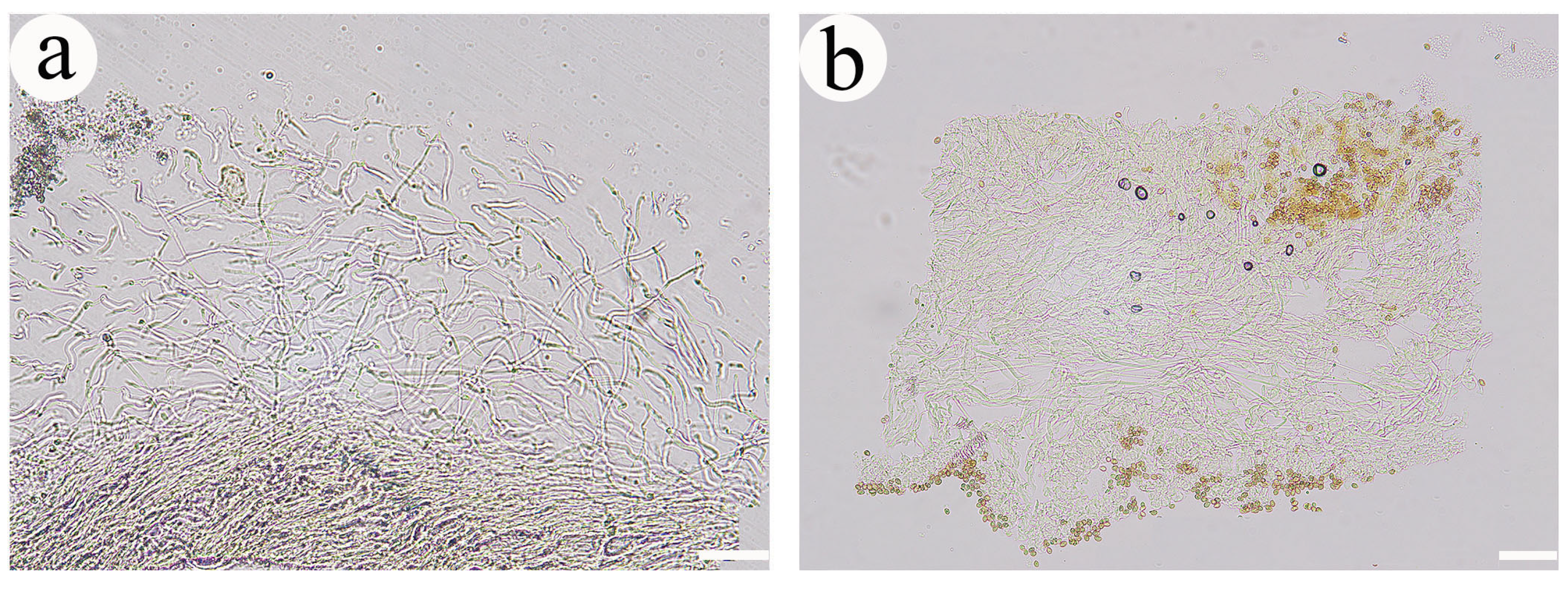
Pileipellis trichoderm-type, none gelatinized, composed of cylindrical interwoven hyphae, hyaline to light clay buff, thin-walled; terminal cells flexuous, not differentiated into distinct pileocystidia; heavily coarsely encrusted over nearly the whole hyphal; hyphal diameter 3–7 µm. Lamellae trama hyphae hyaline, thin-walled, interwoven arranged, 2–4 µm in diam. Basidiospores are hyaline to light brown, ellipsoid, thin-walled, smooth, IKI–, CB–, 5.5–8 × 3.5–4 µm, L = 6.7 µm, W = 3.8 µm, Q = 1.8 (n = 30/1). Basidia clavate to widen clavate, 2- to 4-spored, 21–26.5 × 4.5–5 µm. Cheilocystidia fusiform, thick-walled, 32–57 × 14–17 µm. All hyphae clamped; IKI–, CB–; tissue not discoloring in KOH.
Figure 2.
Basidiocarps of Crepidotus species. (a,b) Crepidotus hainanensis (Holotype); (c,d) Crepidotus subasiaticus (Holotype). Scale bars: 1 cm.
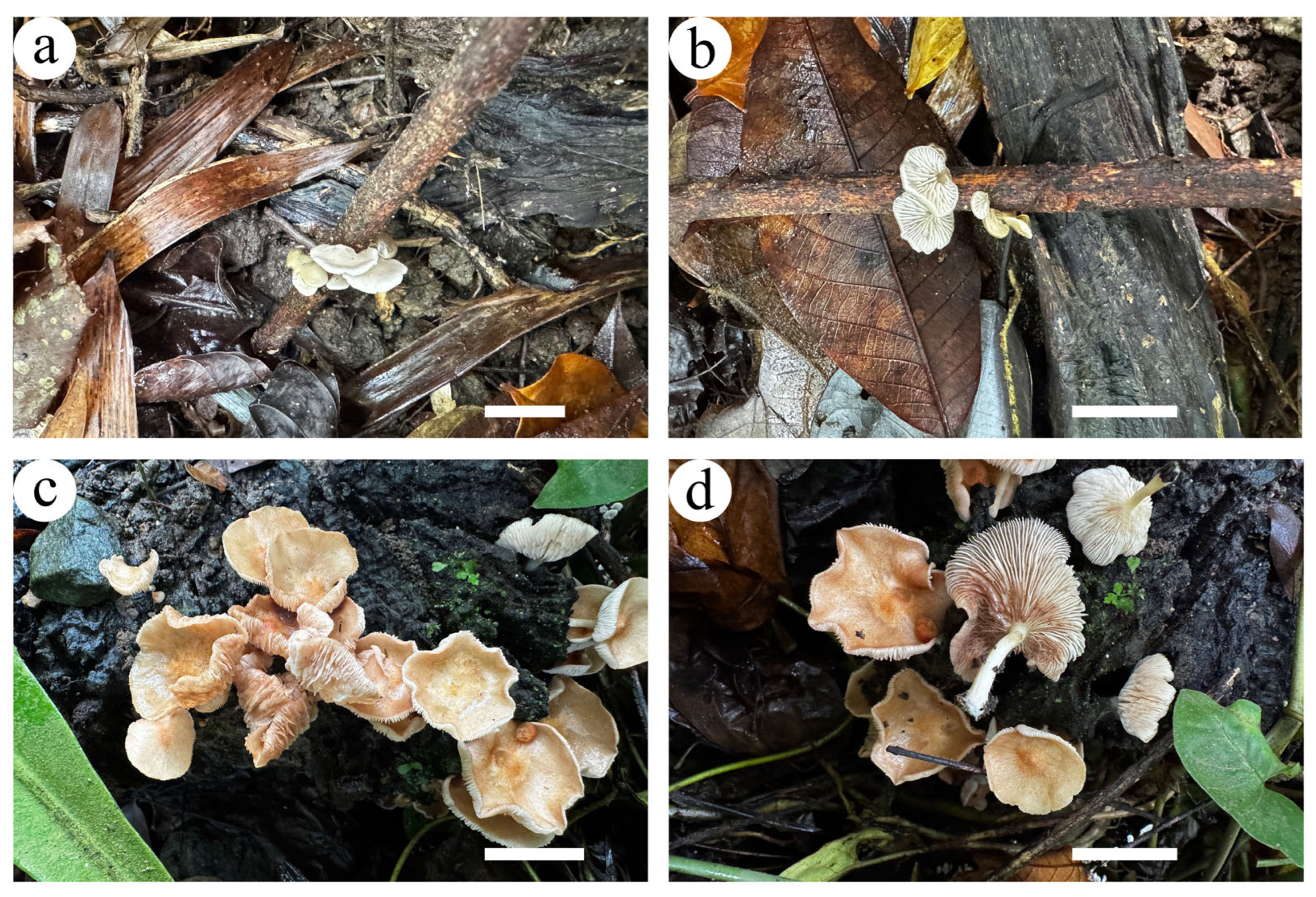
Figure 3.
SEM of basidiospores of Crepidotus species. (a,b) Crepidotus hainanensis; (c,d) Crepidotus subasiaticus. Scale bars: 2 µm.
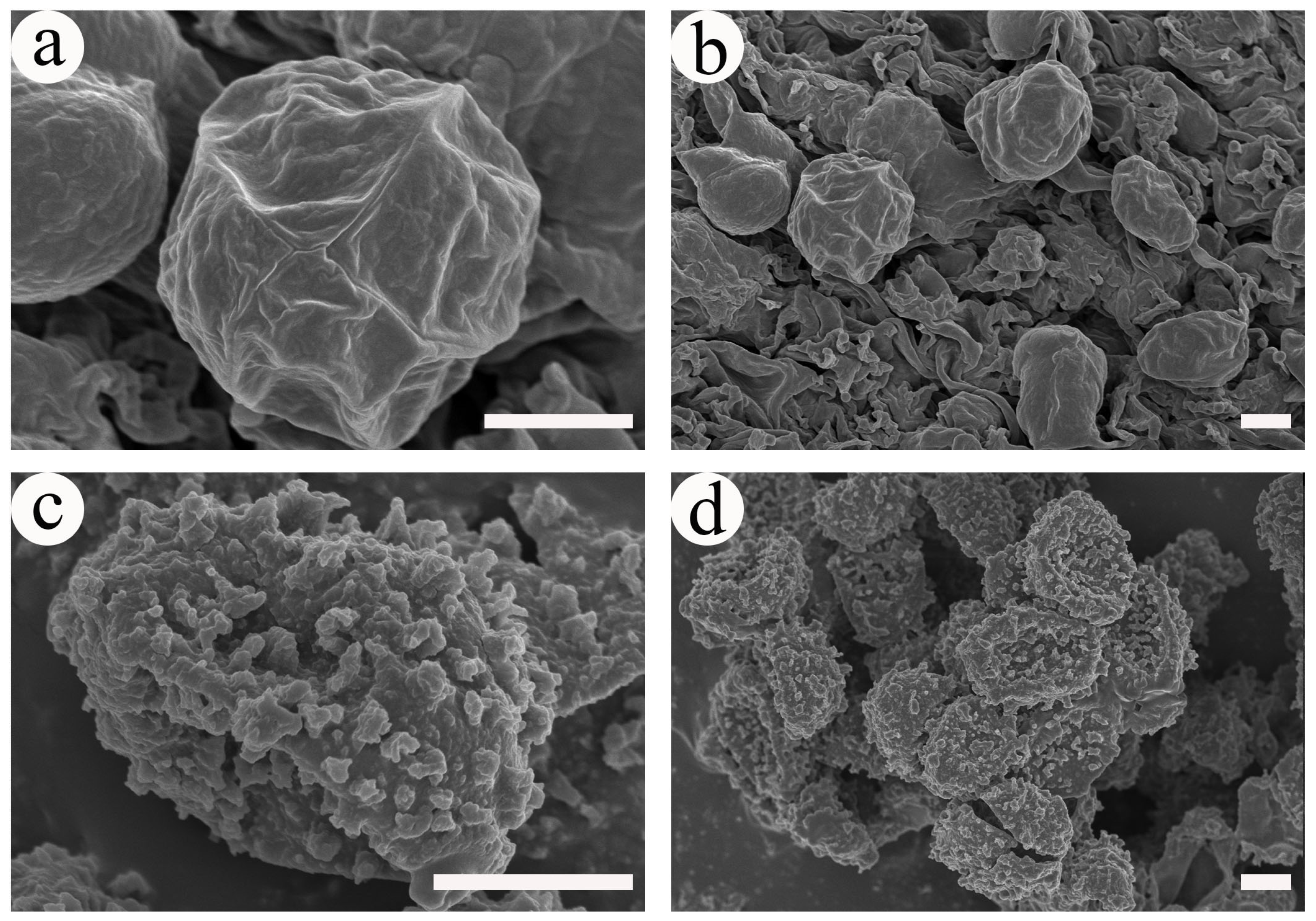
Figure 4.
Pileipellis of Crepidotus species (photographed from the holotype, at ×20 magnification). (a) Crepidotus hainanensis; (b) Crepidotus subasiaticus. Scale bars: 50 µm. Structures were stained in IKI solution.
Crepidotus subasiaticus C.G. Song & Z.F. Jia, sp. nov.
MycoBank no.: 865425
Diagnosis. Differs from other Crepidotus species by its pileate, stipitate basidiomata; pinkish buff, cinnamon buff, clay-pink to yellowish-brown pileal surface; clamped hyphae; ramiferous to digitiform and thin-walled cheilocystidia; and olivaceous buff to yellowish-brown, ellipsoid, thick-walled, finely verrucose basidiospores.
Holotype. Hainan Province, BaoTing County, Yanuoda Tropical Rainforest, on stump, alt. 280 m, 2025.4.15, MHN250188.
Figure 5.
Microscopic structures of Crepidotus hainanensis (photographed from the holotype, at ×100 magnification). (a) Basidiospores; (b) basidia and basidioles; (c) cheilocystidia; (d) hyphae from context. Scale bars: 10 µm. Structures were stained in CB solution.
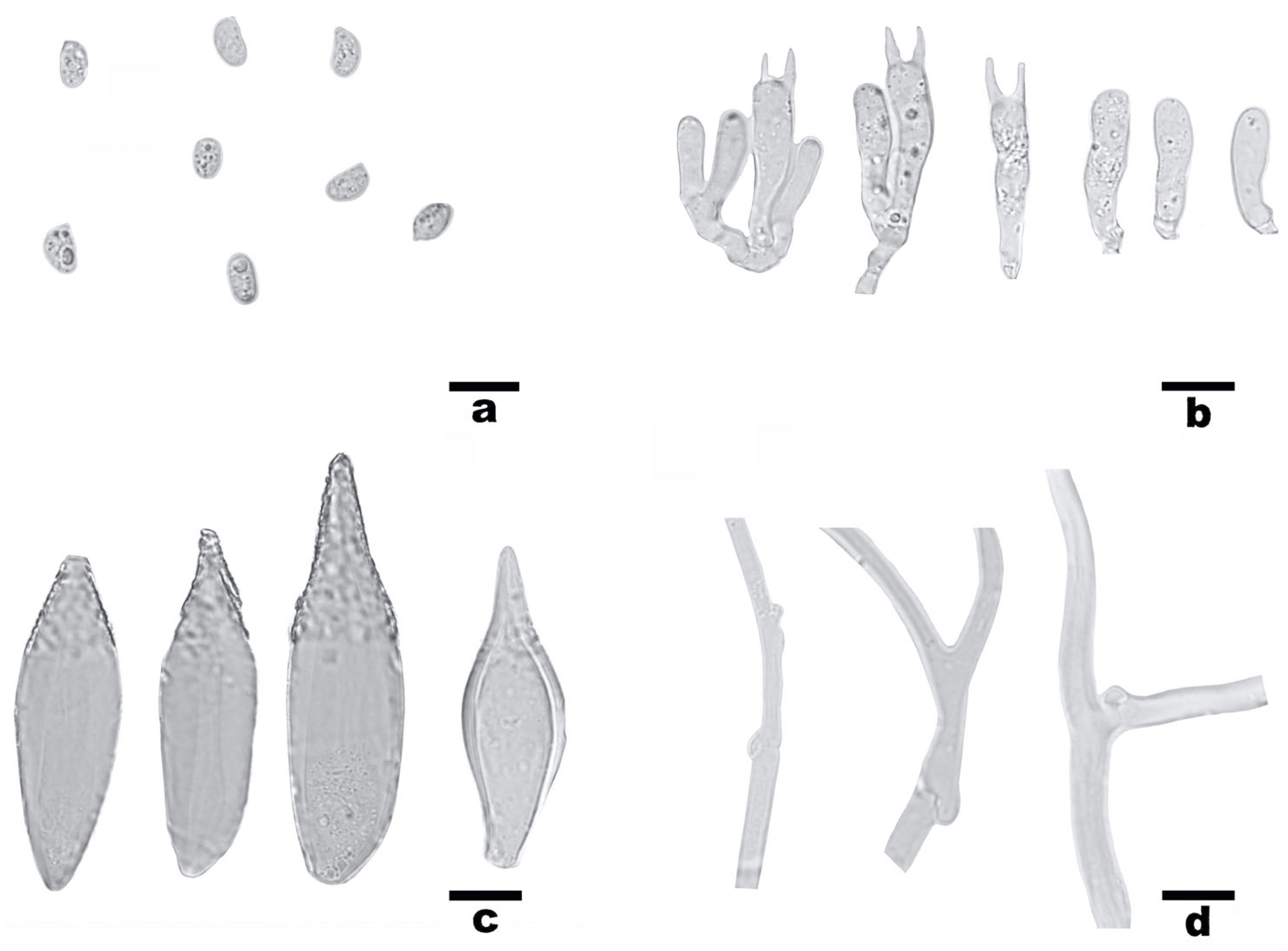
Figure 6.
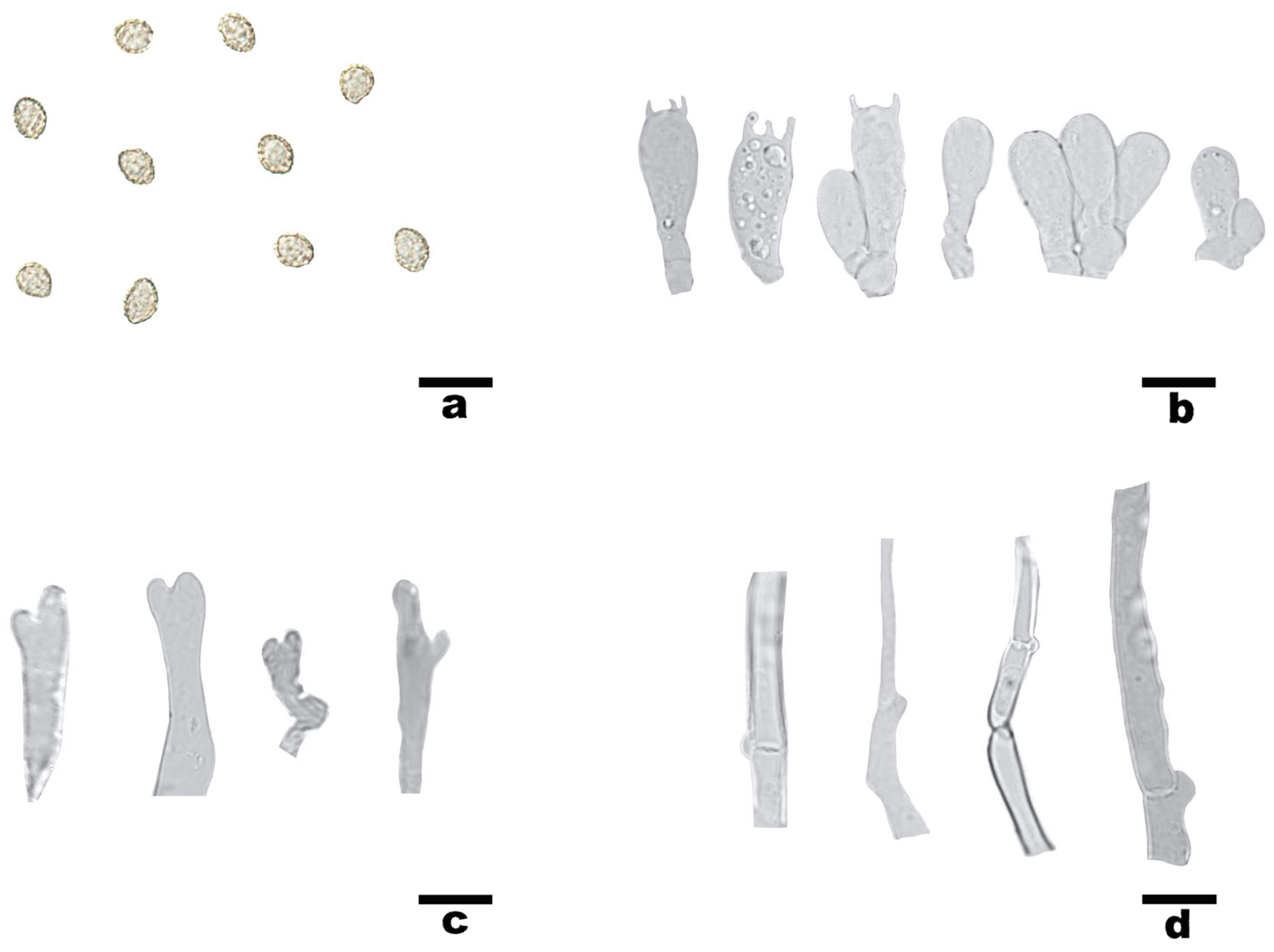
Microscopic structures of Crepidotus subasiaticus (photographed from the holotype, at ×100 magnification). (a) Basidiospores; (b) basidia and basidioles; (c) cheilocystidia; (d) hyphae from context. Scale bars: 10 µm. Structures were stained in CB solution.
Etymology. (Lat.): subasiaticus referring to the species is related to Crepidotus asiaticus.
Basidiomata are annual, pileate, stipitate, fleshy, soft. Pileus circular, initially umbrilliform, becoming plano-convex to applanate with a broad obtuse umbo, disk slightly raised and typically darker in color, with a velutinous tomentum, up to 3 cm in diam. Pileal surface pinkish buff (5A3), cinnamon buff (4B4), clay-pink (6B4) to yellowish-brown (5B7); margin straight to uplifted, wavy, folded, entire. Lamella up to 2.5 mm broad, with 27–40 complete lamellae and 3–6 lamellulae between two complete lamellae, adnate, short decurrent, or with a decurrent tooth, sometimes exhibit forking at their ends, white (1A1) to cream (4A3), sometimes orange-brown, especially when drenched. Context: thin (<5 mm thick), cream. Stipe centrally, cylindrical, solid, white (1A1) to cream (4A3), up to 3.5 cm long, 0.4 cm wide.
Pileipellis trichoderm-type, none gelatinized, composed of cylindrical interwoven hyphae, clay buff to brown, thick-walled; terminal cells sometimes flexuous, not differentiated into distinct pileocystidia; only terminal parts of hyphae bear faint fine encrustations, main hyphal bodies smooth; hyphae 4–9 µm in diam. Lamellae trama hyphae hyaline, thin-walled, regularly arranged, 3–6 µm in diam. Basidiospores are olivaceous buff to yellowish-brown, broadly ellipsoid, thick-walled, finely verrucose, IKI–, CB–, 5–7 × 4–5.5 µm, L = 5.9 µm, W = 4.6 µm, Q = 1–1.49 (n = 60/2). Basidia clavate to widen clavate, 2- to 4-spored, 19–26 × 6.5–9.5 µm. Cheilocystidia are ramiferous to digitiform, thin-walled, 13–32 × 5–12 µm. Generative hyphae clamped; IKI–, CB–; tissue not discoloring in KOH.
Paratypes. Hainan Province, BaoTing County, Yanuoda Tropical Rainforest, on stump, alt. 280 m, 2025.4.15, MHN250187 & MHN250189.
| Key to species of C. subg. Dochmiopus known in China | |
| 1. Basidiospores smooth to nearly smooth | 2 |
| 1. Basidiospores ornamented | 10 |
| 2. Pileocystidia present | C. autochthonus |
| 2. Pileocystidia absent | 3 |
| 3. Pileipellis gelatinous | C. betulae |
| 3. Pileipellis non-gelatinous | 4 |
| 4. Pileus whitish | 5 |
| 4. Pileus not whitish | C. cinereofuscus |
| 5. Basidiospores hyaline | C. hainanensis |
| 5. Basidiospores not hyaline | 6 |
| 6. Cheilocystidia vine-shaped | C. trichocraspedotus |
| 6. Cheilocystidia not vine-shaped | 7 |
| 7. Pileipellis hyphae clampless | C. albidus |
| 7. Pileipellis hyphae clamped | 8 |
| 8. Lamellae edge not fimbriate | 9 |
| 8. Lamellae edge fimbriate | 10 |
| 9. Pileipellis a cutis | C. caspari |
| 9. Pileipellis a trichoderm | C. lamellomaculatus |
| 10. Basidiospores width < 4.5 µm | C. albissimus |
| 10. Basidiospores width ≥ 4.5 µm | 11 |
| 11. Pileipellis a cutis, pileus greyish white to silver-grey | C. occidentalis |
| 11. Pileipellis a trichoderm, pileus white to light orange-yellow | C. ulmicola |
| 12. Inhabitant living plants | C. herbaceus |
| 12. Inhabitation of rotten branches and woods | 13 |
| 13. Pileus reddish | C. reticulatus |
| 13. Pileus not reddish | 14 |
| 14. Pleurocystidia present | C. luteicolor |
| 14. Pleurocystidia absent | 15 |
| 15. Pileocystidia present | 16 |
| 15. Pileocystidia absent | 17 |
| 16. Pileus buff yellow to apricot yellow | C. succineus |
| 16. Pileus white | C. vulgaris |
| 17. Basidiospores subglobose to ellipsoid, Qm ≤ 1.6 | 18 |
| 17. Basidiospores amygdaliform, oblong to cylindrical, Qm > 1.6 | 29 |
| 18. Pileus brown, at point of attachment smooth | C. payettensis |
| 18. Pileus not brown, at point of attachment villous, fibrillose or tomentose | 19 |
| 19. Pileus surface smooth | 20 |
| 19. Pileus surface not smooth | 22 |
| 20. Pileus translucent striate | C. capitatocystidiatus |
| 20. Pileus not translucent striate | 21 |
| 21. Lamellae edge not fimbriate, cheilocystidia apex bifurcate | 22 |
| 21. Lamellae edge fimbriate, cheilocystidia apex branched or stag antlers | C. macedonicus |
| 22. Pileipellis a cutis | C. furcaticystidiosus |
| 22. Pileipellis a trichoderm | C. stenocystis |
| 23. Pileus hygrophanous | 24 |
| 23. Pileus non-hygrophanous | 25 |
| 24. Cheilocystidia lageniform | C. lutescens |
| 24. Cheilocystidia narrowly clavate to narrowly utriform | C. croceotinctus |
| 25. Pileipellis within crusted hyphae | 26 |
| 25. Pileipellis without crusted hyphae | 27 |
| 26. Cheilocystidia clavate or ventricose, sessile | C. kauffmanii |
| 26. Cheilocystidia clavate to narrowly clavate, stipitate | C. clavocystidiatus |
| 27. Pileus orange-yellow to brownish-yellow | 28 |
| 27. Pileus white to cream | 29 |
| 28. Stipe only observed at primordial stages | C. yuanchui |
| 28. Stipe observed at all stages | C. subasiaticus |
| 29. Basidiospores subglobose to broadly ellipsoid, Q ≤ 1.30 | C. cesatii |
| 29. Basidiospores ellipsoid to ovoid, Q > 1.30 | C. subverrucisporus |
| 30. Pileus light, smoky or rusty brown | C. herbarum |
| 30. Pileus white to light yellow or orange | 31 |
| 31. Basidiospores length ≥ 8.0 µm | C. luteolus |
| 31. Basidiospores length < 8.0 µm | 32 |
| 32. Cheilocystidia with a long cylindrical protuberance at the apex | C. tomentellus |
| 32. Cheilocystidia without a long cylindrical protuberance at the apex | 33 |
| 33. Cheilocystidia filiform, narrowly lageniform, sometimes diverticular or knobby | C. neotrichocystis |
| 33. Cheilocystidia clavate, utriform, strangulated, sometimes stag antlers | C. variabilis |
4. Discussion
In the present study, phylogenetic analyses based on the combined ITS and nLSU sequence data were performed to recognize two undescribed species from China as well as their systematic position within Crepidotus.
Due to the presence of clamped hypha and smooth basidiospores, Crepidotus hainanensis was recognized within C. subg. Dochmiopus Sect. Autochthoni (Senn-Irlet) Consiglio & Setti [4,11]. Phylogenetic analyses (Figure 1) revealed that Crepidotus hainanensis formed an independent lineage, clearly differs from other Crepidotus species. Morphologically, C. hainanensis is similar to C. versutus Peck in having hemispherical, tomentose basidiomata, white context, and ellipsoid basidiospores [36]. However, C. versutus can be distinguished by its brownish-yellow lamellae, clampless hyphae, and longer basidiospores (7.8–) 8.7–9.9 (–11.6) × (4.3–) 4.8–5.2 (–5.8) μm [36].
Crepidotus species, such as C. asiaticus Guzm.-Dáv., C.K. Pradeep & T.J. Baroni, C. ibericus (G. Moreno & Esteve-Rav.) Bandala, Esteve-Rav. & Montoya, C. nyssicola (Murrill) Singer, C. subfulviceps (Murrill) Aime, Vila & P.-A. Moreau, and C. thermophilus (Singer) Aime, T.J. Baroni & O.K. Mill. are distinguished by possessing a well-developed stipe [9,16,37,38,39]. In this study, a stipitate Crepidotus species was collected from Hainan Province and described here as C. subasiaticus and belonging to the C. subg. Dochmiopus sect. Dochmiopus on account of its clamped hyphae and verrucose basidiospores [4,11]. Phylogenetically, C. subasiaticus is closely related to C. asiaticus (Figure 1) which can be distinguished by its reddish brown pileal surface and narrower basidia measuring 15–25 (–27.5) × 5.5–7.5 µm [16].
In this study, the combined ITS and nLSU dataset was employed as molecular markers, and previous studies have confirmed that these two gene fragments can clearly delimit species within the genus Crepidotus. Although additional loci such as rpb2 could potentially improve phylogenetic resolution, the scarcity of publicly available rpb2 sequences for most Crepidotus taxa would limit the comparability and taxonomic scope of our analysis. Our results (Figure 1) demonstrate that the ITS + nLSU dataset provides sufficient discriminatory power to clearly resolve the two new species, including C. subasiaticus and its closely related taxon C. asiaticus, with strong support values (ML ≥ 70%, BPP ≥ 0.95), thus validating the robustness of our marker selection for the proposed taxonomic conclusions. It should be noted that Crepidotus hainanensis is currently known only from the type collection, and C. subasiaticus is represented by several specimens. Therefore, the full range of intraspecific morphological variation for both species cannot be fully assessed at present, and further sampling is expected to expand our understanding of their morphological plasticity.
The Crepidotus species described here exhibit a relatively conserved ecological strategy as saprotrophic fungi, occurring predominantly on decaying wood and plant debris across diverse forest ecosystems. Their broad distribution spanning tropical to temperate regions, combined with marked morphological diversity and phylogenetic differentiation, indicates substantial adaptability to varied substrates and environmental conditions. Continued exploration and systematic revision of Crepidotus will not only enhance documentation of global fungal diversity, but also advance understanding of the evolutionary history and ecological roles of agaric fungi within forest ecosystems from China.
5. Conclusions
The Crepidotus species described here are the result of a morphological study integrating morphological characteristics and a phylogenetic molecular analysis combining ITS + nLSU sequence. The discovery of the two new wood-inhabiting species further demonstrates the rich fungal diversity in Hainan’s tropical rainforests and reinforces the need for continued field investigations and systematic revisions of Crepidotus in Asia, while providing fundamental data for understanding the phylogeny, diversity and biogeography of this ecologically important saprotrophic genus.
Author Contributions
Z.-F.J. and C.-G.S. designed the research; C.-G.S. prepared the samples; B.-W.Y. and P.-P.L. conducted the molecular experiments and analyzed the data; C.-G.S. drafted the manuscript. All authors have read and agreed to the published version of the manuscript.
Funding
The research is supported by the National Natural Science Foundation of China (No. 32500002).
Institutional Review Board Statement
Not applicable.
Data Availability Statement
The data and results of this study are available upon reasonable request. Please contact the main author of this publication.
Conflicts of Interest
The authors declare no conflicts of interest. The funders had no role in the design of the study; in the collection, analyses, or interpretation of data; in the writing of the manuscript, or in the decision to publish the results.
References
- Donk, M.A. New and revised nomina generica conservanda proposed for Basidiomycetes. Bull. Jard. Bot. Buitenzorg 1949, 18, 83–168. [Google Scholar]
- Han, M.H.; Na, Q.; Wei, R.X.; Zeng, H.; Hu, Y.P.; Zhang, L.B.; Du, J.H.; Zou, L.; Tang, W.M.; Cheng, X.H.; et al. Phylogenetic and morphological perspectives on Crepidotus subg. Dochmiopus: Exploratively unveiling hidden diversity in China. J. Fungi 2024, 10, 710. [Google Scholar] [CrossRef] [Scilit] [PubMed]
- Pilát, A. Monographie des espèces européennes du genre Crepidotus Fr. Atlas Champ. Eur. 1948, 6, 1–84. [Google Scholar]
- Consiglio, G.; Setti, L. Il genere Crepidotus in Europa; Fondazione Centro Studi Micologici: Vicenza, Italy, 2008. [Google Scholar]
- Hausknecht, A.; Krisai-Greilhuber, I. Die Gattung Crepidotus in Österreich. Österr. Z. Pilzk. 2010, 19, 53–91. [Google Scholar]
- Jančovičová, S.; Adamčíková, K.; Caboň, M.; Adamčík, S. Phylogeny of Crepidotus applanatus look-alikes reveals convergent morphology evolution and a new species C. pini. J. Fungi 2022, 8, 489. [Google Scholar] [CrossRef] [Scilit] [PubMed]
- Senn-Irlet, B. The genus Crepidotus (Fr.) Staude in Europe. Persoonia 1995, 16, 1–80. [Google Scholar]
- Singer, R. Contributions toward a monograph of the genus Crepidotus. Lilloa 1947, 13, 59–95. [Google Scholar]
- Singer, R. The genera Marasmiellus, Crepidotus and Simocybe in the Neotropics. Beitr. Nova Hedwig. 1973, 44, 1–517. [Google Scholar]
- Singer, R. The Agaricales in Modern Taxonomy, 4th ed.; Koeltz Scientific Books: Königstein, Germany, 1986; pp. 1–981. [Google Scholar]
- Hesler, L.R.; Smith, A.H. North American Species of Crepidotus; Hafner Publishing Company: New York, NY, USA, 1965. [Google Scholar]
- Senn-Irlet, B.; de Meijer, A.A.R. The genus Crepidotus from the State of Paraná, Brazil. Mycotaxon 1998, 66, 165–199. [Google Scholar] [CrossRef] [Scilit]
- Bandala, V.M.; Montoya, L.; Moreno, G. Two Crepidotus from Mexico with notes on selected type collections. Mycotaxon 1999, 72, 403–416. [Google Scholar] [CrossRef] [Scilit]
- Bandala, V.M.; Montoya, L. A taxonomic revision of some American Crepidotus. Mycologia 2000, 92, 341–353. [Google Scholar] [CrossRef] [Scilit]
- Ge, Y.P.; Yang, S.; Bau, T. Crepidotus lutescens sp. nov., an ochraceous salmon colored species from northeast China. Phytotaxa 2017, 297, 189. [Google Scholar] [CrossRef] [Scilit]
- Guzmán-Dávalos, L.; Pradeep, C.K.; Vrinda, K.B.; Manoj Kumar, A.; Ramírez-Cruz, V.; Herrera, M.; Villalobos-Arámbula, A.R.; Soytong, K.; Baroni, T.J.; Aime, M.C. A new stipitate species of Crepidotus from India and Thailand, with notes on other tropical species. Mycologia 2017, 109, 804–814. [Google Scholar] [CrossRef] [Scilit] [PubMed]
- Manoj Kumar, A.; Vrinda, K.B.; Pradeep, C.K. Two new species of Crepidotus from peninsular India. Phytotaxa 2018, 372, 67. [Google Scholar] [CrossRef] [Scilit]
- Manoj Kumar, A.; Pradeep, C.K.; Aime, M.C. New species and new records of Crepidotus from India. Mycol. Prog. 2022, 21, 311–326. [Google Scholar] [CrossRef] [Scilit]
- Izhar, A.; Usman, M.; Khalid, A.N. Crepidotus iqbalii, a new stipitate species from Pakistan. Phytotaxa 2021, 500, 95–107. [Google Scholar] [CrossRef] [Scilit]
- Takahashi, H. New species of Clitocybe and Crepidotus from eastern Honshu, Japan. Mycoscience 2003, 44, 103–107. [Google Scholar] [CrossRef] [Scilit]
- Liu, S.L.; Zhao, P.; Cai, L.; Shen, S.; Wei, H.W.; Na, Q.; Han, M.; Wei, R.; Ge, Y.; Ma, H.; et al. Catalogue of fungi in China 1. new taxa of plant-inhabiting fungi. Mycology 2024, 3, 1–58. [Google Scholar] [CrossRef] [Scilit] [PubMed]
- Yang, X.; Han, M.; Wei, T.; Liu, T. New Crepidotus findings in China: Crepidotus cinereofuscus sp. nov. and record of C. stenocystis. Biodivers. Data J. 2025, 13, e153245. [Google Scholar] [CrossRef] [Scilit] [PubMed]
- Ge, Y.P.; Bau, T. Descriptions of six new species of Crepidotus from China. Mycosystema 2020, 39, 238–255. [Google Scholar] [CrossRef]
- Song, C.G.; Xu, T.M.; Xu, Y.H.; Wang, D.; Zeng, L.; Fan, X.P.; Sun, Y.F.; Cui, B.K. Systematic revision, molecular phylogeny and divergence times of Thelephorales. Mycosphere 2025, 16, 296–422. [Google Scholar] [CrossRef] [Scilit]
- Rathnayaka, A.R.; Tennakoon, D.S.; Jones, G.E.; Wanasinghe, D.N.; Bhat, D.J.; Priyashantha, A.H.; Stephenson, S.L.; Tibpromma, S.; Karunarathna, S.C. Significance of precise documentation of hosts and geospatial data of fungal collections, with an emphasis on plant-associated fungi. N. Z. J. Bot. 2025, 63, 462–489. [Google Scholar] [CrossRef] [Scilit]
- Song, C.G.; Gao, J.T.; Yang, B.W.; Jia, Z.F. Dentipellicula hainanensis (Hericiaceae, Agaricomycetes) and Phylloporia rigida (Hymenochaetaceae, Agaricomycetes), two new wood-inhabiting fungi from Hainan. MycoKeys 2026, 128, 351–374. [Google Scholar] [CrossRef] [Scilit] [PubMed]
- Song, C.G.; Sun, Y.F.; Liu, S.; Chen, Y.Y.; Cui, B.K. Phylogenetic analyses and morphological studies reveal four new species of Phellodon from China. J. Fungi 2023, 9, 30. [Google Scholar] [CrossRef] [Scilit] [PubMed]
- Katoh, K.; Standley, D.M. MAFFT multiple sequence alignment software version 7: Improvements in performance and usability. Mol. Biol. Evol. 2013, 30, 772–780. [Google Scholar] [CrossRef] [Scilit] [PubMed]
- Hall, T.A. BioEdit: A user-friendly biological sequence alignment editor and analysis program for windows 95/98/NT. Nucleic Acids Symp. Ser. 1999, 41, 95–98. [Google Scholar] [CrossRef] [Scilit]
- Vizzini, A.; Angelini, C.; Ercole, E. A new Neopaxillus species (Agaricomycetes) from the Dominican Republic and the status of Neopaxillus within the Agaricales. Mycologia 2012, 104, 138–147. [Google Scholar] [CrossRef] [Scilit] [PubMed]
- Posada, D.; Crandall, K.A. Modeltest: Testing the model of DNA substitution. Bioinformatics 1998, 14, 817–818. [Google Scholar] [CrossRef] [Scilit] [PubMed]
- Stamatakis, A. RAxML-VI-HPC: Maximum likelihood-based phylogenetic analyses with thousands of taxa and mixed models. Bioinformatics 2006, 22, 2688–2690. [Google Scholar] [CrossRef] [Scilit] [PubMed]
- Nylander, J.A.A. MrModeltest v2. Program; Uppsala University: Uppsala, Sweden, 2004. [Google Scholar]
- Ronquist, F.; Huelsenbeck, J.P. MrBayes 3: Bayesian phylogenetic inference under mixed models. Bioinformatics 2003, 19, 1572–1574. [Google Scholar] [CrossRef] [Scilit] [PubMed]
- Rambaut, A. FigTree v1.4.4. Available online: https://github.com/rambaut/figtree/releases (accessed on 25 March 2026).
- Yang, S.S. Taxonomy and Molecular Phylogeny of Crepidotus in China. Ph.D. Thesis, Jilin Agricultural University, Changchun, China, 2014. [Google Scholar]
- Aime, M.C.; Baroni, T.J.; Miller, O.K., Jr. Crepidotus thermophilus comb. nov., a reassessment of Melanomphalia thermophila, a rarely collected tropical agaric. Mycologia 2002, 94, 1059–1065. [Google Scholar] [CrossRef] [Scilit]
- Aime, M.C.; Vila, J.; Moreau, P.A. Crepidotus subfulviceps comb. nov., a stipitate Crepidotus from North America and Europe. Mycotaxon 2009, 110, 283–287. [Google Scholar] [CrossRef] [Scilit]
- Bandala, V.M.; Esteve-Raventós, F.; Montoya, L. Two remarkable brown-spored agarics from Spain: Simocybe parvispora sp. nov. and Crepidotus ibericus comb. nov. Sydowia 2008, 60, 181–196. [Google Scholar]

Disclaimer/Publisher’s Note: The statements, opinions and data contained in all publications are solely those of the individual author(s) and contributor(s) and not of MDPI and/or the editor(s). MDPI and/or the editor(s) disclaim responsibility for any injury to people or property resulting from any ideas, methods, instructions or products referred to in the content. |
© 2026 by the authors. Licensee MDPI, Basel, Switzerland. This article is an open access article distributed under the terms and conditions of the Creative Commons Attribution (CC BY) license.